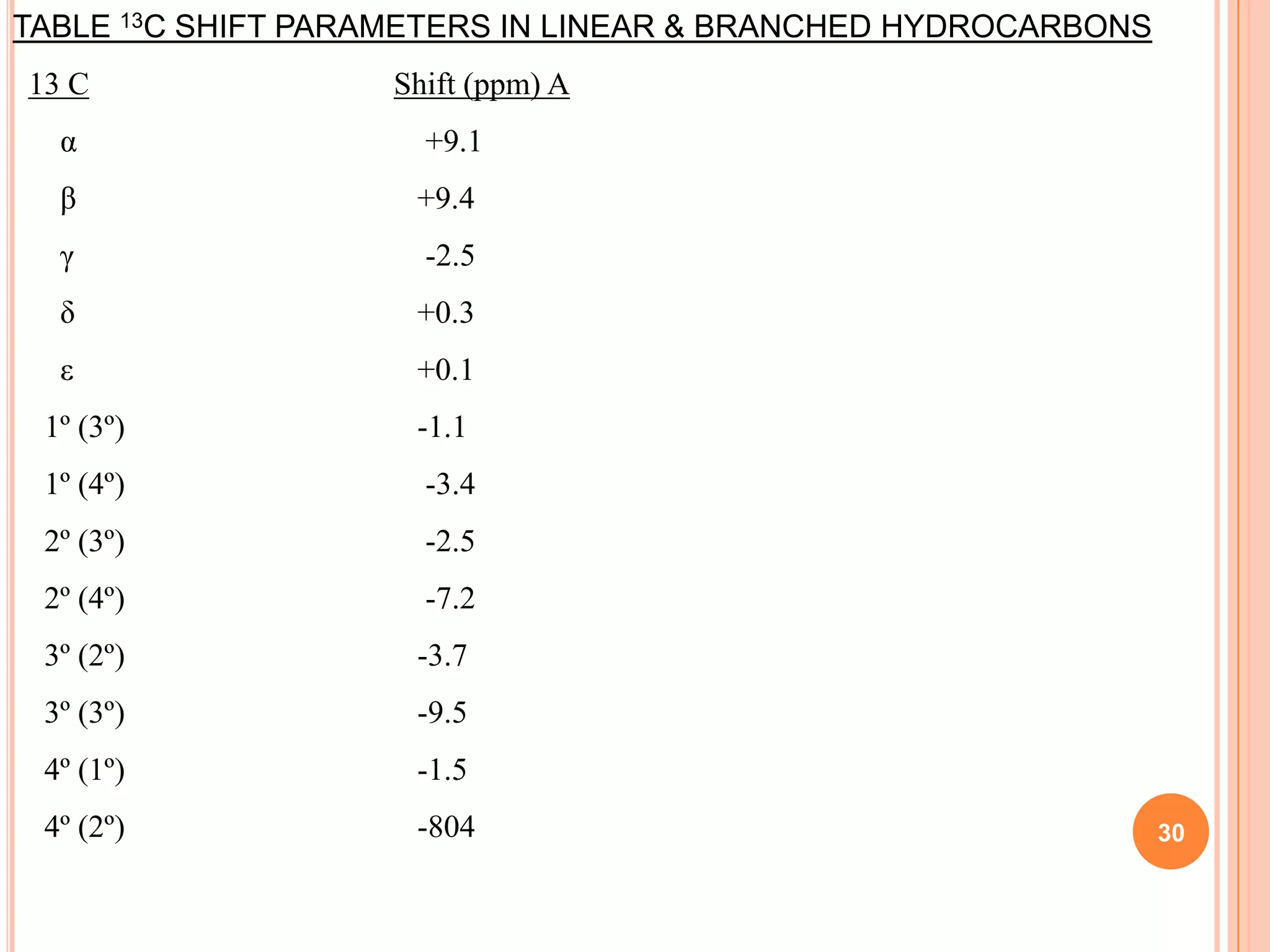
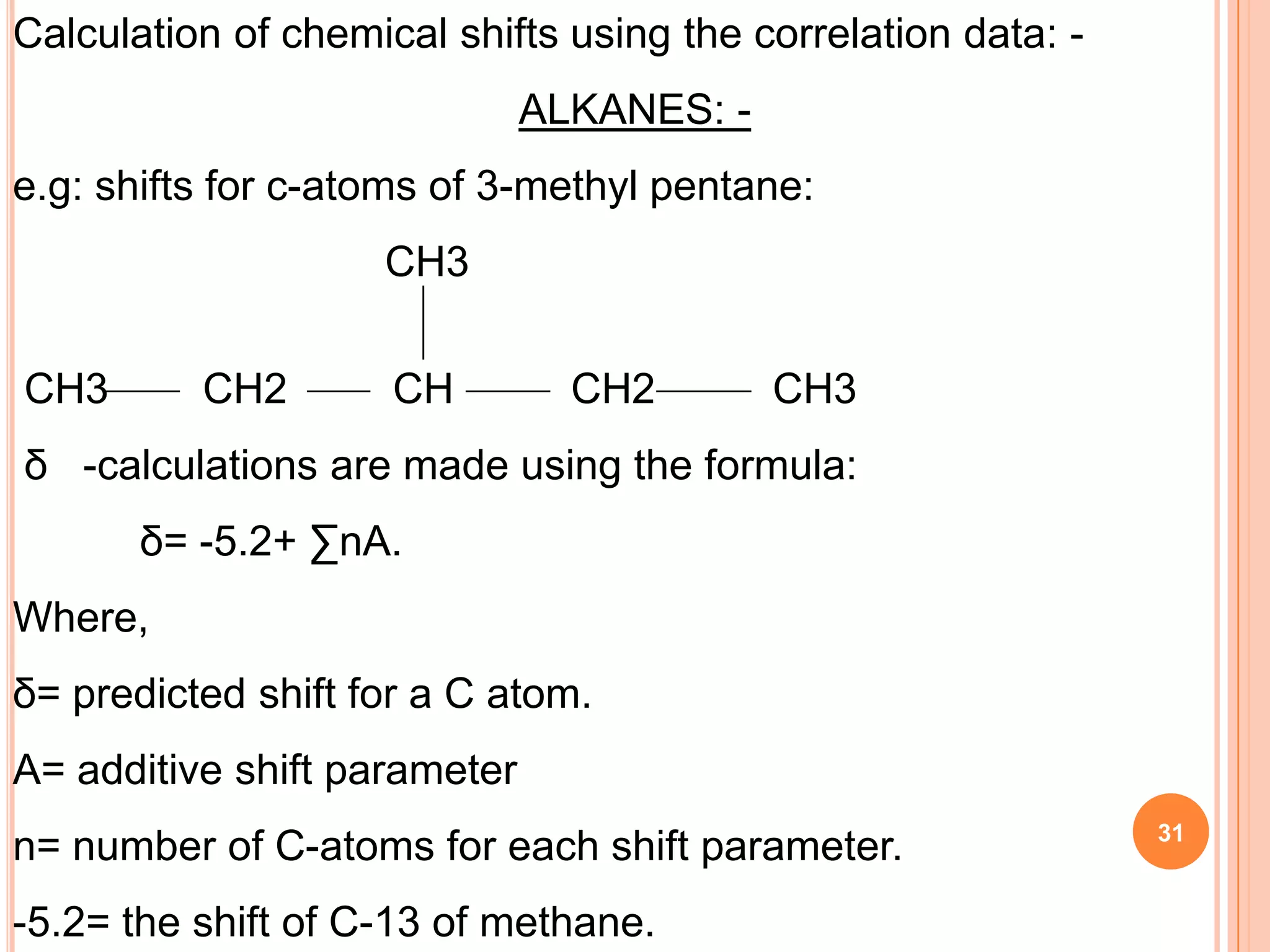
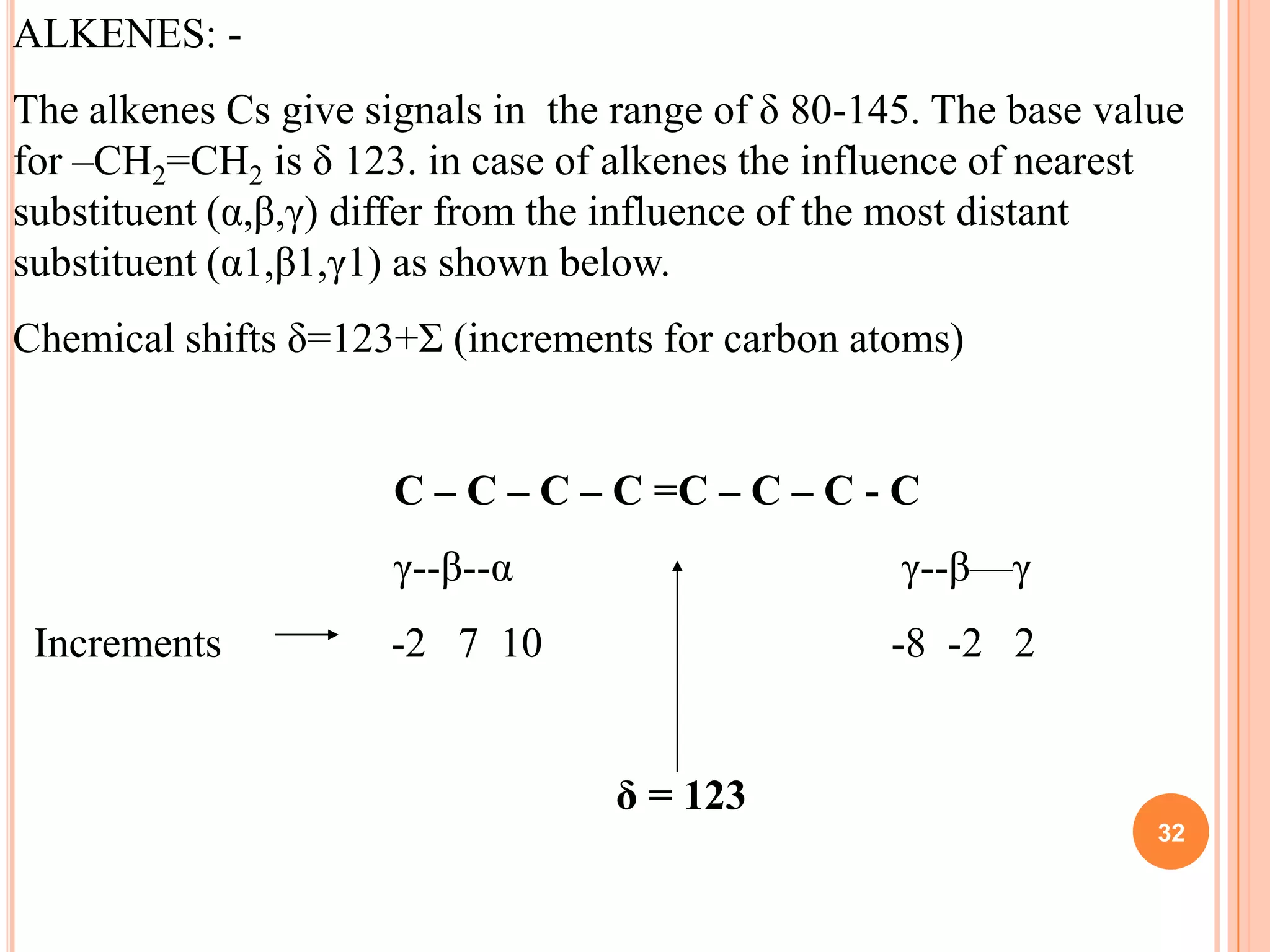
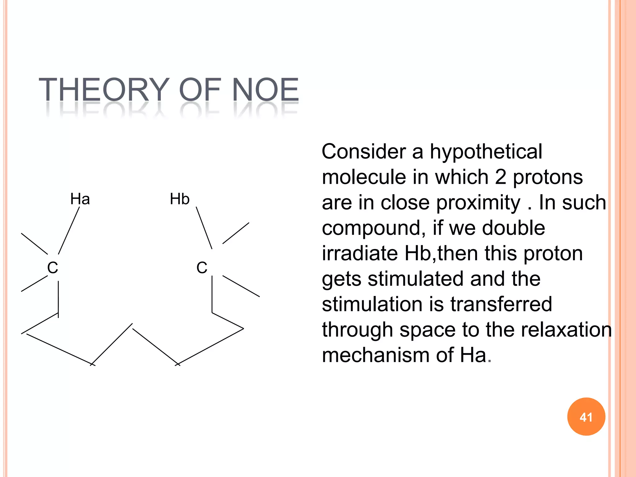
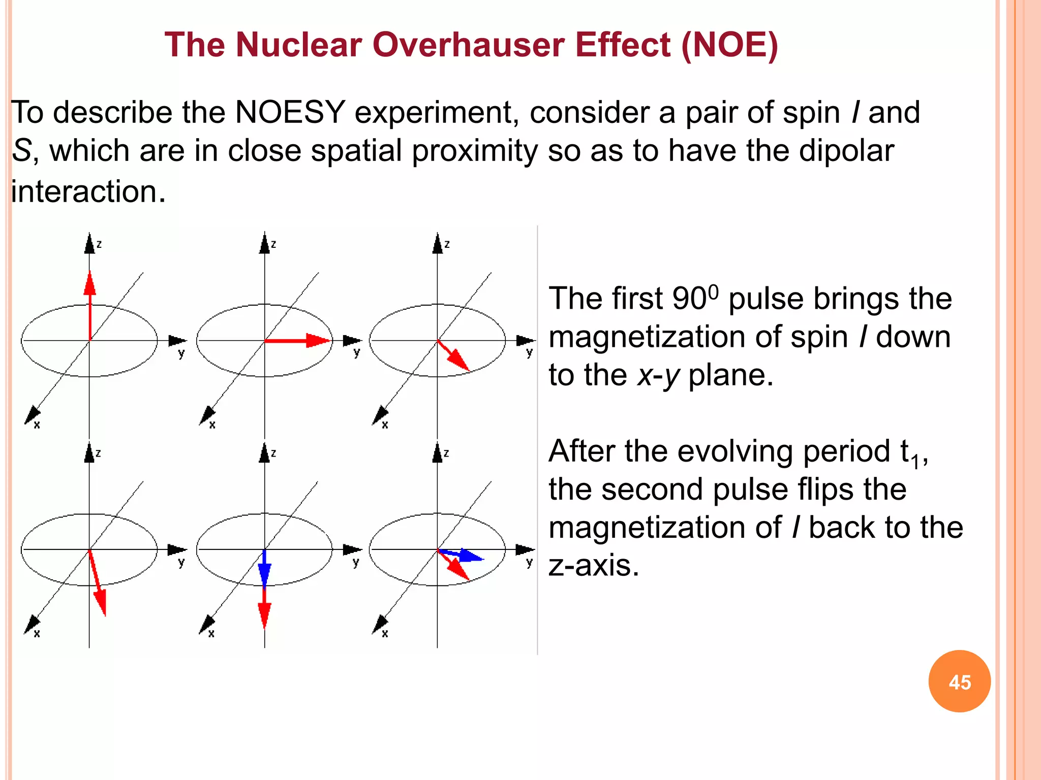
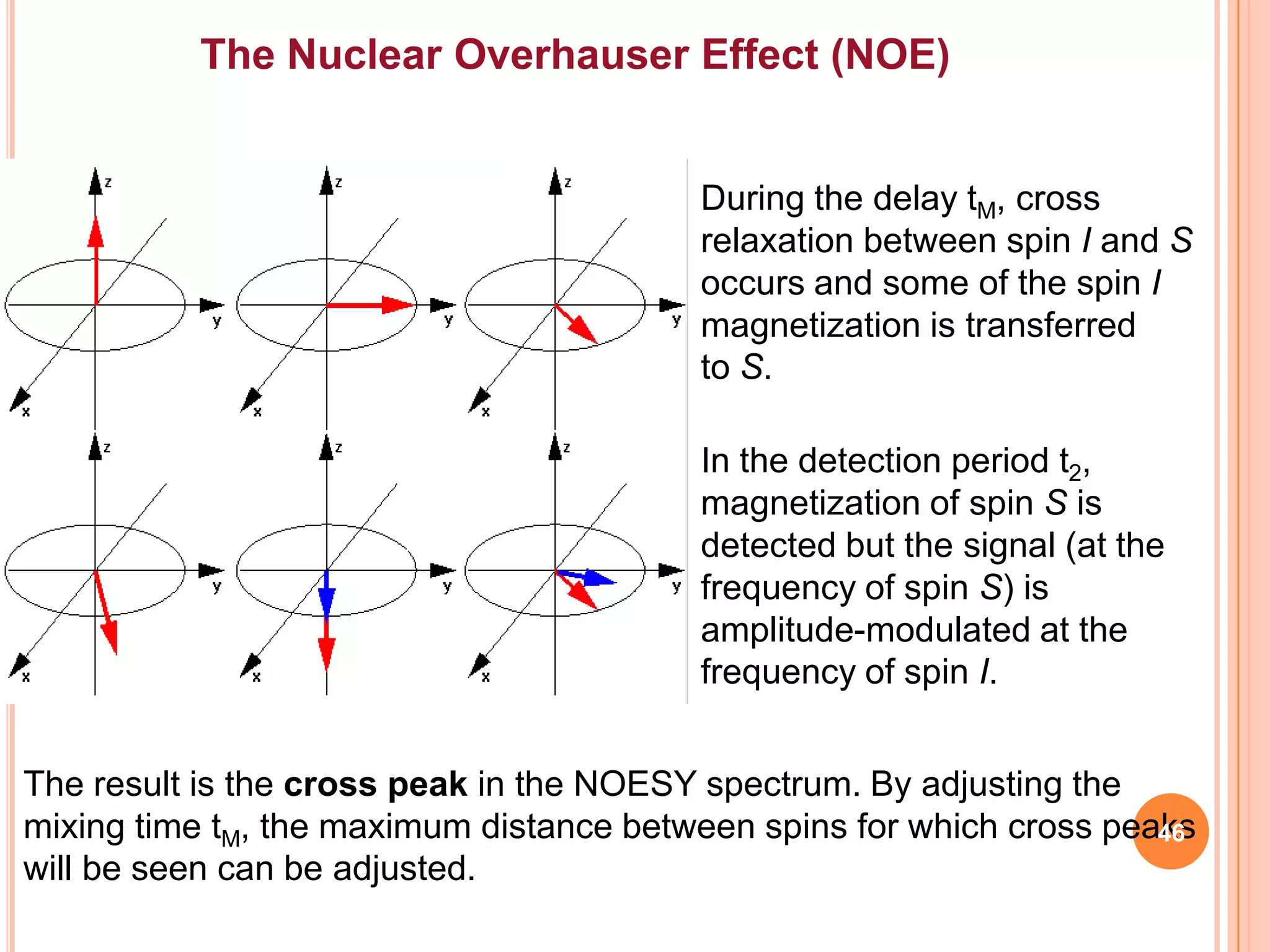
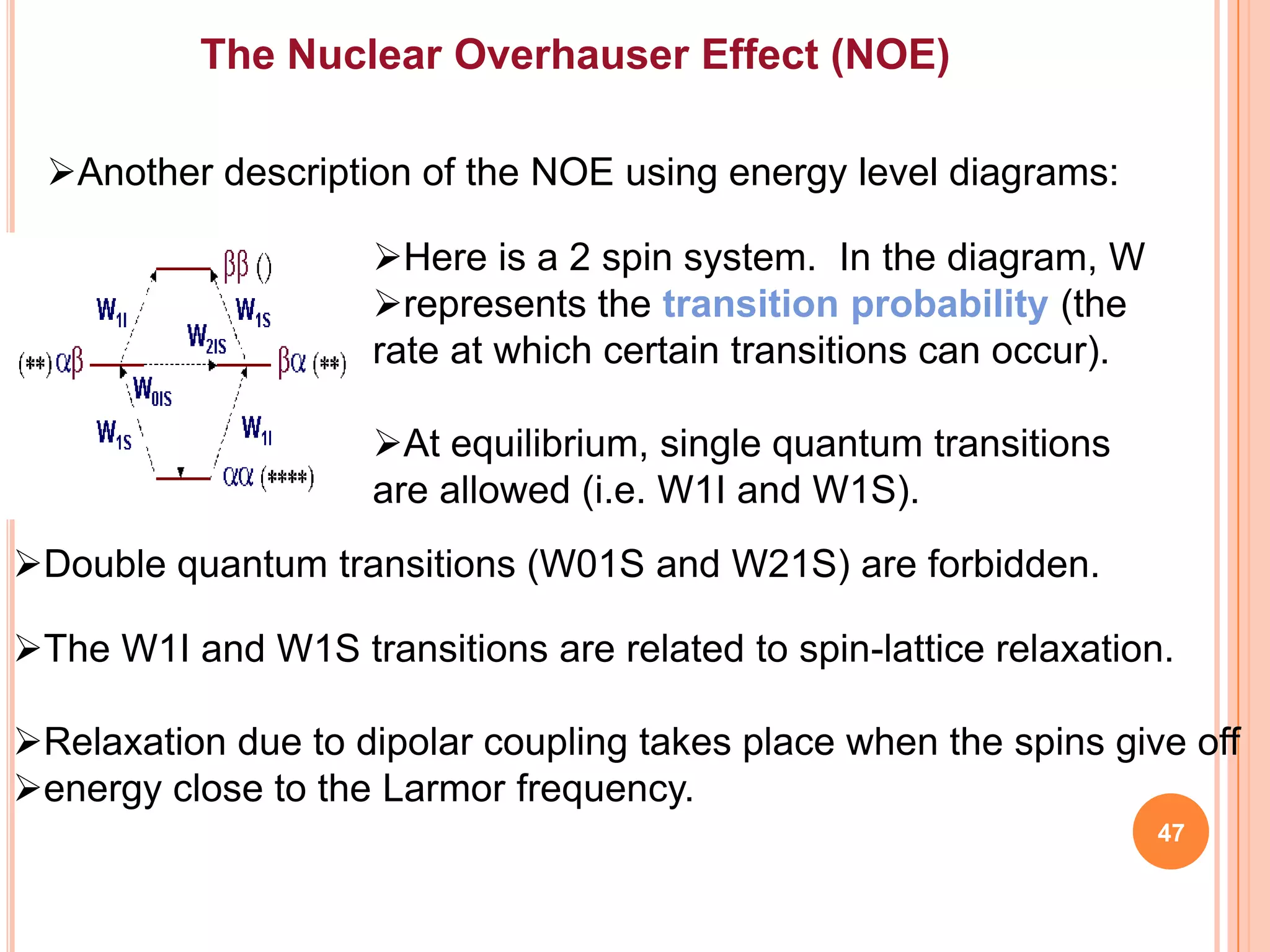
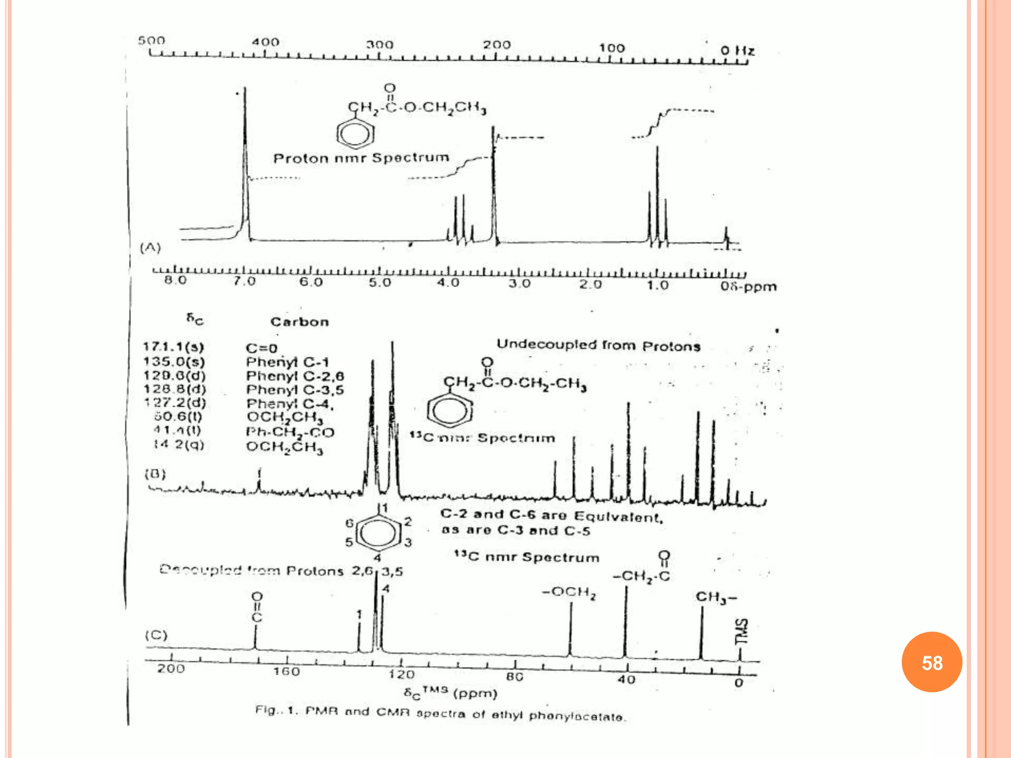
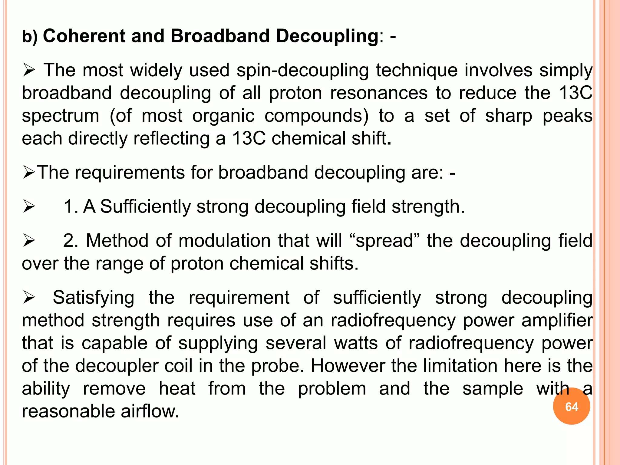
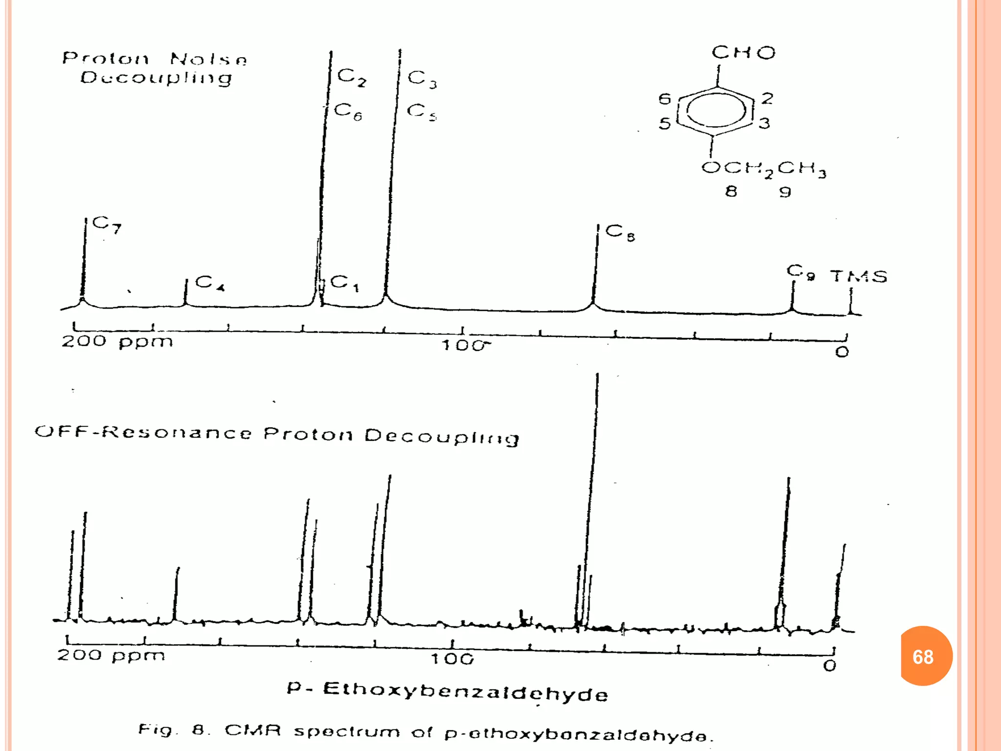
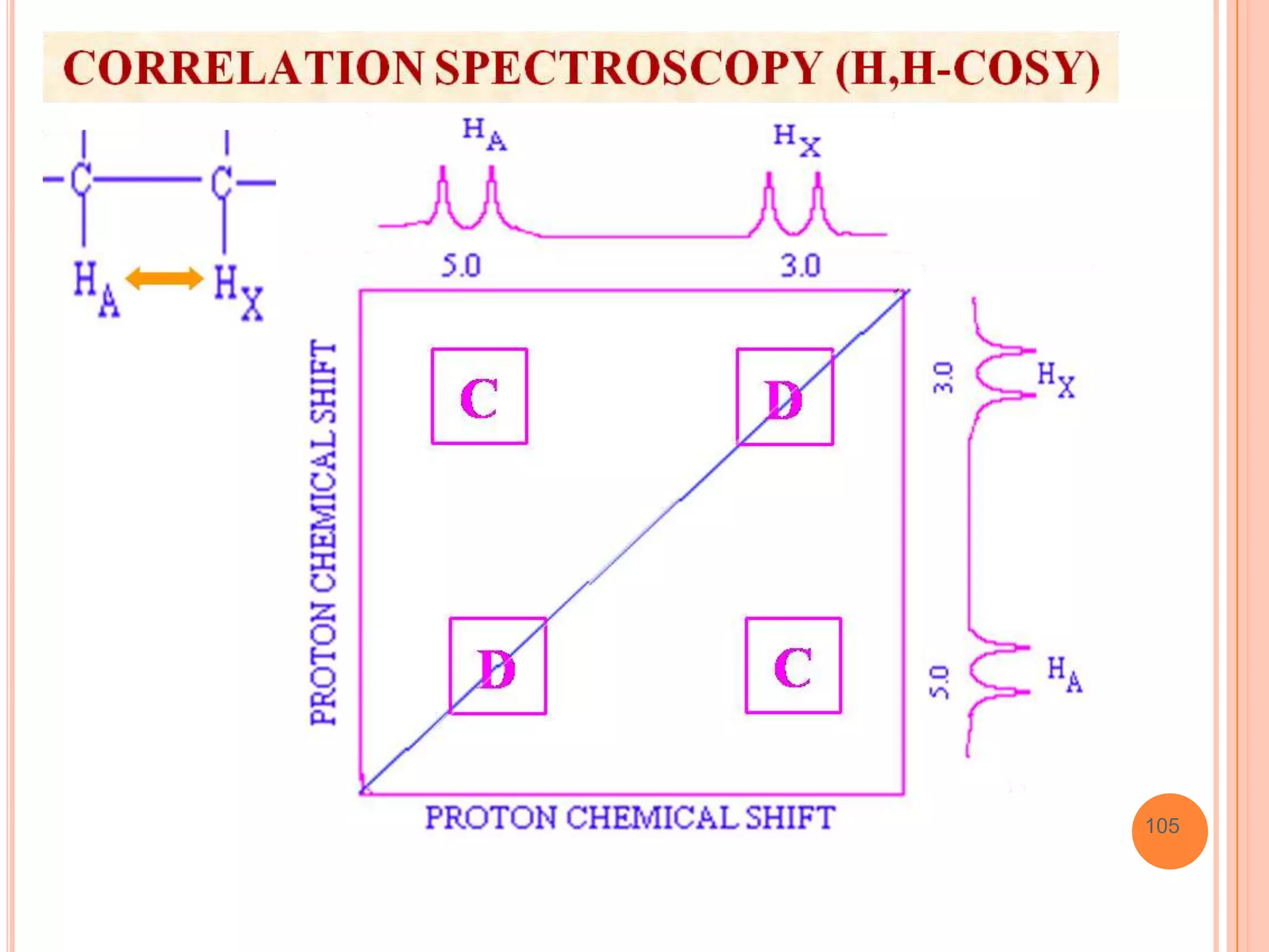
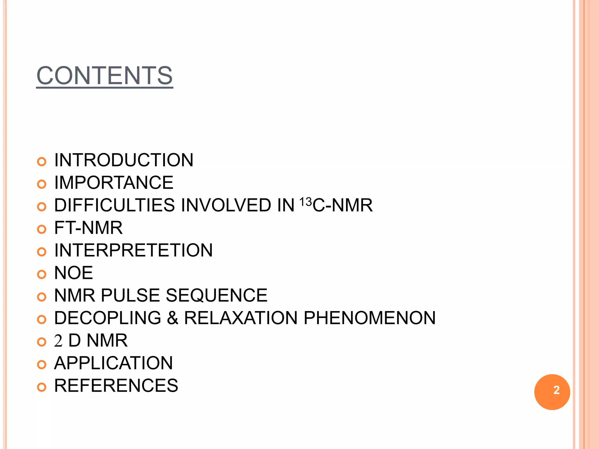
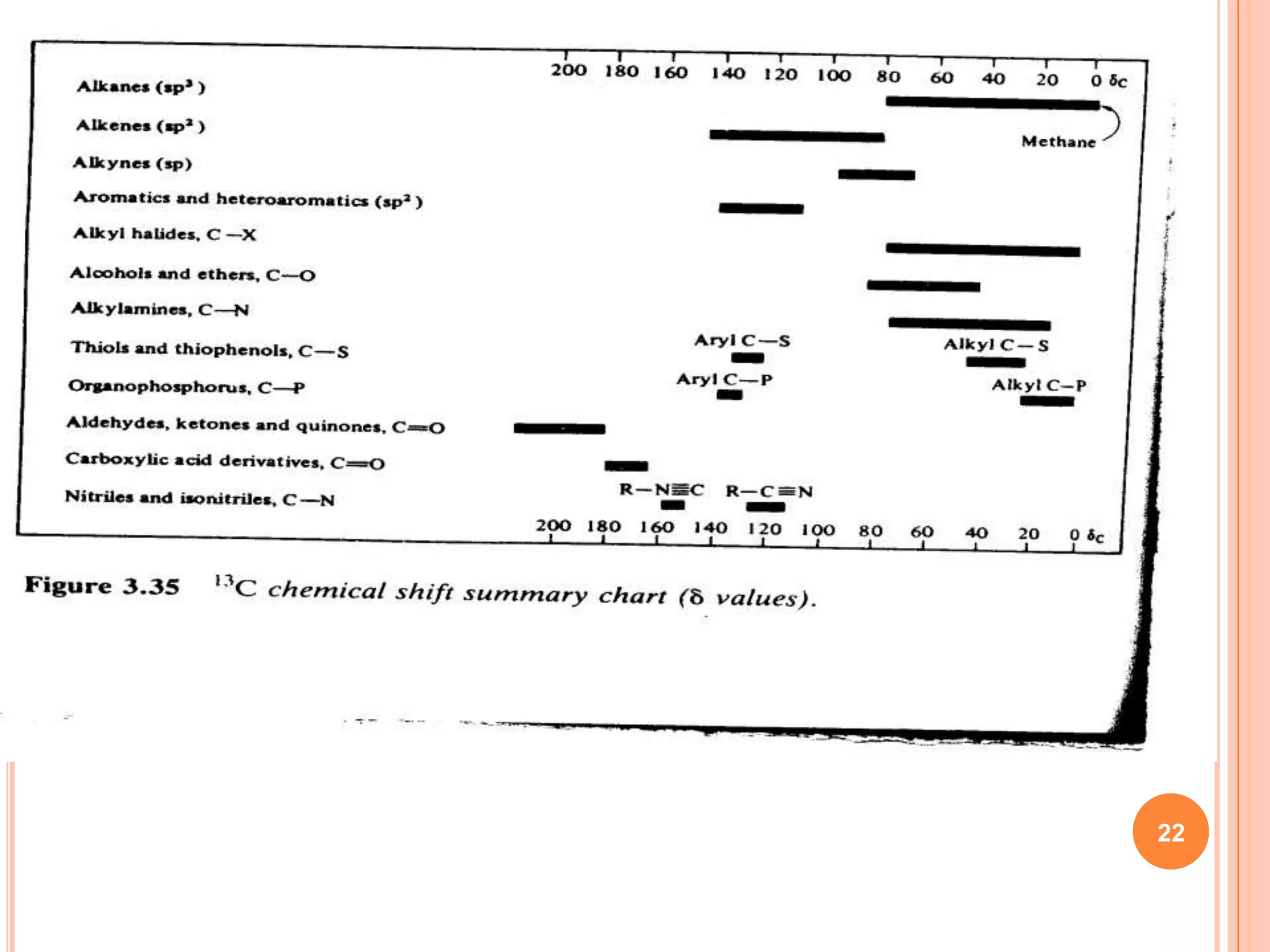
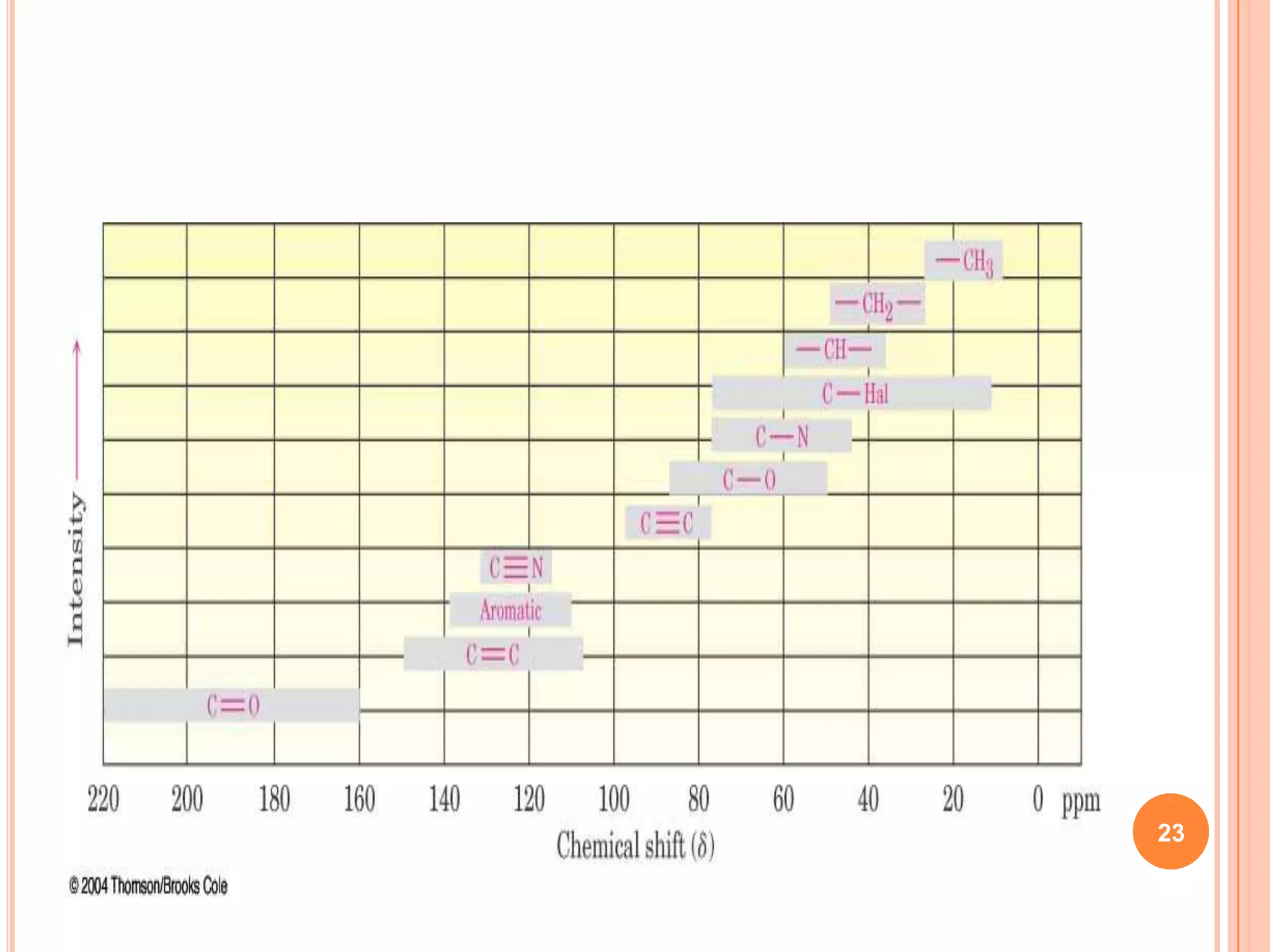
SlideShare a Scribd company logo
13C   NMR


    Presented by
    Pradip Ghori
1
CONTENTS


   INTRODUCTION
   IMPORTANCE
   DIFFICULTIES INVOLVED IN 13C-NMR
   FT-NMR
   INTERPRETETION
   NOE
   NMR PULSE SEQUENCE
   DECOPLING & RELAXATION PHENOMENON
   2 D NMR
   APPLICATION
   REFERENCES                          2
INTRODUCTION:-

   13C   is a natural, stable isotope of carbon.
   13C   NMR is analogous to proton NMR.
 NMR spectroscopy is based on the measurement of
 absorption of EMR in radiofrequency region of
 roughly 4 to 900 MHz with applied magnetic field.
 Nuclei of atoms are involved.
 NMR technique can be classified as
 PMR:- 1H NMR
 Isotopic NMR:- 12C NMR, 19F NMR, 31P NMR.         3
 The 13C-NMR spectra are recorded by the pulsed –FT-NMR
method, with the sensitivity enhanced by several spectra.
 The spin quantum number of 12C =zero, therefore it is non-
magnetic and hence does not give NMR signal.
 Both 13C and 1H have spin quantum number i.e. I = ½, so we
can expect to see coupling in the spectrum between:
   a)   13C   – 13C
   b)   13C   – 1H


                                                         4
 The probability of the two 13C atoms being together in the
same molecule is so low that 13C – 13C coupling are not
observed.
 Only 1.1% of carbon atoms in 13C are magnetic and these
nuclei split the protons in 13C-H groups into doublet and hence
the 13C-H coupling is seen in the spectra.
 However these couplings make the 13C spectra extremely
complex and can be eliminated by decoupling.
 The spectroscopy that is done using this nucleus 13C NMR
gives the information about the carbon chains in the compound.

                                                            5
This information is complimentary                   to   that
obtained from 1H NMR spectroscopy:-
   The number of signals tells us how many different carbons
or different sets of equivalent carbons are present in a
molecule.
 The splitting of a signal tells us how much hydrogen is
attached to each carbon.
 The chemical shift tells us the hybridization (sp3,sp2, & sp)
of each carbon.
 The chemical shift tells about the electronic environment of
each carbon with respect to other, nearby carbon or functional
groups.
                                                            6
Importance of 13C-NMR / why should 13C-NMR be
recorded when PNMR is present:

 13C NMR is a non-destructive and non-invasive
method.
13C NMR can be used in biological systems and
easy assessment of the metabolism of carbon and its
pathway.
 Chemical shift for 13C NMR ranges from (δ = 0-240)
when compared to proton NMR (δ =0-14). Since
chemical shift gives information regarding the physico-
chemical environment of compound, i.e. when
chemically closely related metabolites are under NMR
scan, they are often well separated and resolved to  7

obtain clearly identifiable spectra.
 As 13C nuclei have low abundance, thus tagging the specific
carbon position by selective 13C enrichment, thus 13C labeling
increases the signal intensities and often helps to trace the
cellular metabolism
 Labeling with 13C helps to know the fate of specific carbon
throughout the metabolism with out need for tedious isolation
and purification.
 The danger involved in using radioactive isotopes in tracing is
avoided as 13C nuclei are stable carbon isotope.
 Labeling at multiple carbon sites in the same molecule and
homonucleus 13C-13C spin coupling provides novel biochemical
information.
                                                              8
Difficulties of recording 13C spectra than 1H spectra are
because of the following reasons:
  1.   Natural low abundance of 13C .
  2.   Magnetic movement and Gyro magnetic ratio.
  3.   Chemical shift.
  4.   Decoupling phenomenon pronounced C-13 and H-1
       spin-spin interactions.


                                                       9
1. Natural abundance:-
    The natural abundance of 13C is only 1.1% that of
    12C, which is not detectable by NMR, that renders CMR less

    sensitive that PMR.
2. Magnetic movement and Gyro magnetic ratio: -
 The 13C nucleus has only a weak magnetic movement and
  consequently a small gyromagnetic ratio.
    Sensitivity of CMR is much reduced due to the presence of
    only 1.1% magnetic isotope (13C) in the sample.
   13C sensitivity is 1/4th that of C(Overall sensitivity of   13C

    compared with 1H is about 1/5700).
 The gyromagnetic ratio of      13C   is 1.4043 as compared to
                                                                10
  5.5854 of a proton 1H
Carbon-13 NMR
These factors show that   13C   CMR is much less sensitive than
PMR.
 The weak signals observed in 13C-CMR are therefore
scanned i.e., recorded routinely by Pulse-irradiation, signal-
summation and Fourier transforms.
 The low sensitivity of CMR is overcome by the use of large
samples, upon 2ml in 15mm tubes and by enhancement and
decoupling techniques in conjunction with highly stable
spectrometers operating at high fields


                                                             12
The problems arises during recording CMR can be readily
eliminated by adopting following methods:-
    a) Fourier transform technique
    b) NOE
    C) DECOUPLING


                                                          13
FT NMR to record the spectra:
It is a Fourier Transform NMR Spectroscopy.

TYPES OF FT NMR:
Multi-Dimensional:
The use of pulses of different shapes, frequencies and durations
in specifically designed patterns or pulse sequences allows the
spectroscopist to extract different types of information about the
molecule.
Multi-dimensional nuclear magnetic resonance spectroscopy is
a kind of FT-NMR in which there are at least two pulses and, as
the experiment is repeated, the pulse sequence is varied.
In multidimensional nuclear magnetic resonance there will be a
sequence of pulses and, at least, one variable time period. 14
In three dimensions, two time sequences will be varied. In four
dimensions three will be varied.
2-dimensional and multidimensional FT-NMR into a powerful
technique for studying biochemistry,in particular for the
determination of the structure of biopolymers such as proteins or
even small nucleic acids.
 Pulsed radiofrequency-fourier transforms NMR
spectroscopy:
The NMR spectrometer operates by exciting the nuclei of the
isotope under observation only one type at a time.
 In the case of 1H nuclei each distinct type of proton
(phenyl, methyl, and vinyl) is excited individually and its
resonance peak is observed and recorded independently of all
the others. Scanning is done individually until all types have come
                                                                15
into resonance.
FT NMR is an alternate approach to use powerful but short of
energy called pulse that excites all of the magnetic nuclei in the
molecule simultaneously for example, all of the 1H nuclei are
induced undergo resonance at the same time.
 An instrument with T magnetic fields uses a short burst of 90
MHz energy to accomplish this.
 The source is turned on and off very quickly and generates a
pulse.
 Similarly FT NMR operates in case of carbon also. 12C
nucleus is not magnetically active because spin number I=0 but
the 13C nucleus like the 1H nucleus has a spin number of ½,
however since the natural abundance of 13C is only about 1.1%
that of 12C and its sensitivity is only about 1.6% that of 1H, the
overall sensitivity of 13C compared with 1H is about 1/5700.   16
Pulsed FT NMR permits simultaneously irradiation of all 13C
nuclei and hence 13C spectra.
A pulse is a powerful but short burst of energy. According to
the variation of the Heisenberg Uncertainly principle, even
though the frequency of the oscillator generating this pulse is
set to 90 MHz, if the duration of the pulse is very short, the
frequency content of the pulse is uncertain because the
oscillator was on long enough to establish a solid fundamental
frequency.
 Therefore, the pulse actually contains a range of frequencies
centered about the fundamental. This range of frequencies is
great enough to excite all of the distinct types of the carbons in
the molecule at once with this single burst of energy.


                                                               17
     When pulse is discontinued, the excited nuclei begin to
lose their excitation energy and return to their original spin
state or relax.As each excited nucleus relaxes, it emits the
electromagnetic radiation. Since the molecule contains many
different nuclei, many different frequencies of the
electromagnetic radiation are emitted simultaneously, this
emission is called “free induction decay” signal.
The intensity of FID decays with time as all of the nuclei
eventually lose their excitation. This FID is complex and it is a
superimposed combination of all the frequencies emitted. The
individual frequencies due to different nuclei can be extracted
by using a computer and by Fourier transform analysis.

                                                              18
An Advantages of Fourier Transforms: -
FT NMR spectroscopy is one of the principal techniques used
to obtain physical, chemical, electronic and structural information
about a molecule. It is the only technique that can provide
detailed information on the exact three-dimensional structure of
biological molecules in solution. Also, FT nuclear magnetic
resonance is one of the techniques that have been used to build
elementary quantum computers.
 Fourier transform is more sensitive.
 It takes few seconds to measure FID.
 With computer and fast measurement, it is possible to repeat
and average the measurement of the FID signal.
 This is a real advantage when the sample is small in which19
case the FID is weak in intensity and has a great amount of
noise associated with
NOISE:-
Noise is random electronic signals that are usually visible as
fluctuations of the baseline in the signal. Since noise is
random; it normally cancels out of the spectrum after many
interactions of the spectrum are added together. Using this
procedure one can show that signal to noise ratio improves as
a function of the square root of the number of the scans, n.
     S/N     = f√ n
Therefore pulsed FT-NMR is especially suitable for
examination of the nuclei that are not very abundant in nature.
 Nuclei that is not strongly magnetic.
 Or very dilute sample
                                                            20
INTERPRETATION OF C-13 NMR SPECTRA: -


Chemical shifts in C-13 NMR Spectra: -
The range of shifts generally encountered in routine C-13
studies is about 240 ppm. Therefore C-13 chemical shifts
represent the spread of chemical shifts of about 12 times that
of the proton
 The peak assignment or chemical shifts in CMR are made
on the basis of reference compounds.


                                                            21
22
23
13C NMR spectrum of 2-Amino-5-(4-methylphenyl)-5H-thiazolo[4,3-b]-1,3,4-
thiadiazole (1b)


      H 3C
                         S


                     N

                 N             S


                         NH2


Molecular Formula: C11H11N3S2


                                                        CH3


                                                                           24
13C NMR spectrum of 2-(Alanyl)-Amino-5-(4-methylphenyl)-5H-thiazolo[4,3-b]-1,3,4-
thiadiazole (3c)


                  S


  H 3C                N          S

                      N

                                 NH

            3c        O

                                      CH3

                          H 2N


  Molecular Formula: C14H16N4OS2
                                                             CH3


            C=O                                         CH


                                                                                    25
13C   NMR spectrum of 2-(Alanyl)-Amino-5-(4-chlorophenyl)-1,3,4-thiadiazole (2a)


                             N
                                 N
             Cl
                             S       NH

                                 O
                        2a
                                     H 2N
           Molecular Formula: C11H11ClN4OS


                                                                CH3
                                                       CH
               C=O


                                                                                   26
13C   NMR spectrum of 5-(4-methylphenyl)-N-[(1E)-phenylmethylene][1,3]thiazolo[4,3-
                                  b][1,3,4]thiadiazol-2-amine (3d)


                       S

    H 3C               N      S
                       N
                  3d          N


M olecular F orm ula   = C1 8H 1 5N 3S 2


                                                                    CH3


                                                               CH


                                                                                          27
13C   NMR spectrum of 3-chloro-1-[5-(4-methylphenyl)[1,3]thiazolo[4,3-b][1,3,4]thiadiazol-2-yl]-4-
                                  phenylazetidin-2-one (4d)


                          S
                                       S
                               N               O

                                   N       N

                                                   Cl
           H 3C
                          4d

                                                                           CH3
      M olecular F orm ula = C 20 H 16 C lN 3 O S 2


                                                                   CH-Cl

                     C=O

                                                                 CH


                                                                                            28
Factors Influencing Chemical Shifts :-
 Shifts are mainly related to hybridization and substituent
electronegativity. Solvent effects are also very important as in
proton 1H spectra.
 Chemical shifts for 13C are affected by substituents as far
removed as the δ position. Pronounced shifts for 13C are
caused by substituents at the ortho, Meta, and para positions in
the benzene ring.
 Steric compression causes 13C chemical shifts to move up
field significantly.
 Up field shifts my also occur on dilution.
 Hydrogen bonding effects may cause downfield especially
with polar solvents.
                                                               29
TABLE 13C SHIFT PARAMETERS IN LINEAR & BRANCHED HYDROCARBONS
13 C                Shift (ppm) A
  α                   +9.1
  β                  +9.4
  γ                   -2.5
  δ                  +0.3
  ε                  +0.1
 1º (3º)             -1.1
 1º (4º)              -3.4
 2º (3º)              -2.5
 2º (4º)              -7.2
 3º (2º)             -3.7
 3º (3º)             -9.5
 4º (1º)             -1.5
 4º (2º)             -804                                      30
Calculation of chemical shifts using the correlation data: -
                              ALKANES: -
e.g: shifts for c-atoms of 3-methyl pentane:
                    CH3


CH3       CH2        CH         CH2        CH3
δ -calculations are made using the formula:
       δ= -5.2+ ∑nA.
Where,
δ= predicted shift for a C atom.
A= additive shift parameter
                                                               31
n= number of C-atoms for each shift parameter.
-5.2= the shift of C-13 of methane.
ALKENES: -
The alkenes Cs give signals in the range of δ 80-145. The base value
for –CH2=CH2 is δ 123. in case of alkenes the influence of nearest
substituent (α,β,γ) differ from the influence of the most distant
substituent (α1,β1,γ1) as shown below.
Chemical shifts δ=123+Σ (increments for carbon atoms)


                      C – C – C – C =C – C – C - C
                      γ--β--α                      γ--β—γ
 Increments          -2 7 10                      -8 -2 2


                                   δ = 123
                                                                  32
E.g.: predict the 13C-chemical shift values for the alkenes in 2-
pentene
CH2-CH=CH-CH2-CH3                     base value : δ = 123
C2 1α,1α’,1β’                        C3 1α,1β, 1α’
δ =123+10-8-2=123.                    δ 123+10+7-8=132.
ALKYNES: -
E.g.: - HC ≡ C - O - CH2 - CH3;       H3C - C ≡ C - O - CH3


      23.2    89.4                       28.0   88.4


               Base value to HC ≡ CH is δ =72.
                                                                    33
Influence of Functional Group substituents on Alkene &
Aromatic Chemical Shifts:-
Alkene δ values will be affected by substitution at points
further along the carbon chain (as in allyl alcohol CH2 = CH-
CH2-OH). But, systematic correlations have not been
compiled. However the major influence on the alkene Cs will
be the direct substituent (in case of allyl alcohol, it is the –
CH2 group). So we can say, if a substituent is identifiable as
–CH2X, it should simply be treated as –CH3.
The same principles hold good in predicting the shifts in the
aromatic δ values. The deviation from predicted values is
often due to or associated with H- bonding and steric effects.


                                                               34
E.g.: - compounds related to salicylic acid show such
deviation because of the strong intramolecular H-bonding
between the –OH group and the ortho-carbonyl group.


                                                           35
Using the correlation data:


  BASE             ISOPROPYL(R)       NITRO        TOTAL
C1     128            +21             +1             150
C2     128             0               -5            123
C3     128             0             +20             148
C4     128             -2              -5            121
C5     128             0               +1            129
C6     128             0               +6            134


To predict the δ values of isopropyl group carbon atoms the benzene
ring is considered to be the substituent isopropyl as alkane.
                                                                  36
CARBONYL GROUP CHEMICAL SHIFTS:
Major strength of C-13 NMR is the ability to observe the NMR
characteristics of carbonyl C directly. The carbonyl resonance
is at very high frequency and also, different classes appear
within narrow ranges (advantage). So that quite fine
distinctions can be made, in the knowledge that, the influences
of unaccounted factors will be minimal.
Introduction of alkyl group on the Cs directly attached to- CO
usually shifts the –CO signal by 2-3 ppm. conjugation with –
CO group causes –CO resonance’s shift upfield (lower
frequency). The anions of carboxylic acids are not much
shifted in range from the free acids, inspite of the fact that the
C-O bonding in carboxylate anions is weaker than the true
C=O bond in acid. This theory fails to offer a convincing
explanation.
                                                              37
READING THE C-13 SPECTRUM:
The first steps in deducing the structure of an organic
compound, using the C-13 NMR spectrum are; -
 Count the number of signals in the spectrum; tis is the
number of non-equivalent C environments in the molecule.
(Identify and discount the signals from solvent).
 Use figure (δ values table) to assign signals approximately
the regions δ 0-80, δ 80-150 and δ 160-220(carbonyl carbons).
 Note the intensities of the peaks: non-proton bearing Cs
give lower intensity signals, and groups of two or more
equivalent Cs give higher intensity signals.
 Take account of any multiplicity into (q, t, d or s).
 Use the correlation tables to predict the chemical shifts of all
                                                                38
Cs inn each putative structure.
Use of Correlation Tables: -
Two principle predictable influences that we can quantify in
determining the chemical shift positions of a C atom:
 1. The number of other carbon atoms attached to it (and
whether these are CH3, CH2, CH, and C groups).
 2. Natural of all other substituents attached (or nearby along a
chain of other C atoms). But it is important to compute 1 before 2.
LIMITATIONS OF 13C NMR STUDIES: -
 Sensitivity of C-NMR compared to PMR, chromatography,
spectrophotometry, radiochemical studies, etc. is poor.
 Limitation factors of C-NMR like intrinsically low sensitivity of
magnetic resonance techniques, low gyromagnetic ratio of 13C
and low natural abundance of 13C.
                                                                39
NUCLEAR OVER HAUSER EFFECT (NOE):
In NMR spectroscopy, changes brought about in the energy
populations of one nucleus by the decoupling of a neighboring
nucleus are named the Nuclear Overhauser Effect of (NOE).
 Two conditions that always apply to NOE are
 It arises only during the double irradiation of one nucleus, and
affects another nucleus which must be close but not necessarily
coupled with the irradiated nucleus.
 It is associated with dipolar relaxation mechanisms.Maximum
NOE operates on CH3, CH2 and CH carbons, whereas no
enhancement arises for 4 carbons (includes carbons on aromatic
rings with substituent's attached.)
                                                              40
THEORY OF NOE
                  Consider a hypothetical
                  molecule in which 2 protons
    Ha   Hb       are in close proximity . In such
                  compound, if we double
                  irradiate Hb,then this proton
C             C
                  gets stimulated and the
                  stimulation is transferred
                  through space to the relaxation
                  mechanism of Ha.

                                              41
Thus, due to increase in spin lattice relaxation of Ha, its
signal will appear more intense by 15 to 50%. So, if the
intensity of absorption of Ha signal is increased by double
irradiating Hb, then protons Ha and Hb must be in close
proximity in a molecule


                                                              42
The Nuclear Overhauser Effect (NOE)
The carbon-13 spectrum from CH3I.

 The NMR spectrum from the carbon-13 nucleus will
 yield one absorption peak in the spectrum.
In reality, we see a single line with a relative intensity of 24.

 Adding the nuclear spin from one hydrogen
 will split the carbon-13 peak into two peaks.

 Adding one more hydrogen will split each of
 the two carbon-13 peaks into two, giving a
 1:2:1 ratio.

  The final hydrogen will split each of the previous
  peaks, giving a 1:3:3:1 ratio.
If the hydrogen spin system is saturated, the four lines collapse into a
single line having an intensity which is eight times greater than the outer
                                                                          43
peak in the 1:3:3:1 quartet since 1+3+3+1=8 .
The Nuclear Overhauser Effect (NOE)

If the hydrogen spin system is saturated, the four lines collapse into a
single line having an intensity which is eight times greater than the outer
peak in the 1:3:3:1 quartet since 1+3+3+1=8 .
In reality, we see a single line with a relative intensity of 24.

This is because of the Nuclear Overhauser Effect (NOE).

The NOE is one of the ways that spin system can release energy.

Magnetization transfer between spins is mediated by
dipolar coupling.


                                                                          44
The Nuclear Overhauser Effect (NOE)
To describe the NOESY experiment, consider a pair of spin I and
S, which are in close spatial proximity so as to have the dipolar
interaction.


                                      The first 900 pulse brings the
                                      magnetization of spin I down
                                      to the x-y plane.

                                      After the evolving period t1,
                                      the second pulse flips the
                                      magnetization of I back to the
                                      z-axis.


                                                                    45
The Nuclear Overhauser Effect (NOE)


                                         During the delay tM, cross
                                         relaxation between spin I and S
                                         occurs and some of the spin I
                                         magnetization is transferred
                                         to S.

                                         In the detection period t2,
                                         magnetization of spin S is
                                         detected but the signal (at the
                                         frequency of spin S) is
                                         amplitude-modulated at the
                                         frequency of spin I.


The result is the cross peak in the NOESY spectrum. By adjusting the
mixing time tM, the maximum distance between spins for which cross peaks
                                                                      46
will be seen can be adjusted.
The Nuclear Overhauser Effect (NOE)

 Another description of the NOE using energy level diagrams:

                     Here is a 2 spin system. In the diagram, W
                     represents the transition probability (the
                     rate at which certain transitions can occur).

                     At equilibrium, single quantum transitions
                     are allowed (i.e. W1I and W1S).
Double quantum transitions (W01S and W21S) are forbidden.

The W1I and W1S transitions are related to spin-lattice relaxation.

Relaxation due to dipolar coupling takes place when the spins give off
energy close to the Larmor frequency.
                                                                     47
In pulse acquire experiment the x and y components of the free
induction signal
could be computed by thinking about the evolution of the
magnetization
during the acquisition time. we assumed that the magnetization
started out along the −y axis as this is where it would be rotated to
by
a 90◦ pulse. For the purpose we are going to assume that the
magnetization starts out along x; we will see later that this choice of
starting
position is essentially arbitrary.


                                                                   48
From fig we can easily see that the x and y
components of the magnetization are:

.The signal that we detect is proportional to these magnetizations. The
constant
of proportion depends on all sorts of instrumental factors which need not
concern us here; we will simply write the detected x and y signals, Sx (t ) and


where S0 gives is the overall size of the signal and we have reminded
ourselves
that the signal is a function of time by writing it as Sx (t ) etc.
It is convenient to think of this signal as arising from a vector of length
S0
rotating at frequency ; the x and y components of the vector give
Sx and Sy,
as is illustrated in Fig. 4.3.                                          49
50
NMR Pulse Sequences
The 90o-FID Sequence

In the 90-FID pulse sequence, net
magnetization is rotated down into the
X'Y' plane with a 90o pulse.


The net magnetization vector begins to
precess about the +Z axis.


The magnitude of the vector also
decays with time.


                                             51
NMR Pulse Sequences
                     The 90o-FID Sequence
A timing diagram is a multiple axis plot of some aspect of a pulse
sequence versus time. A timing diagram for a 90-FID pulse sequence
has a plot of RF energy versus time and another for signal versus
time.
                       When this sequence is repeated, for example
                       when signal-to-noise improvement is needed,
                       the amplitude of the signal (S) will depend on
                       T1 and the time between repetitions, called
                       the repetition time (TR), of the sequence.


In the signal equation below, k is a proportionality constant and   is the
density of spins in the sample.

                          S=k     ( 1 - e-TR/T1 )
                                                                     52
NMR Pulse Sequences
The Spin-Echo Sequence

In the spin-echo pulse sequence, a 90o
pulse is first applied to the spin
system.

The 90o degree pulse rotates the
magnetization down into the X'Y' plane.
The transverse magnetization begins to
dephase.

At some point in time after the 90o pulse,
a 180o pulse is applied. This pulse rotates
the magnetization by 180o about the X'
axis.

The 180o pulse causes the magnetization        53
to at least partially rephase and to produce
a signal called an echo.
NMR Pulse Sequences
 The Spin-Echo Sequence


A timing diagram shows the relative positions of the two radio
frequency pulses and the signal.

The signal equation for a repeated spin echo sequence as a
function of the repetition time, TR, and the echo time (TE) defined
as the time between the 90o pulse

                                                                      54
NMR Pulse Sequences

The Inversion Recovery Sequence
In this sequence, a 180o pulse is first
applied. This rotates the net
magnetization down to the -Z axis.
The magnetization undergoes
spin-lattice relaxation and returns
toward its equilibrium position along
the +Z axis.
Before it reaches equilibrium, a 90o
pulse is applied which rotates the
longitudinal magnetization into the
XY plane. In this example, the 90o
pulse is applied shortly after the        55

180o pulse.
NMR Pulse Sequences

  The Inversion Recovery Sequence
Once magnetization is present in the XY plane it rotates about
the Z axis and dephases giving a FID.

The timing diagram shows the relative positions of the two
radio frequency pulses and the signal.


                                                             56
Decoupling phenomenon/spin-decoupling methods:
 Non decoupled (proton coupled) 13C spectra usually show
complex overlapping multiplets that are very difficult to
interpret, but some spectra are simple and can be interpreted
easily.


                                                           57
58
Various decoupling methods are as follows: -
a) Multiplicity & Proton (1H) Decoupling- Noise
  Decoupling.
b) Coherent & Broadband Decoupling.
c) Off-Resonance Decoupling.
d) Selective Proton Decoupling.


                                                  59
a) Multiplicity & Proton (1H) Decoupling- Noise Decoupling: -
Both 13C & 1H have I=1/2, so that we would expect to see
coupling in the spectrum between
   a) 13C-13C
   b) 13C-1H
 However the probability of 2 C13 atoms being together in the
same molecule is so low that 13C-13C couplings are not usually
observed.
The complicating effects of proton coupling in 13C spectra i.e.,
in 13C-1H coupling can be eliminated by decoupling the 1H nuclei
by double irradiation at their resonant frequencies. this is an
example of Heterounuclear De-coupling.

                                                              60
Here specific protons are not decoupled but all
protons are simultaneously decoupled by double
irradiation while recording the 13 C spectrum. A
decoupling signal is used that has all the 1H
frequencies spread around 80-100 Hz & is therefore a
form of radio frequency noise. Spectra derived thus are
1H decoupled or nose decoupled.
The proton-decoupled spectrum is recorded by
irradiating the sample at 2 frequencies.
 The First radio frequency signal is used to effect
carbon magnetic resonance (CMR), while simultaneous
exposure to the second signal causes all the protons in
resonance at the same time and flip their α & β spins
very fast.
                                                          61
In the noise decoupled spectrum of diethyl phthalate:-


                                                         62
63
b) Coherent and Broadband Decoupling: -
 The most widely used spin-decoupling technique involves simply
broadband decoupling of all proton resonances to reduce the 13C
spectrum (of most organic compounds) to a set of sharp peaks
each directly reflecting a 13C chemical shift.
The requirements for broadband decoupling are: -
    1. A Sufficiently strong decoupling field strength.
 2. Method of modulation that will “spread” the decoupling field
over the range of proton chemical shifts.
 Satisfying the requirement of sufficiently strong decoupling
method strength requires use of an radiofrequency power amplifier
that is capable of supplying several watts of radiofrequency power
of the decoupler coil in the probe. However the limitation here is the
ability remove heat from the problem and the sample with a
                                                                   64
reasonable airflow.
Here the decoupling frequency is phrase modulated with a
50% duty cycle, 100Hz square wave. Residual broadening of
decoupled off-resonance 13C peaks is significantly reduced
using this method in comparison to the former method. This
method is now being widely used in broadband (1H)
decoupling.


                                                       65
C) Off Resonance Decoupling: -
 The off-resonance coupling not only simplifies the spectrum but also
retains the residual 13C-H coupling information.
This is a deliberately inefficient double irradiation of the proton
frequencies.
 The decoupler is offset by 1000-2000Hz upfield or about 2000-3000Hz
downfield from the frequency of TMS without using the nose generator.
 In off-resonance decoupling, while recording the CMR spectrum, the
sample is irradiated at a frequency close (but not identical) to the
resonance frequency of protons.
 Consequently, the multiples become narrow and not removed
altogether as in fully decoupled spectra i.e., the weak C-H coupling are
decoupled and strong couplings remain though somewhat distorted.


                                                                     66
The residuals coupling constant Jr is < true J.
        Jr =2 J    /γB2
  = difference between decoupled frequencies and
Resonance frequencies of 1H of interest.
J = true coupling constant.
B2= strength of rotating magnetic field generated by the
decoupler frequencies.
 = gyro-magnetic ratio.


                                                           67
68
d) Selective Proton Decoupling: -
•When a specific proton is irradiated at its exact frequency at a lower
power level than is used for off-resonance decoupling, the absorbance of
the directly bonded 13C becomes a singlet, while the other 13C
absorptions show residual coupling.
                     DEUTERIUM COUPLING: -
The number of orientations, which any magnetic nucleus can adopt in
magnetic field, is (2I+1). I = spin quantum number.
 Thus for 1H & 13C where, I = ½,2 orientations arise either +I or –I. But for
deuterium whose I = 1, 3 orientations arise:
 a) Aligned with the magnetic field most stable will augment Bo
 b)Across the field on a plane
       Deuterium nucleus is précessing on a plane cutting across Bo
(magnetic field) & will not change field strength (1H frequencies
unchanged).
                                                                         69
c) Antiparallel/non-aligned with the Bo    least stable         will diminish Bo
proton frequency reduced


         (a)                         (b)                  (c)


                                                                                   70
Protons coupled with one deuterium nucleus come to resonance at
three different frequencies i.e., the 1H signal appears as a triplet; the
line separation correspond to JH-D .
 If a group of protons signal is coupled to more than one
Deuterium then the Multiplicity of the proton signal is found from the
general formula (2nI=1).
 Thus two (equal) deuterium couplings give rise to quintets, &
three deuterium gives septets & so on.
 Deuteriated solvents (deuteriochloroform CDCL3, deuteriobenzene
C6D6, deuterioacetone CD3COCD3 , or dexadeuteriodimethyl
sulphoxide CD3SOCD3 ) give rise to 13C signals, which are split by
coupling to deuterium.
 Thus in molecules with one deuteron attached to each carbon (as
in CDCL3 & C6D6) the C-13 signal form the solvent are a 1:1:1
triplet. For CD3 groups (CD3COCB, CD3SOCD3 ), the solvent gives
                                                                71
rise to a septet with line intensities 1:3:6:7:6:3:1.
72
Relaxation Phenomenon: -

  What happens when protons absorb energy?
 Nuclei in the lower energy state undergo, transitions to
the higher energy state; the populations of the tow states may
approach equality, and if this arises, no further net absorption
of energy can occur and the observed resonance signal will
fade out saturation of the signal.
However, during a normal NMR run, the populations in the 2
spin states do not become equal, because higher E nuclei are
constantly returning to the lower energy spin state

                                                             73
E

    Opposed


    aligned


              Bo   74
How do the nuclear lose energy and undergo
transition from the high to the low-energy state?
 The energy difference E can be re-emitted as
radio frequency E that is monitored by a radio frequency
detector as evidence of resonance condition having been
reached.
However 2 important radiation-less processed exist,
which enable high-energy nuclei to lose energy.
 Spin-Lattice Relaxation
 Spin-Spin Relaxation
                                                           75
1) Spin-Lattice Relaxation
       The high energy nuclear can undergo energy loss (or
relaxation) by transferring E to some electromagnetic vector
present in the surrounding environment e.g.: a nearby solvent
molecule undergoing continuous vibration and rotational
changes, will have associated electrical and magnetic
changes, which might just be properly oriented and of the
correct dimension to absorb E. since the nuclear may be
surrounded by a whole array of neighboring atoms either in
the same molecule or in solvent molecules, etc., this
relaxation process is termed spin-lattice relaxation.


                                                          76
2) Spin-Spin Relaxation: -
 A 2nd relaxation process involves transferring E to c neighboring
nucleus, provided that the particular value of E is common to both nuclei
this mutual exchange of spin energy is termed spin-spin relaxation. While
one nucleus loses energy, the other nucleus gains energy, so that no net
change in the population of the 2 spin states is involved.
Relaxation phenomenon in terms of magnetization and vectors:-
                                        Aligned with the field
One nucleus is an      either
applied either field                          or
precesses
                                        Opposed to the field


                                                                      77
When the system of nuclear spins relaxes, two different
processes are identified:
(a) the reduced z-axis component eventually increases
back to Mo
(b) the y-axis component reduces to zero.


                                                           78
APPLICATIONS
 13C-NMR is mainly used to study the metabolism in humans
1. Brain function.
2. Glucose metabolism and Glycogen quantification.
3. Glucose metabolism in the muscle.
4. Mechanism of hepatic glycogen repletion.
5. Disease status.
6. Characteristics of body fluids and isolated tissues.


                                                          79
2D NMR
All 2D experiments are a simple series of 1D experiments
collected with different timing.

2D NMR differ from the conventional NMR in that response
intensity would be function of two frequency rather than a single
frequency.

                1D one time variable
                     one intensity variable

                2D two time variables
                     two intensity variables               75
                                                          80
1-D NMR - ONE OR TWO-
        DIMENSIONS?
    1-D NMR COMPRISES TWO
DIMENSIONS (ONE FREQUENCY AND
      ONE INTENSITY AXES)


                           81
2-D NMR
    • 2-D NMR CONSISTS OF TWO
FREQUENCIES AND ONE INTENSITY AXES
      - INTENSITY NOT COUNTED


                               82
The two dimension of NMR based on dimension of time.
One of the dimension is time domain with which we can collect
the free induction decay (FID) output             from the
spectrophotometer and which contain frequency &intensity
information .

The second dimension is refer to the time pass away /
lapsing between application of some distribution to the system
and the onset of collection of data in the first time domain.

The second time period is varied in regular way and series of
FID response collected corresponding to each period chosen .

                                                          83
WHAT…?

Stack of several 1D spectra

Each 1D is different from the
next by a Small Change in the
evolution time t1

Parameters for each
successive experiment in the
series are constant except the
phase of the pulses

FT of the two time                  84
domains provides a map
of spin-spin correlations
WHY 2D-NMR…?
The various 2D-NMR techniques are useful when 1D-NMR
is insufficient, as the signals start overlapping because of
their resonant frequencies are very similar.

2D-NMR techniques can save time especially when
interested in connectivity between different types of nuclei
(e. g., proton and carbon).

This method is useful when the multiplets overlap or when
extensive second order coupling complicates in the 1D
spectrum.
                                                         85
86
2D SPECTRUM-ACTUAL VIEW


                      87
STACKED PLOT   CONTOUR PLOT


                              88
THEORY
The basic 2D NMR experiment consists of a pulse sequence
that excites the nuclei with two pulses or groups of pulses.

The groups of pulses may be purely radiofrequency (rf) or
include magnetic gradient pulses. The acquisition is carried out
many times, incrementing the delay (evolution time - t1) between
the two pulse groups.

The first aim of the system (pulse) will be the preparation of the
spin system.

The variable Td is renamed as evolution time, T1.
                                                             89
Secondly mixing event, in which information from one part
of the spin system is relayed to other parts.

Finally, an acquisition period (T2) as with all 1D
experiments.

 Schematically, it is presented as following:


T1 is the variable delay time, and T2 is the normal
acquisition time.
 This can be envisioned having f1 and f2, for both
                                                  90

frequencies.
BASIC SEQUENCES OF 2D-NMR
                   PREPARATION PERIOD:

During this period, magnetization is prepared by application
of a pulse or a series of pulses (generally 900 pulse and 1800
refocusing pulse) to the spin system for evolution process.

The nuclei is allowed to relax to their equilibrium state.

For this reason, the actual time is usually set to about five
times the average relaxation time of the nuclei(about 2
seconds)

                                                              91
EVOLUTION PERIOD:

The preparation period is followed by evolution phase during
which the spin system evolves, sometimes under the influence
of chosen experimental conditions.

The evolution period is critical as its duration T1 will affect the
FID acquired during the detection time T2.

The time interval serves as a variable whose value changes
the phase and amplitude of the peaks.

The components of magnetization on the Y-axis depends on
the length of time allowed for the evolution of magnetization
before detection.
                                                               92
MIXING:

Evolution phase is followed by mixing phase in which one
or more radio frequency pulse are applied and to generate
observable transverse magnetization.

The mixing period may be of zero or finite duration and
during detection period it do not fixed the FID is acquired.


ACQUISITION TIME:

The essence of 2D experiments is that the time period T1
is used to modulate the FID.

                                                          93
Fourier transformation of the FID acquired during the fixed
time T2 yields a series of spectra, each corresponding to a
different value; a second transformation is then carried out
over the period T1 which gives the two dimensional spectrum.

Finally there is a detection phase in which the correlated
NMR signal is recorded.


                                                        94
95
Classification of 2D NMR

                       2D NMR


       J-resolved 2D                    COSY
            NMR


Homonuclear    Heteronucler   Homonuclear   Heteronucler
                                                   96
J-RESOLVED SPECTROSCOPY (ROSY)
In this technique, the scalar coupling are spread out along one
axis of the plot whereas the other axis represents chemical shift.

This is thus, a useful method for separating crowded spectra
with overlapping multiplets.

In spectra, the chemical shift on one axis is plotted against the
multiplicity on the other axis but the graph obtained indicates that
the mid points of the multiplets lie on the middle row of the stack
plot.

It is represented using stacked plots which representing signal
                                                          97
intensity perpendicular to plan of pages.
ADVANTAGE:


J-Resolved 2D-NMR spectra allow identification of-
     1.chemical shift position
     2.Multiplicity
     3.coupling constant-J


DISADVANTAGE:

It do not necessarily establish proton coupled with proton or
carbons


                                                            98
J-RESOLVED SPECTRUM


                      99
HOMONUCLEAR ROSY

The separate presentation of chemical shift and coupling
information is the basic of homonuclear ROSY.


                                                       100
E.g. Ethyl acetate

1. The normal ROSY spectra for ethyl acetate is at (a) and
   its simplicity does not require 2D treatment although it is
   a representative model.
2. At (b), the chemical shift is plotted at one axis and the
   multiplicity on other.
3. The additional information with its presentation reveals
   the projection spectrum at (c).

ADVANTAGES:
 It helps in separation of overlapping multiplets.
 The decoupled projection spectrum can be much more
  facilitated by ROSY.
                                                            101
HETERONUCLEAR ROSY
•In this spectrum, the multiplicity information for the carbon-
proton coupling is plotted against the carbon is chemical shift.

•E.g. Decalin

•The projection spectrum in the case of trans Decalin would be
the broad band C-H NMR spectrum which is in any event easily
recorded by simpler means.


                                                            102
CORRELATED 2D NMR (COSY)

•Here, correlation is plotted in second dimension with the
classical chemical shift in the other dimension. It is
represented by using counter plot which represents peak
intensity

•COSY help to establish - proton couple with proton
                         - proton couple with carbon

•While determine molecular structure from a high resolution
NMR spectrum . It is important to establish signal which is
comes from nuclei couple via the scalar interaction .
                                                       103
COSY

While determine molecular structure from a high resolution
NMR spectrum . It is important to establish signal which is
comes from nuclei couple via the scaler interaction . The
scaler interaction allows to inter the location of nucei in
molecule because the coupling constant j- depend on
     - the no of chemical bond are separating from those
nuclei
     - whether the bonds are single or double
     - the angle they form with other bands

                                                        104
105
106
APPLICATION OF NMR
QUANTITATIVE ANALYSIS
The concentration of species can be determined directly by
making use of signal area per proton and the area of that
identifiable peak of one of the constituent for e.g. if the solvent
present in known amount were benzene, cyclohexane or
water, the area of single proton peak for these compound could
be used in order to set the required information.

ANALYSIS OF MULTICOMPONENT MIXTURE
Hollis   has described a method for the determination of
aspirin, phenacetin and caffeine in commercial analgesic
preparation.
Chamber lain and kolthoff have described a method for the
rapid analysis of benzene, ethylene glycol and water in
mixture.

ELEMENTAL ANALYSIS

The total concentration of a given kind of magnetic nucleus
in sample can also be determine by NMR for e.g. the
integrated NMR intensities of
Proton peak for a large no. of organic compound have
successfully determined by Jungnikel and forbes.


                                                      108
IDENTIFICATION OF COMPOUND


 The structure of unknown compound from its NMR can be
easily decided by certain principles, some of them are
The no. of main NMR signal should be equal to the no of
equivalent protons in interested compound.

The type of methylene hydrogen atom, methyl group
hydrogen, ether hydrogen etc. is indicated by chemical shift.

The possible arrangement of group in the molecule is
indicated by spin-spin splitting.

The area under NMR is directly proportional to the no. of
nuclei present in each group.
                                                           109
HYDROGEN BONDING

Hydrogen bonding        causes a decreasing the electron
shielding on the proton. Breaking of intermolecular hydrogen
bond is indicated by an up field shift of the signal.

The downfield shift depends upon the strength of hydrogen
bonding.


KETOENOL TAUTOMERISM

The keto-enol tautomerism has also been studied by NMR
spectroscopy.
                                                         110
STRUCTURAL DETERMINATION

 NMR spectroscopy is very helpful in studying and
establishing the structure of complexes, organic and
inorganic compounds.
For e.g.

 A) structure of SOF4 - only one resolution field signal is
obtained while 19F spectrum of SOF4 is recorded
indicating that all the four fluorine in the molecule of SOF4
are equivalent.

B) Structure of HF2 if 19F magnetic resonance spectrum
of HF2 is recorded, only one signal is recorded showing
that HF2 has linear structure.
                                                                111
INTERMOLECULAR CONVERSION

EXCHANGE EFFECTS

The physical state of the sample and the type of nucleus are
two important factors upon which the width of absorption
band in NMR depends.


The width is small (2-3Hz) for most of the liquids: although
broad bands have also been observed in the NMR spectra
of liquids and this fact may be accounted for in terms of
exchange effects.

                                                               112
QUESTION
                           20 MARK
 1(A).Explain the techniques used for decoupling its
  interpretation between 13C NMR & 1H NMR interaction in
  carbon-13 NMR.
   1.(B)Describe the concept of NMR Spectroscopy. What are the
    factor affecting Chemical Shifts. (April 2008, Sept 2007)
   2.What are Decoupling methods? What is significance in 13C
    NMR Spectroscopy?
                        10 MARK
 1.What is Decoupling? What is its significance in 13C NMR
  Spectroscopy?(May 2010)
 2.Discuss 13C NMR Spectroscopy & its application (May           113
  2012)
5 MARK
 1.Give Principles of 13C NMR Spectroscopy?(OCT
  2010).
 2.Explain Chemical Shifts in NMR.(2004)

 3.Explain Brief account on 2-D NMR(May 2011)

 4.Explain brief account on Nuclear overhouser
  effect.(2006,2008,April 2009)
 Give detail on NMR pulse sequense.(1996,2003,2006)


                                                       114
REFERENCES
1. James Keeper. In: Understanding of NMR spectroscopy;
   Wiley VCH, NY.2002
2. Joseph B. Lambert, Eugene P. Mazzola. In: NMR
   spectroscopy; Pearson Education Inc. NJ.
3. Jag Mohan. In: Organic spectroscopy; Narosa publication
   house.
4. Skoog, Holler, Nieman. In: Principles of instrumental
   analysis; Harcourt asia pte ltd.
5. G. Ganglitz, T. Vo-Dinh. In: Handbook of spectroscopy;
   Wiley VCH, NY.2003.
6. Sharma BK. Instrumental methods of chemical analysis;
   GOEL publishing House, Meerut                          115
7. Some internet sources
References : -
8.Organic spectroscopy by William Kemp.
9.Spectroscopy of organic compounds by P.S.Kalsi.
10.Spectrometer identification of organic compounds by
   Silverstein.
11.Elementary organic spectroscopy by Y.R.Sharma.


                                                         116
Nmr pradip

More Related Content

What's hot

13 C NMR Spectroscopy by Dr Anthony Melvin Crasto
13 C NMR Spectroscopy by Dr Anthony Melvin Crasto13 C NMR Spectroscopy by Dr Anthony Melvin Crasto
13 C NMR Spectroscopy by Dr Anthony Melvin Crasto
Anthony Melvin Crasto Ph.D
 
Nmr chemical shift, By Dr. UMESH KUMAR SHARMA AND ARATHY S A
Nmr chemical shift, By Dr. UMESH KUMAR SHARMA AND ARATHY S ANmr chemical shift, By Dr. UMESH KUMAR SHARMA AND ARATHY S A
Nmr chemical shift, By Dr. UMESH KUMAR SHARMA AND ARATHY S A
Dr. UMESH KUMAR SHARMA
 
Sujit 1 h nmr ppt
Sujit 1 h nmr pptSujit 1 h nmr ppt
Sujit 1 h nmr ppt
Sujitlal Bhakta
 
1H NMR Spectroscopy
1H NMR Spectroscopy1H NMR Spectroscopy
1H NMR Spectroscopy
Tushar Naik
 
theory and principles of ft nmr
theory and principles of ft nmrtheory and principles of ft nmr
theory and principles of ft nmr
chaithra B
 
C-13 NMR
C-13 NMRC-13 NMR
Nuclear magnetic resonance
Nuclear magnetic resonanceNuclear magnetic resonance
Nuclear magnetic resonance
School of Pharmacy DAVV Indore
 
Factors and applications of IR Spectroscopy
Factors and applications of IR SpectroscopyFactors and applications of IR Spectroscopy
Factors and applications of IR Spectroscopy
Kasturi Banerjee
 
Dept nmr
Dept nmrDept nmr
Dept nmr
SidruAkhtar
 
Overtone &amp; fermi resonance in IR
Overtone &amp; fermi resonance in IROvertone &amp; fermi resonance in IR
Overtone &amp; fermi resonance in IR
Mahgull Syed
 
Proton NMR
Proton NMRProton NMR
Proton NMR
Rafa Zubair
 
NMR SPECTROSCOPY .ppt
NMR SPECTROSCOPY .pptNMR SPECTROSCOPY .ppt
NMR SPECTROSCOPY .ppt
Priyanka Yadav
 
NMR spectroscopy- Spin-lattice & spin-spin relaxation, signal splitting & sig...
NMR spectroscopy- Spin-lattice & spin-spin relaxation, signal splitting & sig...NMR spectroscopy- Spin-lattice & spin-spin relaxation, signal splitting & sig...
NMR spectroscopy- Spin-lattice & spin-spin relaxation, signal splitting & sig...
AYESHA NAZEER
 
C13 NMR spectroscopy
C13 NMR spectroscopyC13 NMR spectroscopy
C13 NMR spectroscopy
chemnidhi
 
Nmr spin spin coupling
Nmr spin spin couplingNmr spin spin coupling
Nmr spin spin coupling
ArpitSuralkar
 
M.Sc. 2D NMR.pptx
M.Sc. 2D NMR.pptxM.Sc. 2D NMR.pptx
NMR spectroscopy
NMR spectroscopy NMR spectroscopy
NMR spectroscopy
AnukrittiMehra
 
Proton nmr by Bhushan Chavan
Proton nmr by Bhushan ChavanProton nmr by Bhushan Chavan
Proton nmr by Bhushan Chavan
Bhushan Chavan
 
Comparison of 1H-NMR and 13C-NMR
Comparison of 1H-NMR and 13C-NMRComparison of 1H-NMR and 13C-NMR
Comparison of 1H-NMR and 13C-NMR
Vrushali Tambe
 
13C-NMR SPECTROSCOPY
13C-NMR SPECTROSCOPY13C-NMR SPECTROSCOPY
13C-NMR SPECTROSCOPY
ramanbrar09
 

What's hot (20)

13 C NMR Spectroscopy by Dr Anthony Melvin Crasto
13 C NMR Spectroscopy by Dr Anthony Melvin Crasto13 C NMR Spectroscopy by Dr Anthony Melvin Crasto
13 C NMR Spectroscopy by Dr Anthony Melvin Crasto
 
Nmr chemical shift, By Dr. UMESH KUMAR SHARMA AND ARATHY S A
Nmr chemical shift, By Dr. UMESH KUMAR SHARMA AND ARATHY S ANmr chemical shift, By Dr. UMESH KUMAR SHARMA AND ARATHY S A
Nmr chemical shift, By Dr. UMESH KUMAR SHARMA AND ARATHY S A
 
Sujit 1 h nmr ppt
Sujit 1 h nmr pptSujit 1 h nmr ppt
Sujit 1 h nmr ppt
 
1H NMR Spectroscopy
1H NMR Spectroscopy1H NMR Spectroscopy
1H NMR Spectroscopy
 
theory and principles of ft nmr
theory and principles of ft nmrtheory and principles of ft nmr
theory and principles of ft nmr
 
C-13 NMR
C-13 NMRC-13 NMR
C-13 NMR
 
Nuclear magnetic resonance
Nuclear magnetic resonanceNuclear magnetic resonance
Nuclear magnetic resonance
 
Factors and applications of IR Spectroscopy
Factors and applications of IR SpectroscopyFactors and applications of IR Spectroscopy
Factors and applications of IR Spectroscopy
 
Dept nmr
Dept nmrDept nmr
Dept nmr
 
Overtone &amp; fermi resonance in IR
Overtone &amp; fermi resonance in IROvertone &amp; fermi resonance in IR
Overtone &amp; fermi resonance in IR
 
Proton NMR
Proton NMRProton NMR
Proton NMR
 
NMR SPECTROSCOPY .ppt
NMR SPECTROSCOPY .pptNMR SPECTROSCOPY .ppt
NMR SPECTROSCOPY .ppt
 
NMR spectroscopy- Spin-lattice & spin-spin relaxation, signal splitting & sig...
NMR spectroscopy- Spin-lattice & spin-spin relaxation, signal splitting & sig...NMR spectroscopy- Spin-lattice & spin-spin relaxation, signal splitting & sig...
NMR spectroscopy- Spin-lattice & spin-spin relaxation, signal splitting & sig...
 
C13 NMR spectroscopy
C13 NMR spectroscopyC13 NMR spectroscopy
C13 NMR spectroscopy
 
Nmr spin spin coupling
Nmr spin spin couplingNmr spin spin coupling
Nmr spin spin coupling
 
M.Sc. 2D NMR.pptx
M.Sc. 2D NMR.pptxM.Sc. 2D NMR.pptx
M.Sc. 2D NMR.pptx
 
NMR spectroscopy
NMR spectroscopy NMR spectroscopy
NMR spectroscopy
 
Proton nmr by Bhushan Chavan
Proton nmr by Bhushan ChavanProton nmr by Bhushan Chavan
Proton nmr by Bhushan Chavan
 
Comparison of 1H-NMR and 13C-NMR
Comparison of 1H-NMR and 13C-NMRComparison of 1H-NMR and 13C-NMR
Comparison of 1H-NMR and 13C-NMR
 
13C-NMR SPECTROSCOPY
13C-NMR SPECTROSCOPY13C-NMR SPECTROSCOPY
13C-NMR SPECTROSCOPY
 

Similar to Nmr pradip

Nmr spectroscopy by dr. umesh kumar sharma and susan jacob
Nmr spectroscopy by dr. umesh kumar sharma and susan jacobNmr spectroscopy by dr. umesh kumar sharma and susan jacob
Nmr spectroscopy by dr. umesh kumar sharma and susan jacob
Dr. UMESH KUMAR SHARMA
 
BT631-18-NMR_3
BT631-18-NMR_3BT631-18-NMR_3
BT631-18-NMR_3
Rajesh G
 
Chemical shift with c13 nmr
Chemical shift with c13 nmrChemical shift with c13 nmr
Chemical shift with c13 nmr
N K
 
Nuclear magnetic resonance effect, introduction, principles, applications
Nuclear magnetic resonance effect, introduction, principles, applicationsNuclear magnetic resonance effect, introduction, principles, applications
Nuclear magnetic resonance effect, introduction, principles, applications
nivedithag131
 
Seminar on c-13 Nuclear magnetic resonance Spectroscopy
Seminar on c-13 Nuclear magnetic resonance SpectroscopySeminar on c-13 Nuclear magnetic resonance Spectroscopy
Seminar on c-13 Nuclear magnetic resonance Spectroscopy
nivedithag131
 
Analysis.pptx
Analysis.pptxAnalysis.pptx
Analysis.pptx
SrijitaDhara
 
c13 nmr.pptx
c13 nmr.pptxc13 nmr.pptx
c13 nmr.pptx
DR. Farah Naz
 
Sana 13 nmr
Sana 13 nmrSana 13 nmr
Sana 13 nmr
sanjay patil
 
NMR
NMRNMR
PRINCIPLES of FT-NMR & 13C NMR
PRINCIPLES of FT-NMR & 13C NMRPRINCIPLES of FT-NMR & 13C NMR
PRINCIPLES of FT-NMR & 13C NMR
Aditya Sharma
 
C 13 NMR
C 13 NMRC 13 NMR
C 13 NMR
keerthi Rangana
 
C-13 NMR Spectroscopy ppt(10 Minute explanation)
C-13 NMR Spectroscopy ppt(10 Minute explanation)C-13 NMR Spectroscopy ppt(10 Minute explanation)
C-13 NMR Spectroscopy ppt(10 Minute explanation)
Madhav Ingalageri
 
Nmr good
Nmr goodNmr good
Nmr spectroscopy
Nmr spectroscopyNmr spectroscopy
Nmr spectroscopy
keyurbhuva2
 
Introductory discussion to 13 C NMR
Introductory discussion to 13 C NMRIntroductory discussion to 13 C NMR
Introductory discussion to 13 C NMR
Sheama Farheen Savanur
 
COMPARISION BETWEEN 1 H & 13 C NMR
COMPARISION  BETWEEN  1 H  & 13 C NMRCOMPARISION  BETWEEN  1 H  & 13 C NMR
COMPARISION BETWEEN 1 H & 13 C NMR
Raju Sanghvi
 
Nuclear magnetic resonance
Nuclear magnetic resonance Nuclear magnetic resonance
Nuclear magnetic resonance
Rudresh H M
 
Carbon 13
Carbon 13Carbon 13
Carbon 13
arif iqbal
 
Nuclear magnetic resonance spectroscopy
Nuclear magnetic resonance spectroscopyNuclear magnetic resonance spectroscopy
Nuclear magnetic resonance spectroscopy
dhruvkd786
 
CARBON 13 NMR
CARBON 13 NMRCARBON 13 NMR
CARBON 13 NMR
Tejaswini Chandra
 

Similar to Nmr pradip (20)

Nmr spectroscopy by dr. umesh kumar sharma and susan jacob
Nmr spectroscopy by dr. umesh kumar sharma and susan jacobNmr spectroscopy by dr. umesh kumar sharma and susan jacob
Nmr spectroscopy by dr. umesh kumar sharma and susan jacob
 
BT631-18-NMR_3
BT631-18-NMR_3BT631-18-NMR_3
BT631-18-NMR_3
 
Chemical shift with c13 nmr
Chemical shift with c13 nmrChemical shift with c13 nmr
Chemical shift with c13 nmr
 
Nuclear magnetic resonance effect, introduction, principles, applications
Nuclear magnetic resonance effect, introduction, principles, applicationsNuclear magnetic resonance effect, introduction, principles, applications
Nuclear magnetic resonance effect, introduction, principles, applications
 
Seminar on c-13 Nuclear magnetic resonance Spectroscopy
Seminar on c-13 Nuclear magnetic resonance SpectroscopySeminar on c-13 Nuclear magnetic resonance Spectroscopy
Seminar on c-13 Nuclear magnetic resonance Spectroscopy
 
Analysis.pptx
Analysis.pptxAnalysis.pptx
Analysis.pptx
 
c13 nmr.pptx
c13 nmr.pptxc13 nmr.pptx
c13 nmr.pptx
 
Sana 13 nmr
Sana 13 nmrSana 13 nmr
Sana 13 nmr
 
NMR
NMRNMR
NMR
 
PRINCIPLES of FT-NMR & 13C NMR
PRINCIPLES of FT-NMR & 13C NMRPRINCIPLES of FT-NMR & 13C NMR
PRINCIPLES of FT-NMR & 13C NMR
 
C 13 NMR
C 13 NMRC 13 NMR
C 13 NMR
 
C-13 NMR Spectroscopy ppt(10 Minute explanation)
C-13 NMR Spectroscopy ppt(10 Minute explanation)C-13 NMR Spectroscopy ppt(10 Minute explanation)
C-13 NMR Spectroscopy ppt(10 Minute explanation)
 
Nmr good
Nmr goodNmr good
Nmr good
 
Nmr spectroscopy
Nmr spectroscopyNmr spectroscopy
Nmr spectroscopy
 
Introductory discussion to 13 C NMR
Introductory discussion to 13 C NMRIntroductory discussion to 13 C NMR
Introductory discussion to 13 C NMR
 
COMPARISION BETWEEN 1 H & 13 C NMR
COMPARISION  BETWEEN  1 H  & 13 C NMRCOMPARISION  BETWEEN  1 H  & 13 C NMR
COMPARISION BETWEEN 1 H & 13 C NMR
 
Nuclear magnetic resonance
Nuclear magnetic resonance Nuclear magnetic resonance
Nuclear magnetic resonance
 
Carbon 13
Carbon 13Carbon 13
Carbon 13
 
Nuclear magnetic resonance spectroscopy
Nuclear magnetic resonance spectroscopyNuclear magnetic resonance spectroscopy
Nuclear magnetic resonance spectroscopy
 
CARBON 13 NMR
CARBON 13 NMRCARBON 13 NMR
CARBON 13 NMR
 

Nmr pradip

  • 1. 13C NMR Presented by Pradip Ghori 1
  • 2. CONTENTS  INTRODUCTION  IMPORTANCE  DIFFICULTIES INVOLVED IN 13C-NMR  FT-NMR  INTERPRETETION  NOE  NMR PULSE SEQUENCE  DECOPLING & RELAXATION PHENOMENON  2 D NMR  APPLICATION  REFERENCES 2
  • 3. INTRODUCTION:-  13C is a natural, stable isotope of carbon.  13C NMR is analogous to proton NMR.  NMR spectroscopy is based on the measurement of absorption of EMR in radiofrequency region of roughly 4 to 900 MHz with applied magnetic field.  Nuclei of atoms are involved.  NMR technique can be classified as  PMR:- 1H NMR  Isotopic NMR:- 12C NMR, 19F NMR, 31P NMR. 3
  • 4.  The 13C-NMR spectra are recorded by the pulsed –FT-NMR method, with the sensitivity enhanced by several spectra.  The spin quantum number of 12C =zero, therefore it is non- magnetic and hence does not give NMR signal.  Both 13C and 1H have spin quantum number i.e. I = ½, so we can expect to see coupling in the spectrum between:  a) 13C – 13C  b) 13C – 1H 4
  • 5.  The probability of the two 13C atoms being together in the same molecule is so low that 13C – 13C coupling are not observed.  Only 1.1% of carbon atoms in 13C are magnetic and these nuclei split the protons in 13C-H groups into doublet and hence the 13C-H coupling is seen in the spectra.  However these couplings make the 13C spectra extremely complex and can be eliminated by decoupling.  The spectroscopy that is done using this nucleus 13C NMR gives the information about the carbon chains in the compound. 5
  • 6. This information is complimentary to that obtained from 1H NMR spectroscopy:-  The number of signals tells us how many different carbons or different sets of equivalent carbons are present in a molecule.  The splitting of a signal tells us how much hydrogen is attached to each carbon.  The chemical shift tells us the hybridization (sp3,sp2, & sp) of each carbon.  The chemical shift tells about the electronic environment of each carbon with respect to other, nearby carbon or functional groups. 6
  • 7. Importance of 13C-NMR / why should 13C-NMR be recorded when PNMR is present:  13C NMR is a non-destructive and non-invasive method. 13C NMR can be used in biological systems and easy assessment of the metabolism of carbon and its pathway.  Chemical shift for 13C NMR ranges from (δ = 0-240) when compared to proton NMR (δ =0-14). Since chemical shift gives information regarding the physico- chemical environment of compound, i.e. when chemically closely related metabolites are under NMR scan, they are often well separated and resolved to 7 obtain clearly identifiable spectra.
  • 8.  As 13C nuclei have low abundance, thus tagging the specific carbon position by selective 13C enrichment, thus 13C labeling increases the signal intensities and often helps to trace the cellular metabolism  Labeling with 13C helps to know the fate of specific carbon throughout the metabolism with out need for tedious isolation and purification.  The danger involved in using radioactive isotopes in tracing is avoided as 13C nuclei are stable carbon isotope.  Labeling at multiple carbon sites in the same molecule and homonucleus 13C-13C spin coupling provides novel biochemical information. 8
  • 9. Difficulties of recording 13C spectra than 1H spectra are because of the following reasons: 1. Natural low abundance of 13C . 2. Magnetic movement and Gyro magnetic ratio. 3. Chemical shift. 4. Decoupling phenomenon pronounced C-13 and H-1 spin-spin interactions. 9
  • 10. 1. Natural abundance:- The natural abundance of 13C is only 1.1% that of 12C, which is not detectable by NMR, that renders CMR less sensitive that PMR. 2. Magnetic movement and Gyro magnetic ratio: -  The 13C nucleus has only a weak magnetic movement and consequently a small gyromagnetic ratio.  Sensitivity of CMR is much reduced due to the presence of only 1.1% magnetic isotope (13C) in the sample.  13C sensitivity is 1/4th that of C(Overall sensitivity of 13C compared with 1H is about 1/5700).  The gyromagnetic ratio of 13C is 1.4043 as compared to 10 5.5854 of a proton 1H
  • 12. These factors show that 13C CMR is much less sensitive than PMR.  The weak signals observed in 13C-CMR are therefore scanned i.e., recorded routinely by Pulse-irradiation, signal- summation and Fourier transforms.  The low sensitivity of CMR is overcome by the use of large samples, upon 2ml in 15mm tubes and by enhancement and decoupling techniques in conjunction with highly stable spectrometers operating at high fields 12
  • 13. The problems arises during recording CMR can be readily eliminated by adopting following methods:- a) Fourier transform technique b) NOE C) DECOUPLING 13
  • 14. FT NMR to record the spectra: It is a Fourier Transform NMR Spectroscopy. TYPES OF FT NMR: Multi-Dimensional: The use of pulses of different shapes, frequencies and durations in specifically designed patterns or pulse sequences allows the spectroscopist to extract different types of information about the molecule. Multi-dimensional nuclear magnetic resonance spectroscopy is a kind of FT-NMR in which there are at least two pulses and, as the experiment is repeated, the pulse sequence is varied. In multidimensional nuclear magnetic resonance there will be a sequence of pulses and, at least, one variable time period. 14
  • 15. In three dimensions, two time sequences will be varied. In four dimensions three will be varied. 2-dimensional and multidimensional FT-NMR into a powerful technique for studying biochemistry,in particular for the determination of the structure of biopolymers such as proteins or even small nucleic acids.  Pulsed radiofrequency-fourier transforms NMR spectroscopy: The NMR spectrometer operates by exciting the nuclei of the isotope under observation only one type at a time.  In the case of 1H nuclei each distinct type of proton (phenyl, methyl, and vinyl) is excited individually and its resonance peak is observed and recorded independently of all the others. Scanning is done individually until all types have come 15 into resonance.
  • 16. FT NMR is an alternate approach to use powerful but short of energy called pulse that excites all of the magnetic nuclei in the molecule simultaneously for example, all of the 1H nuclei are induced undergo resonance at the same time.  An instrument with T magnetic fields uses a short burst of 90 MHz energy to accomplish this.  The source is turned on and off very quickly and generates a pulse.  Similarly FT NMR operates in case of carbon also. 12C nucleus is not magnetically active because spin number I=0 but the 13C nucleus like the 1H nucleus has a spin number of ½, however since the natural abundance of 13C is only about 1.1% that of 12C and its sensitivity is only about 1.6% that of 1H, the overall sensitivity of 13C compared with 1H is about 1/5700. 16 Pulsed FT NMR permits simultaneously irradiation of all 13C nuclei and hence 13C spectra.
  • 17. A pulse is a powerful but short burst of energy. According to the variation of the Heisenberg Uncertainly principle, even though the frequency of the oscillator generating this pulse is set to 90 MHz, if the duration of the pulse is very short, the frequency content of the pulse is uncertain because the oscillator was on long enough to establish a solid fundamental frequency.  Therefore, the pulse actually contains a range of frequencies centered about the fundamental. This range of frequencies is great enough to excite all of the distinct types of the carbons in the molecule at once with this single burst of energy. 17
  • 18. When pulse is discontinued, the excited nuclei begin to lose their excitation energy and return to their original spin state or relax.As each excited nucleus relaxes, it emits the electromagnetic radiation. Since the molecule contains many different nuclei, many different frequencies of the electromagnetic radiation are emitted simultaneously, this emission is called “free induction decay” signal. The intensity of FID decays with time as all of the nuclei eventually lose their excitation. This FID is complex and it is a superimposed combination of all the frequencies emitted. The individual frequencies due to different nuclei can be extracted by using a computer and by Fourier transform analysis. 18
  • 19. An Advantages of Fourier Transforms: - FT NMR spectroscopy is one of the principal techniques used to obtain physical, chemical, electronic and structural information about a molecule. It is the only technique that can provide detailed information on the exact three-dimensional structure of biological molecules in solution. Also, FT nuclear magnetic resonance is one of the techniques that have been used to build elementary quantum computers.  Fourier transform is more sensitive.  It takes few seconds to measure FID.  With computer and fast measurement, it is possible to repeat and average the measurement of the FID signal.  This is a real advantage when the sample is small in which19 case the FID is weak in intensity and has a great amount of noise associated with
  • 20. NOISE:- Noise is random electronic signals that are usually visible as fluctuations of the baseline in the signal. Since noise is random; it normally cancels out of the spectrum after many interactions of the spectrum are added together. Using this procedure one can show that signal to noise ratio improves as a function of the square root of the number of the scans, n.  S/N = f√ n Therefore pulsed FT-NMR is especially suitable for examination of the nuclei that are not very abundant in nature.  Nuclei that is not strongly magnetic.  Or very dilute sample 20
  • 21. INTERPRETATION OF C-13 NMR SPECTRA: - Chemical shifts in C-13 NMR Spectra: - The range of shifts generally encountered in routine C-13 studies is about 240 ppm. Therefore C-13 chemical shifts represent the spread of chemical shifts of about 12 times that of the proton  The peak assignment or chemical shifts in CMR are made on the basis of reference compounds. 21
  • 22. 22
  • 23. 23
  • 24. 13C NMR spectrum of 2-Amino-5-(4-methylphenyl)-5H-thiazolo[4,3-b]-1,3,4- thiadiazole (1b) H 3C S N N S NH2 Molecular Formula: C11H11N3S2 CH3 24
  • 25. 13C NMR spectrum of 2-(Alanyl)-Amino-5-(4-methylphenyl)-5H-thiazolo[4,3-b]-1,3,4- thiadiazole (3c) S H 3C N S N NH 3c O CH3 H 2N Molecular Formula: C14H16N4OS2 CH3 C=O CH 25
  • 26. 13C NMR spectrum of 2-(Alanyl)-Amino-5-(4-chlorophenyl)-1,3,4-thiadiazole (2a) N N Cl S NH O 2a H 2N Molecular Formula: C11H11ClN4OS CH3 CH C=O 26
  • 27. 13C NMR spectrum of 5-(4-methylphenyl)-N-[(1E)-phenylmethylene][1,3]thiazolo[4,3- b][1,3,4]thiadiazol-2-amine (3d) S H 3C N S N 3d N M olecular F orm ula = C1 8H 1 5N 3S 2 CH3 CH 27
  • 28. 13C NMR spectrum of 3-chloro-1-[5-(4-methylphenyl)[1,3]thiazolo[4,3-b][1,3,4]thiadiazol-2-yl]-4- phenylazetidin-2-one (4d) S S N O N N Cl H 3C 4d CH3 M olecular F orm ula = C 20 H 16 C lN 3 O S 2 CH-Cl C=O CH 28
  • 29. Factors Influencing Chemical Shifts :-  Shifts are mainly related to hybridization and substituent electronegativity. Solvent effects are also very important as in proton 1H spectra.  Chemical shifts for 13C are affected by substituents as far removed as the δ position. Pronounced shifts for 13C are caused by substituents at the ortho, Meta, and para positions in the benzene ring.  Steric compression causes 13C chemical shifts to move up field significantly.  Up field shifts my also occur on dilution.  Hydrogen bonding effects may cause downfield especially with polar solvents. 29
  • 30. TABLE 13C SHIFT PARAMETERS IN LINEAR & BRANCHED HYDROCARBONS 13 C Shift (ppm) A α +9.1 β +9.4 γ -2.5 δ +0.3 ε +0.1 1º (3º) -1.1 1º (4º) -3.4 2º (3º) -2.5 2º (4º) -7.2 3º (2º) -3.7 3º (3º) -9.5 4º (1º) -1.5 4º (2º) -804 30
  • 31. Calculation of chemical shifts using the correlation data: - ALKANES: - e.g: shifts for c-atoms of 3-methyl pentane: CH3 CH3 CH2 CH CH2 CH3 δ -calculations are made using the formula: δ= -5.2+ ∑nA. Where, δ= predicted shift for a C atom. A= additive shift parameter 31 n= number of C-atoms for each shift parameter. -5.2= the shift of C-13 of methane.
  • 32. ALKENES: - The alkenes Cs give signals in the range of δ 80-145. The base value for –CH2=CH2 is δ 123. in case of alkenes the influence of nearest substituent (α,β,γ) differ from the influence of the most distant substituent (α1,β1,γ1) as shown below. Chemical shifts δ=123+Σ (increments for carbon atoms) C – C – C – C =C – C – C - C γ--β--α γ--β—γ Increments -2 7 10 -8 -2 2 δ = 123 32
  • 33. E.g.: predict the 13C-chemical shift values for the alkenes in 2- pentene CH2-CH=CH-CH2-CH3 base value : δ = 123 C2 1α,1α’,1β’ C3 1α,1β, 1α’ δ =123+10-8-2=123. δ 123+10+7-8=132. ALKYNES: - E.g.: - HC ≡ C - O - CH2 - CH3; H3C - C ≡ C - O - CH3 23.2 89.4 28.0 88.4 Base value to HC ≡ CH is δ =72. 33
  • 34. Influence of Functional Group substituents on Alkene & Aromatic Chemical Shifts:- Alkene δ values will be affected by substitution at points further along the carbon chain (as in allyl alcohol CH2 = CH- CH2-OH). But, systematic correlations have not been compiled. However the major influence on the alkene Cs will be the direct substituent (in case of allyl alcohol, it is the – CH2 group). So we can say, if a substituent is identifiable as –CH2X, it should simply be treated as –CH3. The same principles hold good in predicting the shifts in the aromatic δ values. The deviation from predicted values is often due to or associated with H- bonding and steric effects. 34
  • 35. E.g.: - compounds related to salicylic acid show such deviation because of the strong intramolecular H-bonding between the –OH group and the ortho-carbonyl group. 35
  • 36. Using the correlation data: BASE ISOPROPYL(R) NITRO TOTAL C1 128 +21 +1 150 C2 128 0 -5 123 C3 128 0 +20 148 C4 128 -2 -5 121 C5 128 0 +1 129 C6 128 0 +6 134 To predict the δ values of isopropyl group carbon atoms the benzene ring is considered to be the substituent isopropyl as alkane. 36
  • 37. CARBONYL GROUP CHEMICAL SHIFTS: Major strength of C-13 NMR is the ability to observe the NMR characteristics of carbonyl C directly. The carbonyl resonance is at very high frequency and also, different classes appear within narrow ranges (advantage). So that quite fine distinctions can be made, in the knowledge that, the influences of unaccounted factors will be minimal. Introduction of alkyl group on the Cs directly attached to- CO usually shifts the –CO signal by 2-3 ppm. conjugation with – CO group causes –CO resonance’s shift upfield (lower frequency). The anions of carboxylic acids are not much shifted in range from the free acids, inspite of the fact that the C-O bonding in carboxylate anions is weaker than the true C=O bond in acid. This theory fails to offer a convincing explanation. 37
  • 38. READING THE C-13 SPECTRUM: The first steps in deducing the structure of an organic compound, using the C-13 NMR spectrum are; -  Count the number of signals in the spectrum; tis is the number of non-equivalent C environments in the molecule. (Identify and discount the signals from solvent).  Use figure (δ values table) to assign signals approximately the regions δ 0-80, δ 80-150 and δ 160-220(carbonyl carbons).  Note the intensities of the peaks: non-proton bearing Cs give lower intensity signals, and groups of two or more equivalent Cs give higher intensity signals.  Take account of any multiplicity into (q, t, d or s).  Use the correlation tables to predict the chemical shifts of all 38 Cs inn each putative structure.
  • 39. Use of Correlation Tables: - Two principle predictable influences that we can quantify in determining the chemical shift positions of a C atom:  1. The number of other carbon atoms attached to it (and whether these are CH3, CH2, CH, and C groups).  2. Natural of all other substituents attached (or nearby along a chain of other C atoms). But it is important to compute 1 before 2. LIMITATIONS OF 13C NMR STUDIES: -  Sensitivity of C-NMR compared to PMR, chromatography, spectrophotometry, radiochemical studies, etc. is poor.  Limitation factors of C-NMR like intrinsically low sensitivity of magnetic resonance techniques, low gyromagnetic ratio of 13C and low natural abundance of 13C. 39
  • 40. NUCLEAR OVER HAUSER EFFECT (NOE): In NMR spectroscopy, changes brought about in the energy populations of one nucleus by the decoupling of a neighboring nucleus are named the Nuclear Overhauser Effect of (NOE).  Two conditions that always apply to NOE are  It arises only during the double irradiation of one nucleus, and affects another nucleus which must be close but not necessarily coupled with the irradiated nucleus.  It is associated with dipolar relaxation mechanisms.Maximum NOE operates on CH3, CH2 and CH carbons, whereas no enhancement arises for 4 carbons (includes carbons on aromatic rings with substituent's attached.) 40
  • 41. THEORY OF NOE Consider a hypothetical molecule in which 2 protons Ha Hb are in close proximity . In such compound, if we double irradiate Hb,then this proton C C gets stimulated and the stimulation is transferred through space to the relaxation mechanism of Ha. 41
  • 42. Thus, due to increase in spin lattice relaxation of Ha, its signal will appear more intense by 15 to 50%. So, if the intensity of absorption of Ha signal is increased by double irradiating Hb, then protons Ha and Hb must be in close proximity in a molecule 42
  • 43. The Nuclear Overhauser Effect (NOE) The carbon-13 spectrum from CH3I. The NMR spectrum from the carbon-13 nucleus will yield one absorption peak in the spectrum. In reality, we see a single line with a relative intensity of 24. Adding the nuclear spin from one hydrogen will split the carbon-13 peak into two peaks. Adding one more hydrogen will split each of the two carbon-13 peaks into two, giving a 1:2:1 ratio. The final hydrogen will split each of the previous peaks, giving a 1:3:3:1 ratio. If the hydrogen spin system is saturated, the four lines collapse into a single line having an intensity which is eight times greater than the outer 43 peak in the 1:3:3:1 quartet since 1+3+3+1=8 .
  • 44. The Nuclear Overhauser Effect (NOE) If the hydrogen spin system is saturated, the four lines collapse into a single line having an intensity which is eight times greater than the outer peak in the 1:3:3:1 quartet since 1+3+3+1=8 . In reality, we see a single line with a relative intensity of 24. This is because of the Nuclear Overhauser Effect (NOE). The NOE is one of the ways that spin system can release energy. Magnetization transfer between spins is mediated by dipolar coupling. 44
  • 45. The Nuclear Overhauser Effect (NOE) To describe the NOESY experiment, consider a pair of spin I and S, which are in close spatial proximity so as to have the dipolar interaction. The first 900 pulse brings the magnetization of spin I down to the x-y plane. After the evolving period t1, the second pulse flips the magnetization of I back to the z-axis. 45
  • 46. The Nuclear Overhauser Effect (NOE) During the delay tM, cross relaxation between spin I and S occurs and some of the spin I magnetization is transferred to S. In the detection period t2, magnetization of spin S is detected but the signal (at the frequency of spin S) is amplitude-modulated at the frequency of spin I. The result is the cross peak in the NOESY spectrum. By adjusting the mixing time tM, the maximum distance between spins for which cross peaks 46 will be seen can be adjusted.
  • 47. The Nuclear Overhauser Effect (NOE) Another description of the NOE using energy level diagrams: Here is a 2 spin system. In the diagram, W represents the transition probability (the rate at which certain transitions can occur). At equilibrium, single quantum transitions are allowed (i.e. W1I and W1S). Double quantum transitions (W01S and W21S) are forbidden. The W1I and W1S transitions are related to spin-lattice relaxation. Relaxation due to dipolar coupling takes place when the spins give off energy close to the Larmor frequency. 47
  • 48. In pulse acquire experiment the x and y components of the free induction signal could be computed by thinking about the evolution of the magnetization during the acquisition time. we assumed that the magnetization started out along the −y axis as this is where it would be rotated to by a 90◦ pulse. For the purpose we are going to assume that the magnetization starts out along x; we will see later that this choice of starting position is essentially arbitrary. 48
  • 49. From fig we can easily see that the x and y components of the magnetization are: .The signal that we detect is proportional to these magnetizations. The constant of proportion depends on all sorts of instrumental factors which need not concern us here; we will simply write the detected x and y signals, Sx (t ) and where S0 gives is the overall size of the signal and we have reminded ourselves that the signal is a function of time by writing it as Sx (t ) etc. It is convenient to think of this signal as arising from a vector of length S0 rotating at frequency ; the x and y components of the vector give Sx and Sy, as is illustrated in Fig. 4.3. 49
  • 50. 50
  • 51. NMR Pulse Sequences The 90o-FID Sequence In the 90-FID pulse sequence, net magnetization is rotated down into the X'Y' plane with a 90o pulse. The net magnetization vector begins to precess about the +Z axis. The magnitude of the vector also decays with time. 51
  • 52. NMR Pulse Sequences The 90o-FID Sequence A timing diagram is a multiple axis plot of some aspect of a pulse sequence versus time. A timing diagram for a 90-FID pulse sequence has a plot of RF energy versus time and another for signal versus time. When this sequence is repeated, for example when signal-to-noise improvement is needed, the amplitude of the signal (S) will depend on T1 and the time between repetitions, called the repetition time (TR), of the sequence. In the signal equation below, k is a proportionality constant and is the density of spins in the sample. S=k ( 1 - e-TR/T1 ) 52
  • 53. NMR Pulse Sequences The Spin-Echo Sequence In the spin-echo pulse sequence, a 90o pulse is first applied to the spin system. The 90o degree pulse rotates the magnetization down into the X'Y' plane. The transverse magnetization begins to dephase. At some point in time after the 90o pulse, a 180o pulse is applied. This pulse rotates the magnetization by 180o about the X' axis. The 180o pulse causes the magnetization 53 to at least partially rephase and to produce a signal called an echo.
  • 54. NMR Pulse Sequences The Spin-Echo Sequence A timing diagram shows the relative positions of the two radio frequency pulses and the signal. The signal equation for a repeated spin echo sequence as a function of the repetition time, TR, and the echo time (TE) defined as the time between the 90o pulse 54
  • 55. NMR Pulse Sequences The Inversion Recovery Sequence In this sequence, a 180o pulse is first applied. This rotates the net magnetization down to the -Z axis. The magnetization undergoes spin-lattice relaxation and returns toward its equilibrium position along the +Z axis. Before it reaches equilibrium, a 90o pulse is applied which rotates the longitudinal magnetization into the XY plane. In this example, the 90o pulse is applied shortly after the 55 180o pulse.
  • 56. NMR Pulse Sequences The Inversion Recovery Sequence Once magnetization is present in the XY plane it rotates about the Z axis and dephases giving a FID. The timing diagram shows the relative positions of the two radio frequency pulses and the signal. 56
  • 57. Decoupling phenomenon/spin-decoupling methods: Non decoupled (proton coupled) 13C spectra usually show complex overlapping multiplets that are very difficult to interpret, but some spectra are simple and can be interpreted easily. 57
  • 58. 58
  • 59. Various decoupling methods are as follows: - a) Multiplicity & Proton (1H) Decoupling- Noise Decoupling. b) Coherent & Broadband Decoupling. c) Off-Resonance Decoupling. d) Selective Proton Decoupling. 59
  • 60. a) Multiplicity & Proton (1H) Decoupling- Noise Decoupling: - Both 13C & 1H have I=1/2, so that we would expect to see coupling in the spectrum between  a) 13C-13C  b) 13C-1H  However the probability of 2 C13 atoms being together in the same molecule is so low that 13C-13C couplings are not usually observed. The complicating effects of proton coupling in 13C spectra i.e., in 13C-1H coupling can be eliminated by decoupling the 1H nuclei by double irradiation at their resonant frequencies. this is an example of Heterounuclear De-coupling. 60
  • 61. Here specific protons are not decoupled but all protons are simultaneously decoupled by double irradiation while recording the 13 C spectrum. A decoupling signal is used that has all the 1H frequencies spread around 80-100 Hz & is therefore a form of radio frequency noise. Spectra derived thus are 1H decoupled or nose decoupled. The proton-decoupled spectrum is recorded by irradiating the sample at 2 frequencies.  The First radio frequency signal is used to effect carbon magnetic resonance (CMR), while simultaneous exposure to the second signal causes all the protons in resonance at the same time and flip their α & β spins very fast. 61
  • 62. In the noise decoupled spectrum of diethyl phthalate:- 62
  • 63. 63
  • 64. b) Coherent and Broadband Decoupling: -  The most widely used spin-decoupling technique involves simply broadband decoupling of all proton resonances to reduce the 13C spectrum (of most organic compounds) to a set of sharp peaks each directly reflecting a 13C chemical shift. The requirements for broadband decoupling are: -  1. A Sufficiently strong decoupling field strength.  2. Method of modulation that will “spread” the decoupling field over the range of proton chemical shifts.  Satisfying the requirement of sufficiently strong decoupling method strength requires use of an radiofrequency power amplifier that is capable of supplying several watts of radiofrequency power of the decoupler coil in the probe. However the limitation here is the ability remove heat from the problem and the sample with a 64 reasonable airflow.
  • 65. Here the decoupling frequency is phrase modulated with a 50% duty cycle, 100Hz square wave. Residual broadening of decoupled off-resonance 13C peaks is significantly reduced using this method in comparison to the former method. This method is now being widely used in broadband (1H) decoupling. 65
  • 66. C) Off Resonance Decoupling: -  The off-resonance coupling not only simplifies the spectrum but also retains the residual 13C-H coupling information. This is a deliberately inefficient double irradiation of the proton frequencies.  The decoupler is offset by 1000-2000Hz upfield or about 2000-3000Hz downfield from the frequency of TMS without using the nose generator.  In off-resonance decoupling, while recording the CMR spectrum, the sample is irradiated at a frequency close (but not identical) to the resonance frequency of protons.  Consequently, the multiples become narrow and not removed altogether as in fully decoupled spectra i.e., the weak C-H coupling are decoupled and strong couplings remain though somewhat distorted. 66
  • 67. The residuals coupling constant Jr is < true J. Jr =2 J /γB2 = difference between decoupled frequencies and Resonance frequencies of 1H of interest. J = true coupling constant. B2= strength of rotating magnetic field generated by the decoupler frequencies. = gyro-magnetic ratio. 67
  • 68. 68
  • 69. d) Selective Proton Decoupling: - •When a specific proton is irradiated at its exact frequency at a lower power level than is used for off-resonance decoupling, the absorbance of the directly bonded 13C becomes a singlet, while the other 13C absorptions show residual coupling. DEUTERIUM COUPLING: - The number of orientations, which any magnetic nucleus can adopt in magnetic field, is (2I+1). I = spin quantum number. Thus for 1H & 13C where, I = ½,2 orientations arise either +I or –I. But for deuterium whose I = 1, 3 orientations arise: a) Aligned with the magnetic field most stable will augment Bo b)Across the field on a plane Deuterium nucleus is précessing on a plane cutting across Bo (magnetic field) & will not change field strength (1H frequencies unchanged). 69
  • 70. c) Antiparallel/non-aligned with the Bo least stable will diminish Bo proton frequency reduced (a) (b) (c) 70
  • 71. Protons coupled with one deuterium nucleus come to resonance at three different frequencies i.e., the 1H signal appears as a triplet; the line separation correspond to JH-D .  If a group of protons signal is coupled to more than one Deuterium then the Multiplicity of the proton signal is found from the general formula (2nI=1).  Thus two (equal) deuterium couplings give rise to quintets, & three deuterium gives septets & so on.  Deuteriated solvents (deuteriochloroform CDCL3, deuteriobenzene C6D6, deuterioacetone CD3COCD3 , or dexadeuteriodimethyl sulphoxide CD3SOCD3 ) give rise to 13C signals, which are split by coupling to deuterium.  Thus in molecules with one deuteron attached to each carbon (as in CDCL3 & C6D6) the C-13 signal form the solvent are a 1:1:1 triplet. For CD3 groups (CD3COCB, CD3SOCD3 ), the solvent gives 71 rise to a septet with line intensities 1:3:6:7:6:3:1.
  • 72. 72
  • 73. Relaxation Phenomenon: - What happens when protons absorb energy?  Nuclei in the lower energy state undergo, transitions to the higher energy state; the populations of the tow states may approach equality, and if this arises, no further net absorption of energy can occur and the observed resonance signal will fade out saturation of the signal. However, during a normal NMR run, the populations in the 2 spin states do not become equal, because higher E nuclei are constantly returning to the lower energy spin state 73
  • 74. E Opposed aligned Bo 74
  • 75. How do the nuclear lose energy and undergo transition from the high to the low-energy state?  The energy difference E can be re-emitted as radio frequency E that is monitored by a radio frequency detector as evidence of resonance condition having been reached. However 2 important radiation-less processed exist, which enable high-energy nuclei to lose energy.  Spin-Lattice Relaxation  Spin-Spin Relaxation 75
  • 76. 1) Spin-Lattice Relaxation The high energy nuclear can undergo energy loss (or relaxation) by transferring E to some electromagnetic vector present in the surrounding environment e.g.: a nearby solvent molecule undergoing continuous vibration and rotational changes, will have associated electrical and magnetic changes, which might just be properly oriented and of the correct dimension to absorb E. since the nuclear may be surrounded by a whole array of neighboring atoms either in the same molecule or in solvent molecules, etc., this relaxation process is termed spin-lattice relaxation. 76
  • 77. 2) Spin-Spin Relaxation: - A 2nd relaxation process involves transferring E to c neighboring nucleus, provided that the particular value of E is common to both nuclei this mutual exchange of spin energy is termed spin-spin relaxation. While one nucleus loses energy, the other nucleus gains energy, so that no net change in the population of the 2 spin states is involved. Relaxation phenomenon in terms of magnetization and vectors:- Aligned with the field One nucleus is an either applied either field or precesses Opposed to the field 77
  • 78. When the system of nuclear spins relaxes, two different processes are identified: (a) the reduced z-axis component eventually increases back to Mo (b) the y-axis component reduces to zero. 78
  • 79. APPLICATIONS 13C-NMR is mainly used to study the metabolism in humans 1. Brain function. 2. Glucose metabolism and Glycogen quantification. 3. Glucose metabolism in the muscle. 4. Mechanism of hepatic glycogen repletion. 5. Disease status. 6. Characteristics of body fluids and isolated tissues. 79
  • 80. 2D NMR All 2D experiments are a simple series of 1D experiments collected with different timing. 2D NMR differ from the conventional NMR in that response intensity would be function of two frequency rather than a single frequency. 1D one time variable one intensity variable 2D two time variables two intensity variables 75 80
  • 81. 1-D NMR - ONE OR TWO- DIMENSIONS? 1-D NMR COMPRISES TWO DIMENSIONS (ONE FREQUENCY AND ONE INTENSITY AXES) 81
  • 82. 2-D NMR • 2-D NMR CONSISTS OF TWO FREQUENCIES AND ONE INTENSITY AXES - INTENSITY NOT COUNTED 82
  • 83. The two dimension of NMR based on dimension of time. One of the dimension is time domain with which we can collect the free induction decay (FID) output from the spectrophotometer and which contain frequency &intensity information . The second dimension is refer to the time pass away / lapsing between application of some distribution to the system and the onset of collection of data in the first time domain. The second time period is varied in regular way and series of FID response collected corresponding to each period chosen . 83
  • 84. WHAT…? Stack of several 1D spectra Each 1D is different from the next by a Small Change in the evolution time t1 Parameters for each successive experiment in the series are constant except the phase of the pulses FT of the two time 84 domains provides a map of spin-spin correlations
  • 85. WHY 2D-NMR…? The various 2D-NMR techniques are useful when 1D-NMR is insufficient, as the signals start overlapping because of their resonant frequencies are very similar. 2D-NMR techniques can save time especially when interested in connectivity between different types of nuclei (e. g., proton and carbon). This method is useful when the multiplets overlap or when extensive second order coupling complicates in the 1D spectrum. 85
  • 86. 86
  • 88. STACKED PLOT CONTOUR PLOT 88
  • 89. THEORY The basic 2D NMR experiment consists of a pulse sequence that excites the nuclei with two pulses or groups of pulses. The groups of pulses may be purely radiofrequency (rf) or include magnetic gradient pulses. The acquisition is carried out many times, incrementing the delay (evolution time - t1) between the two pulse groups. The first aim of the system (pulse) will be the preparation of the spin system. The variable Td is renamed as evolution time, T1. 89
  • 90. Secondly mixing event, in which information from one part of the spin system is relayed to other parts. Finally, an acquisition period (T2) as with all 1D experiments.  Schematically, it is presented as following: T1 is the variable delay time, and T2 is the normal acquisition time.  This can be envisioned having f1 and f2, for both 90 frequencies.
  • 91. BASIC SEQUENCES OF 2D-NMR PREPARATION PERIOD: During this period, magnetization is prepared by application of a pulse or a series of pulses (generally 900 pulse and 1800 refocusing pulse) to the spin system for evolution process. The nuclei is allowed to relax to their equilibrium state. For this reason, the actual time is usually set to about five times the average relaxation time of the nuclei(about 2 seconds) 91
  • 92. EVOLUTION PERIOD: The preparation period is followed by evolution phase during which the spin system evolves, sometimes under the influence of chosen experimental conditions. The evolution period is critical as its duration T1 will affect the FID acquired during the detection time T2. The time interval serves as a variable whose value changes the phase and amplitude of the peaks. The components of magnetization on the Y-axis depends on the length of time allowed for the evolution of magnetization before detection. 92
  • 93. MIXING: Evolution phase is followed by mixing phase in which one or more radio frequency pulse are applied and to generate observable transverse magnetization. The mixing period may be of zero or finite duration and during detection period it do not fixed the FID is acquired. ACQUISITION TIME: The essence of 2D experiments is that the time period T1 is used to modulate the FID. 93
  • 94. Fourier transformation of the FID acquired during the fixed time T2 yields a series of spectra, each corresponding to a different value; a second transformation is then carried out over the period T1 which gives the two dimensional spectrum. Finally there is a detection phase in which the correlated NMR signal is recorded. 94
  • 95. 95
  • 96. Classification of 2D NMR 2D NMR J-resolved 2D COSY NMR Homonuclear Heteronucler Homonuclear Heteronucler 96
  • 97. J-RESOLVED SPECTROSCOPY (ROSY) In this technique, the scalar coupling are spread out along one axis of the plot whereas the other axis represents chemical shift. This is thus, a useful method for separating crowded spectra with overlapping multiplets. In spectra, the chemical shift on one axis is plotted against the multiplicity on the other axis but the graph obtained indicates that the mid points of the multiplets lie on the middle row of the stack plot. It is represented using stacked plots which representing signal 97 intensity perpendicular to plan of pages.
  • 98. ADVANTAGE: J-Resolved 2D-NMR spectra allow identification of- 1.chemical shift position 2.Multiplicity 3.coupling constant-J DISADVANTAGE: It do not necessarily establish proton coupled with proton or carbons 98
  • 100. HOMONUCLEAR ROSY The separate presentation of chemical shift and coupling information is the basic of homonuclear ROSY. 100
  • 101. E.g. Ethyl acetate 1. The normal ROSY spectra for ethyl acetate is at (a) and its simplicity does not require 2D treatment although it is a representative model. 2. At (b), the chemical shift is plotted at one axis and the multiplicity on other. 3. The additional information with its presentation reveals the projection spectrum at (c). ADVANTAGES:  It helps in separation of overlapping multiplets.  The decoupled projection spectrum can be much more facilitated by ROSY. 101
  • 102. HETERONUCLEAR ROSY •In this spectrum, the multiplicity information for the carbon- proton coupling is plotted against the carbon is chemical shift. •E.g. Decalin •The projection spectrum in the case of trans Decalin would be the broad band C-H NMR spectrum which is in any event easily recorded by simpler means. 102
  • 103. CORRELATED 2D NMR (COSY) •Here, correlation is plotted in second dimension with the classical chemical shift in the other dimension. It is represented by using counter plot which represents peak intensity •COSY help to establish - proton couple with proton - proton couple with carbon •While determine molecular structure from a high resolution NMR spectrum . It is important to establish signal which is comes from nuclei couple via the scalar interaction . 103
  • 104. COSY While determine molecular structure from a high resolution NMR spectrum . It is important to establish signal which is comes from nuclei couple via the scaler interaction . The scaler interaction allows to inter the location of nucei in molecule because the coupling constant j- depend on - the no of chemical bond are separating from those nuclei - whether the bonds are single or double - the angle they form with other bands 104
  • 105. 105
  • 106. 106
  • 107. APPLICATION OF NMR QUANTITATIVE ANALYSIS The concentration of species can be determined directly by making use of signal area per proton and the area of that identifiable peak of one of the constituent for e.g. if the solvent present in known amount were benzene, cyclohexane or water, the area of single proton peak for these compound could be used in order to set the required information. ANALYSIS OF MULTICOMPONENT MIXTURE Hollis has described a method for the determination of aspirin, phenacetin and caffeine in commercial analgesic preparation.
  • 108. Chamber lain and kolthoff have described a method for the rapid analysis of benzene, ethylene glycol and water in mixture. ELEMENTAL ANALYSIS The total concentration of a given kind of magnetic nucleus in sample can also be determine by NMR for e.g. the integrated NMR intensities of Proton peak for a large no. of organic compound have successfully determined by Jungnikel and forbes. 108
  • 109. IDENTIFICATION OF COMPOUND The structure of unknown compound from its NMR can be easily decided by certain principles, some of them are The no. of main NMR signal should be equal to the no of equivalent protons in interested compound. The type of methylene hydrogen atom, methyl group hydrogen, ether hydrogen etc. is indicated by chemical shift. The possible arrangement of group in the molecule is indicated by spin-spin splitting. The area under NMR is directly proportional to the no. of nuclei present in each group. 109
  • 110. HYDROGEN BONDING Hydrogen bonding causes a decreasing the electron shielding on the proton. Breaking of intermolecular hydrogen bond is indicated by an up field shift of the signal. The downfield shift depends upon the strength of hydrogen bonding. KETOENOL TAUTOMERISM The keto-enol tautomerism has also been studied by NMR spectroscopy. 110
  • 111. STRUCTURAL DETERMINATION NMR spectroscopy is very helpful in studying and establishing the structure of complexes, organic and inorganic compounds. For e.g. A) structure of SOF4 - only one resolution field signal is obtained while 19F spectrum of SOF4 is recorded indicating that all the four fluorine in the molecule of SOF4 are equivalent. B) Structure of HF2 if 19F magnetic resonance spectrum of HF2 is recorded, only one signal is recorded showing that HF2 has linear structure. 111
  • 112. INTERMOLECULAR CONVERSION EXCHANGE EFFECTS The physical state of the sample and the type of nucleus are two important factors upon which the width of absorption band in NMR depends. The width is small (2-3Hz) for most of the liquids: although broad bands have also been observed in the NMR spectra of liquids and this fact may be accounted for in terms of exchange effects. 112
  • 113. QUESTION 20 MARK  1(A).Explain the techniques used for decoupling its interpretation between 13C NMR & 1H NMR interaction in carbon-13 NMR.  1.(B)Describe the concept of NMR Spectroscopy. What are the factor affecting Chemical Shifts. (April 2008, Sept 2007)  2.What are Decoupling methods? What is significance in 13C NMR Spectroscopy? 10 MARK  1.What is Decoupling? What is its significance in 13C NMR Spectroscopy?(May 2010)  2.Discuss 13C NMR Spectroscopy & its application (May 113 2012)
  • 114. 5 MARK  1.Give Principles of 13C NMR Spectroscopy?(OCT 2010).  2.Explain Chemical Shifts in NMR.(2004)  3.Explain Brief account on 2-D NMR(May 2011)  4.Explain brief account on Nuclear overhouser effect.(2006,2008,April 2009)  Give detail on NMR pulse sequense.(1996,2003,2006) 114
  • 115. REFERENCES 1. James Keeper. In: Understanding of NMR spectroscopy; Wiley VCH, NY.2002 2. Joseph B. Lambert, Eugene P. Mazzola. In: NMR spectroscopy; Pearson Education Inc. NJ. 3. Jag Mohan. In: Organic spectroscopy; Narosa publication house. 4. Skoog, Holler, Nieman. In: Principles of instrumental analysis; Harcourt asia pte ltd. 5. G. Ganglitz, T. Vo-Dinh. In: Handbook of spectroscopy; Wiley VCH, NY.2003. 6. Sharma BK. Instrumental methods of chemical analysis; GOEL publishing House, Meerut 115 7. Some internet sources
  • 116. References : - 8.Organic spectroscopy by William Kemp. 9.Spectroscopy of organic compounds by P.S.Kalsi. 10.Spectrometer identification of organic compounds by Silverstein. 11.Elementary organic spectroscopy by Y.R.Sharma. 116