SlideShare a Scribd company logo
1 of 76
JULY 2007 TUMOURS 1
BONE TUMOURS &
TUMOUR-LIKE CONDITIONS
Dr. Munshya
17/06/2021
TUMOURS 2
Learning Outcomes
By the end of this lecture you should be able to:
1. Classify bone tumours
2. Describe clinical, radiological and histological
features of benign & malignant tumours &
tumour-like conditions
3. Enumerate the common tumours in Zambia
4. Outline the principles of management of most
common tumours
TUMOURS 3
Introduction
 Primary bone tumours are
generally rare
 Most common bone tumour is a
secondary (metastatic) deposit
 The most common malignant
primary bone tumour in Zambia is
Osteosarcoma
TUMOURS 4
Nomenclature
Primary bone tumours are of
three varieties:
1. Benign
2. Malignant
3. Lesions that simulate bone
tumours
TUMOURS 5
Classification
Histology Type Benign Malignant
Haematopoietic Myeloma
• Lymphoma
Chondrogenic Osteochondroma Chondrosarcoma
Chondroma Chondroblastoma
Chondromyxoid Fibroma
Osteogenic Osteiod Osteoma Osteosarcoma
Osteoblastoma
Fibrogenic Fibroma Fibrosarcoma
Malignant fibrous Histiocytom
Lipogenic Lipoma Liposarcoma
Neurogenic Neurilemoma
Notochordal Chordoma
Vascular Haemangioma Haemangioendothelioma
Unknown Origin Giant Cell Tumour (GCT) Ewings Tumour (Sarcoma)
Histiocytoma Malignant
Adamantinoma
TUMOURS 6
Tumour-like Conditions
(simulators)
Young Patient Adult Elderly Patient
Eosinophilic Granuloma
Osteomyelitis
Avulsion Fractures
Aneurysmal Bone Cyst (ABC)
Fibrous Dysplasia
Osteofibrous Dysplasia
Heterotopic osssification
Unicameral Bone Cyst (UBC)
Giant cell reparative
granuloma
Exuberant callus
Synovial Chondromatosis
Pigmented villonodular
synovitis (PVNS)
Stress fracture
Heterotopic Ossification
Ganglion cyst
Metastatic bone disease
Mastocytosis
Hyperparathyroidism
Paget’s disease
Bone infarcts
Ganglion cyst
Cyst secondary to joint
disease
Epidermoid cyst
TUMOURS 7
PRINCIPLES
HISTORY & EXAMINATION
STAGING
WORKUP
RADIOLOGY
BIOPSY
PROCEDURES
CHEMOTHERAPY
RADIOTHERAPY
TUMOURS 8
HISTORY
 Presenting complaint
 Pain and / or swelling
 Character / duration of
symptoms
(distinguish benign / malignant
clinically)
 Past and family history
 Loss of weight
 Other
TUMOURS 9
AGE
 Probability of OS in 12-25 yrs
 Mets in > 50 yrs
 Chondroblastoma vs GCT
10-20 yrs vs 17-30 yrs
vs
TUMOURS 10
CLINICAL
EXAMINATION
 General health
 Anemia , wasting , spleen ?
 Skin lesions
 Precocious puberty
 Exophthalmos ( EG , FD )
TUMOURS 11
CLINICAL
EXAMINATION
(age,sex,site,past history)
 Breasts
 Thyroid
 Chest
 Liver
 Kidney
 Rectal
TUMOURS 12
LOCAL
EXAMINATION
 Location ( epi , meta , diaphysis )
( solitary or multiple )
 Tumor size
 Consistency ( bone or soft tissue )
 Fixed or mobile
 Solitary or multiple
 N/V status
 Lymph nodes ?
TUMOURS 13
TUMOUR WORKUP
 Bloods
 Urinalysis
 CXR
 Plain X-rays
 Bonescan
 MRI
 CT (lesion/chest)
 Angiography
 Biopsy
TUMOURS 14
STAGING
(ENNEKING)
ENNEKING'S SURGICAL STAGES
STAGE GRADE SITE METASTASES
1A
1B
Low(G1)
Low(G1)
Intracompartmental(T1)
Extracompartmental(T2)
None(M0)
None(M0)
2A
2B
High(G2)
High(G2)
Intracompartmental(T1)
Extracompartmental(T2)
None(M0)
None(M0)
3 Low(G1) or High(G2)
Intracompartmental(T1)
or
Extracompartmental(T2)
Yes(M1)
TUMOURS 15
GRADING
 G0= Histologically benign ( well differentiated and
low cell to matrix ratio )
 G1= Low grade malignant (few mitoses, moderate
differentiation and local spread only )
 G2= High grade malignant (frequent mitoses, poorly
differentiated, high risk for metastases )
TUMOURS 16
Features of
aggressive tumours
 Cellular atypia
 Frequent mitoses
 Extensive necrosis
 Significant vascularity
 Small amounts of immature
matrix
TUMOURS 17
Enneking’s 5 Questions
1. Age?
2. Site?
3. What is the tumour doing to the bone?
4. What is the bone doing to the tumour?
5. Any complications?
TUMOURS 18
Osteoid Osteoma
Incidence
• Accounts for 10% of benign bone tumours
• Male : Female 2:1
• Peak age 5 - 25 years (85% in this range)
• Rare over 40 years
Location:
 Any bone, rarely multifocal
tibia & femur in 50%
spine - posterior elements
Only occurs in bones formed by endochondral ossification
May affect any part of a bone but is usually intracortical
TUMOURS 19
Osteoid Osteoma
Clinical Presentation
 Pain is the commonest presentation
 Pain often worse at night and relieved by aspirin (more likely
NSAIDs)
 10% occur in the spine and may -> scoliosis
 Other sites may -> joint effusion, synovitis
 Runs a self limiting course but usually -> surgery for pain relief
 Pain usually decreases as the lesion matures lasting 18 - 30 months
TUMOURS 20
Osteoid Osteoma
X-Rays
 Lytic nidus
surrounded by
sclerotic bone
(which may mask the
nidus)
 Centre of nidus may
be calcified
 CT or tomograms ->
diagnosis
 Hot spot on bone
scan
TUMOURS 21
Osteoid Osteoma
Differential Diagnosis
• Brodie's abscess
• Osteoblastoma
• Stress fracture
Treatment
 NSAIDs
 relieves symptoms
 may take 3-4 years for symptoms to resolve
 Surgical:
 Nidus excision -> no recurrence (need only intact rim of reactive bone around
the nidus to ensure complete excision)
 Intraoperative localisation with:
• Bone scan
• Tetracycline (4mg tetracycline per kg qid 1-2 days pre operatively -> specimen excised
under UV light)
• CT
• X-Ray excised tissue -> contains nidus
TUMOURS 22
Osteochondroma
(Exostosis)
 Cartilage capped bony projection /
exostosis
 Commonest benign tumour of bone
 Developmental abnormality of the
metaphyseal area of any bone formed
in cartilage (endochondral ossification)
 Probably arise from aberrant cartilage
from the perichondral ring of the
growth plate
TUMOURS 23
Osteochondroma
(Exostosis)
Incidence
 Accounts for 45% of benign bone tumours
 12% of all bone tumours
 most become evident under 20 years
 May be solitary or multiple (diaphyseal aclasis)
 Any bone developing by endochondral
ossification may be involved
TUMOURS 24
Osteochondroma
(Exostosis)
 Present with lump or interference of
tendon function
 50% are distal femur, upper tibia or
proximal humerus
 May be sessile or pedunculated
 Active growth during skeletal growth -
>become latent
 Move towards the diaphysis with
growth and usually angle away from the
growth plate
 During growth period bone scan -
>activity at the tip
 Increased activity on bone scan
after maturity suggests malignant
change
TUMOURS 25
Osteochondroma
(Exostosis)
Radiology
 x-ray hallmark is blending of
tumour into underlying metaphysis
 flat, sessile lesion or a peduculated
(stalk like) process
 pedunculated osteochondromas
tend to point away from the physis
 look for a well defined metaphyseal
excrescence of bone with a mottled
density
 Cartilaginous cap displays irregular
areas of calcification
TUMOURS 26
Osteochondroma
(Exostosis)
Pathology
 Normal bone covered by a
cap of normal cartilage
 Cartilage cap resembles
layers of the normal
growth plate
 The cartilage is more
disorganized than normal
 Binucleate chondrocytes in
lacunae
 Covered with a thin layer
of periosteum
TUMOURS 27
Osteochondroma
(Exostosis)
Treatment
 Nil required unless symptomatic (persistent
irritation (from bursitis or tendon) or neurovascular
compromise)
 Extra capsular marginal excision
 Including the cartilaginous cap & overlying
perichondrium
 Deep bony base has minimal activity & may be
removed piecemeal
 Recurrence = < 5%
 Decreased risk of recurrence if excised after
maturity
TUMOURS 28
Giant Cell Tumour
(GCT)
 Benign, usually solitary and locally aggressive
 10% of benign bone lesions
 Can undergo malignant transformation (5-10%)
 Not seen until after the growth plate closes
 Rarely metastasises (<1% to lungs)
 Age 20 - 40 years
 More common in females
 Most commonly seen in the distal femur, proximal tibia and the distal radius
 Nearly always located at the very end of a long bone (Subchondral)
 Pathological fracture occurs in 10 - 15%
 Neighbouring joint often irritated (effusion)
TUMOURS 29
Giant Cell Tumour
(GCT)
Presentation
 Pain, swelling
 Pathological
fracture
TUMOURS 30
Giant Cell Tumour
(GCT)
X-Rays
 Usually well defined
lesion in the epiphysis
extending up to the
joint surface
(subchondral) without
marginal sclerosis,
cortex thinned and
sometimes ballooned
 soap bubble appearance
 Junction with normal
bone poorly defined
TUMOURS 31
Giant Cell Tumour
(GCT)
Pathology
 Soft, friable tumour
 Cut surface tan in colour, with
areas of necrosis and
haemorrhage
 Numerous multinucleated giant
cells. The stromal cells are
homogenous mononuclear
round/ovoid with large nuclei
 The nuclei of the stromal cells
are identical to the nuclei of the
giant cells, a feature which
distinguishes giant cell tumour
from other conditions containing
giant cells
 Up to 50% have soft tissue
extension but does not indicate
malignancy
TUMOURS 32
Giant Cell Tumour
(GCT)
Treatment
 Intralesional excision by "extended" curettage
 Curettage alone has a high local recurrence rate (50%) and the
curettage is "extended" into the bone by a few millimetres by
either using a burr, liquid nitrogen or phenol
 The resulting cavity can be filled with bone graft or cement
 En-bloc resection is possible if the bone is expendable e.g. proximal
fibula, proximal radius
 Amputation reserved for massive local recurrence, malignant change
or infection
 Radiotherapy reserved rare cases of unresectable tumours because
of increased risk of secondary malignancy
TUMOURS 33
Osteosarcoma
 Primary malignant tumour arising from bone and producing bone
Incidence
 Male : Female 2:1
 Bimodal age distribution: -
 Peak 1: 10 - 20 years (age of rapid growth)
 Peak 2 : 50 - 70 years (80% less than 30 and those more than 40 years usually
secondary to Pagets)
 Most common primary bone tumour in adolescents,
 75% occur in the distal femur or around the knee
 25% occur in the humerus
 90% are metaphyseal in long bones
 10% present with macroscopic metastatic disease
TUMOURS 34
Osteosarcoma
Types
 Intramedullary (classical or ordinary)
osteosarcoma
 Surface osteosarcomas:
 Parosteal osteosarcoma
 Periosteal osteosarcoma
 Secondary osteosarcomas:
 Paget’s
 Postradiation

 Telangiectatic osteosarcoma
TUMOURS 35
Osteosarcoma
TUMOURS 36
Osteosarcoma
Clinical Presentation
 Pain which is constant and worse at
night
 Pathological fracture is rare
 May have a tender lump which may lack
a definite edge and may be attached
to muscle
 If vascular may pulsate and feel warm
TUMOURS 37
Osteosarcoma
X-Rays
 Variable with combination of
bone destruction and bone
formation
 Sun ray spicules (Radial
ossification) and
 Codmans triangle (lifting of
periosteum) may be evident
 Cortical breach common
 Adjacent soft tissue mass
 Joint space rarely involved
 25% Lytic
 35% Sclerotic
 40% Mixed
TUMOURS 38
Osteosarcoma
Pathology
 Pleomorphic and
anaplastic cell population
with abundant fibrous
and chondroid matrix
 Stroma of spindle cells
with numerous mitoses.
 Most are high grade
aggressive tumours
usually about 10cm
diameter at diagnosis (~
10/12 growth)
 50% -> osteoblastic
 25% -> chondroid
 25% -> fibroblastic
TUMOURS 39
Osteosarcoma
Differential Diagnosis
 Post traumatic callus or myositis
ossificans
 Stress fracture - pathology may look
similar
 Osteomyelitis
 Ewings
TUMOURS 40
Osteosarcoma
Consider osteosarcoma as a systemic disease therefore
remove the primary site by en bloc excision and treat
microscopic disease by chemotherapy
Chemotherapy T10 regimen (methotrexate, vincristine,
adriamycin)-> 60-75% survival
Chemotherapy continues for 12 months in 4/52 cycles
Radiotherapy -> palliation of local pain and to treat surgically
inaccessible lesions and painful metastatic deposits
radiotherapy may also be used pre-operatively to decrease
the size and vascularity of the tumour.
Surgery:
Wide resection / Amputation
TUMOURS 41
Chondrosarcoma
 Primary malignant tumour whose
cells produce cartilage matrix
 May arise de novo or secondarily
to an existing benign
cartilaginous tumour
TUMOURS 42
Chondrosarcoma
Incidence
 17% of primary malignant
bone tumours
 Peak incidence 30 - 60
years
 Male : Female 2:1
 Sites:
 Pelvis 30%
 Femur 20%
 Femoral head 10%
 Ribs 10%
TUMOURS 43
Chondrosarcoma
Clinical Presentation
 Usually occurs in the metaphysis or
diaphysis
 Presents with constant ache or
increased size of a pre-existing lump
 Metastatic deposits are infrequent
and usually go to lung
TUMOURS 44
Chondrosarcoma
X-Rays
 Variable appearance with 60 - 70% have calcification and
50% have sub periosteal new bone
 May be a large cystic lesion with cortical destruction and
central calcification, endosteal scalloping and cortical
expansion
 Chondrosarcoma can also be classified as:
 Intramedullary, which generally arise from
enchondromas
 Patients with Ollier's disease (multiple
enchondromatosis) or Maffucci's syndrome (multiple
enchondromas and hemangiomas) are at much higher risk
of chondrosarcoma than the normal population
 Surface which arise from osteochondromas/ exostoses.
 Malignant change in an osteochondroma: increased size,
fuzzy outline, cartilage cap more than 1cm thick, base
more than 6cm diameter
TUMOURS 45
Chondrosarcoma
TUMOURS 46
Chondrosarcoma
Pathology
 Cellular
pleomorphism
and increased
cellularity with
focally calcified
matrix
TUMOURS 47
Chondrosarcoma
Treatment
 These tumours tend to metastasise late
therefore attempt wide local excision initially
 Radiotherapy useful for the treatment of
surgically inaccessible sites however are
relatively resistant to chemotherapy and
radiotherapy
Prognosis
 Dependant on grade:
 Low grade -> 65 - 85% 5 year survival
 High grade -> 15 - 25% 5 year survival
TUMOURS 48
Multiple Myeloma
(MM)
 Malignant tumor of plasma cells that
causes widespread osteolytic bone
damage
 May affect any bone with
haematopoietic red marrow (spine,
skull, ribs, sternum and pelvis)
 Age 50-80 year
 M:F 2:1
TUMOURS 49
Multiple Myeloma
(MM)
Presentation
 Bone pain related to the deposits
 Pathological fractures
 Constitutional symptoms related to anaemia, thombocytopenia
and renal failure
 Other symptoms may include cachexia, spinal cord compression
 Amyloidosis in 20%
 Bacterial infections are common because of a lack of normal
immunoglobulin production.
TUMOURS 50
Multiple Myeloma
(MM)
Investigations
 FBC normochromic, normocytic anaemia
 ESR raised ++ (Often >100mm/hour)
 Hypercalcaemia (20-40%)
 Monoclonal immunoglobulin found on serum
electrophoresis (90%)
 Bence Jones proteins (light chain subunits of
immunoglobulin) prese
TUMOURS 51
Multiple Myeloma
(MM)
Radiology
 The radiological appearance of multiple
myeloma is characterised by irregular lytic
defects of different sizes, often described as
"punched out" and have no periosteal reaction
 Skeletal survey is the most sensitive
investigation as a bone scan can fail to have
increased uptake in 25%
 MRI is useful for delineating spinal lesions
TUMOURS 52
Multiple Myeloma
(MM)
Histology
 Biopsy reveals sheets of
densely packed plasma
cells
 The degree of cytological
atypia of these cells has
no prognostic value
 The osteolytic lesions are
caused by increased
osteoclastic resorption
that is stimulated by
cytokines released by the
plasma cells
TUMOURS 53
Multiple Myeloma
(MM)
Treatment
 Radiotherapy
 MM is sensitive to Radiotherapy, and reossification of tumour
defects may occur within several months
 Radiotherapy is recommended for intractable bone pain, it can be
dramatically effective in relieving symptoms
 Chemotherapy
 Palliative only
 Bisphosphonates useful in the treatment of hypercalcaemia.
 Surgery
 Prophylactic IM nails for femoral, humeral deposits
 ORIF of other pathological fractures
 15% may need spinal decompression due to deposits or fracture
TUMOURS 54
Multiple Myeloma
(MM)
Prognosis
 Untreated, a patient with bony lesions will only survive an
average of 6-12 months with the cause of death usually by
infection or haemorrhage
 Improved survival following chemotherapy
 With treatment a survival time of 3-5 years is not uncommon
 Solitary lesions - 60% 5 year survival
 Multiple lesions - 5% 5 year survival
TUMOURS 55
Unicameral (Simple)
Bone Cyst (UBC)
 Benign lesion which occurs during growth
 20% of benign bone lesions
 Age 5-15 years
 Not found in adults
 Sex male to female is 3:1
 The most common location is the proximal humerus (67%) followed by
the proximal femur (15%)
 UBC's may be found in unusual sites (e.g. calcaneum, pelvis) in patients
>17 years
 Cysts may be Active or Latent: Active cysts are located near the
growth plate,
but they move further away as the child grows and become inactive
(latent)
TUMOURS 56
Unicameral (Simple)
Bone Cyst (UBC)
TUMOURS 57
Unicameral (Simple)
Bone Cyst (UBC)
Presentation
 Asymptomatic
 Usually presents as a pathological
fracture (~ 65%)
Aetiology
 Unknown
 Venous obstruction leading to a
transudate of fluid
 Fluid contains high levels of IL-1 &
IL-6, which stimulate osteoclasts
TUMOURS 58
Unicameral (Simple)
Bone Cyst (UBC)
Radiology
 Well defined, central
osteolytic area with a
thin sclerotic margin
(‘Fallen Leaf’ appearance)
 Metaphyseal in young -
moves towards diaphysis
with growth
 It fills and slightly
expands the juxta
epiphyseal metaphysis

 CT not helpful unless the
UBC is in the pelvis
TUMOURS 59
Unicameral (Simple)
Bone Cyst (UBC)
Pathology
 Histologically UBC's are thin walled
cavities filled with blood tinged fluid.
 The lining cells are cuboidal, but are
not an endothelium
 There is endosteal osteoclastic
activity and periosteal new bone
formation
TUMOURS 60
Unicameral (Simple)
Bone Cyst (UBC)
Treatment
 Treatment goal is to minimise fracture risk until the cyst heals
(but this can take years)
 Steroid injection
1-3 percutaneous injections repeated at 2 monthly intervals
60-80% success rate
 Curettage and bone graft - 50% recurrence rate and
possibility of damage to the growth plate
 Bone marrow aspirate has recently been used
 Intramedullary fixation / stabilization with flexible nails may
help stabilize the lesion and may also promote resolution
TUMOURS 61
Aneurysmal Bone
Cyst (ABC)
 Benign solitary, expansile and erosive
lesion of bone
 1% of benign bone lesions
 Age most frequent in children (85%
cases <20 years old)
 Sex female to male is 2:1
 ABC's can be found in any bone in
the body
 The most common location is the
metaphysis of the lower extremity
long bones, more so than the upper
extremity
 The vertebral bodies or arches of
the spine may be involved
 Approximately one-half of lesions in
flat bones occur in the pelvis
TUMOURS 62
Aneurysmal Bone
Cyst (ABC)
Presentation
 Swelling, tenderness and pain
 Occasionally there is limited range of motion
due to joint obstruction
 Spinal lesions can cause neurological symptoms
secondary to cord compression
 Pathological fractures are rare due to the
eccentric location of the lesion
TUMOURS 63
Aneurysmal Bone
Cyst (ABC)
Differential diagnosis
Depending on the location, the
differential includes
 UBC,
 chondromyxoid fibroma,
 giant cell tumor,
 osteoblastoma
 highly malignant telangiectatic
osteosarcoma.
TUMOURS 64
Aneurysmal Bone
Cyst (ABC)
Radiographic features
 On plain film, an ABC is normally placed
eccentrically in the metaphysis and
appears osteolytic
 The periosteum is elevated and the
cortex is eroded to a thin margin
 The expansile nature of the lesion is
often reflected by a "blow-out" or
"soap bubble" appearance
 CT scan can help delineate lesions in the
pelvis or spine where plain film imaging
may be inadequate
 CT scan can narrow the differential
diagnosis of ABC by demonstrating
multiple fluid-fluid levels within the
cystic spaces
 MRI can also confirm the multiple fluid-
fluid levels
TUMOURS 65
Aneurysmal Bone
Cyst (ABC)
Pathology
 Macroscopically, an ABC is like a blood filled sponge
with a thin periosteal membrane. Soft, fibrous walls
separate spaces filled with friable blood clot.
 Microscopically, the ABC has cystic spaces filled
with blood. The fibrous septa have immature woven
bone trabeculae as well as macrophages filled with
haemosiderin, fibroblasts, capillaries and giant
cells.
TUMOURS 66
Aneurysmal Bone
Cyst (ABC)
Treatment
 The treatment approach will vary depending of the location and
aggressiveness of the lesion
 A slow growing, indolent ABC has been observed to regress
spontaneously
 Most lesions can be treated with curettage and application of a high-
speed burr
 Local recurrence rates vary widely,
 Recurrence statistically related to young age and open growth
plates, and may be less likely following wide excision than following
curettage
TUMOURS 67
Fibrous Dysplasia
(FD)
Normal medullary bone is
replaced by variable amounts of
structurally weak fibrous &
osseous tissue
TUMOURS 68
Fibrous Dysplasia
(FD)
Aetiology
 ? developmental hamartoma
Incidence
 5 - 20% benign bone lesions
 Relatively common and usually monostotic
 Affects children and adolescents
 Median age at onset 8 years
 Male > Female ( Albrights - Female > Male)
TUMOURS 69
Fibrous Dysplasia
(FD)
Sites
 Ribs commonest (40%)
 Lower limbs more than upper limbs
 Craniofacial -> skull deformity
 Epiphyses usually spared
 Polyostotic -> pain, fracture (85%), deformity and skin
pigmentation
TUMOURS 70
Fibrous Dysplasia
(FD)
X-Rays
 Lucent lesion in
medullary space
 Sclerotic margin with
no discernable matrix.
 Ground glass
appearance typical
 No periosteal reaction
 Shepherds crook
deformity of proximal
femur caused by
repeated # through
proximal femur lesions
TUMOURS 71
Fibrous Dysplasia
(FD)
Pathology
 Bone replaced by firm, whitish tissue of gritty
consistency
 Vascular tumour with poorly orientated bone
trabeculae separated by fibrous tissue.
 Bone is woven rather than lamellar
 lack of osteoblastic rimming of trabeculae
TUMOURS 72
Fibrous Dysplasia
(FD)
Differential Diagnosis
 Hyperparathyroidism
 osteoblastoma
 osteosarcoma
TUMOURS 73
Fibrous Dysplasia
(FD)
Treatment
 Monostotic -> curettage and grafting if
symptomatic
 Polyostotic -> symptomatic treatment
 May require osteotomy for deformity or
lengthening / shortening procedures
TUMOURS 74
Fibrous Dysplasia
Prognosis
 Monostotic lesions cease activity at
puberty but may be reactivated by
pregnancy
 Polyostotic - 85% -> pathological
fracture
 malignant change occurs after
radiotherapy
TUMOURS 75
Summary
 Primary bone tumours are relatively rare
 Osteosarcoma is the Commonest malignant primary
bone tumour
 Exostoses are the most common benign bone
tumours
 Patients with bone tumours need a thorough work
up.
JULY 2007 TUMOURS 76

More Related Content

Similar to 10. CBU- bone tumours.ppt

Radiological and pathological correlation of bone tumours Dr.Argha Baruah
Radiological and pathological correlation of bone tumours  Dr.Argha BaruahRadiological and pathological correlation of bone tumours  Dr.Argha Baruah
Radiological and pathological correlation of bone tumours Dr.Argha BaruahArgha Baruah
 
Tumours of the bones &amp;joints
Tumours of the bones &amp;jointsTumours of the bones &amp;joints
Tumours of the bones &amp;jointsDr KAMBLE
 
malignantbonetumours-170608150210.pptx
malignantbonetumours-170608150210.pptxmalignantbonetumours-170608150210.pptx
malignantbonetumours-170608150210.pptxasdgja
 
Benign aggressive bone tumor
Benign aggressive bone tumorBenign aggressive bone tumor
Benign aggressive bone tumormanoj das
 
benign tumors of bone 1.pptx
benign tumors of bone 1.pptxbenign tumors of bone 1.pptx
benign tumors of bone 1.pptxasdgja
 
Benign Bone Tumors and Tumor Like Conditions
Benign Bone Tumors and Tumor Like Conditions Benign Bone Tumors and Tumor Like Conditions
Benign Bone Tumors and Tumor Like Conditions priyanka rana
 
Bone Tumor (Benign and malignant)
Bone Tumor (Benign and malignant)Bone Tumor (Benign and malignant)
Bone Tumor (Benign and malignant)Nassr ALBarhi
 
Malignant bone tumours
Malignant bone tumoursMalignant bone tumours
Malignant bone tumoursArchana Koshy
 

Similar to 10. CBU- bone tumours.ppt (20)

Radiological and pathological correlation of bone tumours Dr.Argha Baruah
Radiological and pathological correlation of bone tumours  Dr.Argha BaruahRadiological and pathological correlation of bone tumours  Dr.Argha Baruah
Radiological and pathological correlation of bone tumours Dr.Argha Baruah
 
Tumours of the bones &amp;joints
Tumours of the bones &amp;jointsTumours of the bones &amp;joints
Tumours of the bones &amp;joints
 
malignantbonetumours-170608150210.pptx
malignantbonetumours-170608150210.pptxmalignantbonetumours-170608150210.pptx
malignantbonetumours-170608150210.pptx
 
Benign aggressive bone tumor
Benign aggressive bone tumorBenign aggressive bone tumor
Benign aggressive bone tumor
 
Malignant bone tumor
Malignant bone tumorMalignant bone tumor
Malignant bone tumor
 
Benign bone tumors
Benign bone tumorsBenign bone tumors
Benign bone tumors
 
benign tumors of bone 1.pptx
benign tumors of bone 1.pptxbenign tumors of bone 1.pptx
benign tumors of bone 1.pptx
 
Bone tumours
Bone tumoursBone tumours
Bone tumours
 
Bone tumours
Bone tumoursBone tumours
Bone tumours
 
Bone tumors
Bone tumorsBone tumors
Bone tumors
 
Benign Bone Tumors and Tumor Like Conditions
Benign Bone Tumors and Tumor Like Conditions Benign Bone Tumors and Tumor Like Conditions
Benign Bone Tumors and Tumor Like Conditions
 
Giant cell tumor
Giant cell tumorGiant cell tumor
Giant cell tumor
 
Bone Tumor (Benign and malignant)
Bone Tumor (Benign and malignant)Bone Tumor (Benign and malignant)
Bone Tumor (Benign and malignant)
 
Radiology of Bone Tumours
Radiology of Bone TumoursRadiology of Bone Tumours
Radiology of Bone Tumours
 
Benign bone tumors
Benign bone tumorsBenign bone tumors
Benign bone tumors
 
osteosarcoma
 osteosarcoma osteosarcoma
osteosarcoma
 
malignantbonetumours-170608150210.pdf
malignantbonetumours-170608150210.pdfmalignantbonetumours-170608150210.pdf
malignantbonetumours-170608150210.pdf
 
Malignant bone tumours
Malignant bone tumoursMalignant bone tumours
Malignant bone tumours
 
osteosarcoma.pptx
osteosarcoma.pptxosteosarcoma.pptx
osteosarcoma.pptx
 
Bone tumors
Bone tumorsBone tumors
Bone tumors
 

More from AngetileKasanga

CYSTS OF THE ORAL FACIAL REGION.pptx
CYSTS OF THE ORAL FACIAL REGION.pptxCYSTS OF THE ORAL FACIAL REGION.pptx
CYSTS OF THE ORAL FACIAL REGION.pptxAngetileKasanga
 
FIBRO-OSSEOUS LESIONS 2023.pptx
FIBRO-OSSEOUS LESIONS 2023.pptxFIBRO-OSSEOUS LESIONS 2023.pptx
FIBRO-OSSEOUS LESIONS 2023.pptxAngetileKasanga
 
2023 FIBROUS DYSPLASIA DENTAL.pptx
2023 FIBROUS DYSPLASIA  DENTAL.pptx2023 FIBROUS DYSPLASIA  DENTAL.pptx
2023 FIBROUS DYSPLASIA DENTAL.pptxAngetileKasanga
 
HISTOLOGY OF THE URINARY SYSTEM.pptx
HISTOLOGY OF THE URINARY SYSTEM.pptxHISTOLOGY OF THE URINARY SYSTEM.pptx
HISTOLOGY OF THE URINARY SYSTEM.pptxAngetileKasanga
 
[final] - TB DIAGNOSIS ECHO - 20TH MARCH 2023.pptx
[final] - TB DIAGNOSIS  ECHO - 20TH MARCH 2023.pptx[final] - TB DIAGNOSIS  ECHO - 20TH MARCH 2023.pptx
[final] - TB DIAGNOSIS ECHO - 20TH MARCH 2023.pptxAngetileKasanga
 
Anaemia in pregnancy- Dr Ngwira.pptx
Anaemia in pregnancy- Dr Ngwira.pptxAnaemia in pregnancy- Dr Ngwira.pptx
Anaemia in pregnancy- Dr Ngwira.pptxAngetileKasanga
 
3i.a. Pleural tap- marking key.pdf
3i.a. Pleural tap- marking key.pdf3i.a. Pleural tap- marking key.pdf
3i.a. Pleural tap- marking key.pdfAngetileKasanga
 
Arterial Blood Gases interpretation .pptx
Arterial Blood Gases interpretation .pptxArterial Blood Gases interpretation .pptx
Arterial Blood Gases interpretation .pptxAngetileKasanga
 
1 PNEUMONIA-.. [YR 4] APRIL 2022_102703.pptx
1 PNEUMONIA-.. [YR 4] APRIL 2022_102703.pptx1 PNEUMONIA-.. [YR 4] APRIL 2022_102703.pptx
1 PNEUMONIA-.. [YR 4] APRIL 2022_102703.pptxAngetileKasanga
 
ASSISTED VAGINAL DELIVERY 2022-1 - Copy.pdf
ASSISTED VAGINAL DELIVERY 2022-1 - Copy.pdfASSISTED VAGINAL DELIVERY 2022-1 - Copy.pdf
ASSISTED VAGINAL DELIVERY 2022-1 - Copy.pdfAngetileKasanga
 
2022DIABETES MELLITUS.pptx
2022DIABETES MELLITUS.pptx2022DIABETES MELLITUS.pptx
2022DIABETES MELLITUS.pptxAngetileKasanga
 

More from AngetileKasanga (16)

PERIODONTICS.pptx
PERIODONTICS.pptxPERIODONTICS.pptx
PERIODONTICS.pptx
 
CYSTS OF THE ORAL FACIAL REGION.pptx
CYSTS OF THE ORAL FACIAL REGION.pptxCYSTS OF THE ORAL FACIAL REGION.pptx
CYSTS OF THE ORAL FACIAL REGION.pptx
 
FIBRO-OSSEOUS LESIONS 2023.pptx
FIBRO-OSSEOUS LESIONS 2023.pptxFIBRO-OSSEOUS LESIONS 2023.pptx
FIBRO-OSSEOUS LESIONS 2023.pptx
 
2023 FIBROUS DYSPLASIA DENTAL.pptx
2023 FIBROUS DYSPLASIA  DENTAL.pptx2023 FIBROUS DYSPLASIA  DENTAL.pptx
2023 FIBROUS DYSPLASIA DENTAL.pptx
 
HISTOLOGY OF THE URINARY SYSTEM.pptx
HISTOLOGY OF THE URINARY SYSTEM.pptxHISTOLOGY OF THE URINARY SYSTEM.pptx
HISTOLOGY OF THE URINARY SYSTEM.pptx
 
1. BLOOD Part 2.pptx
1. BLOOD Part 2.pptx1. BLOOD Part 2.pptx
1. BLOOD Part 2.pptx
 
1. BLOOD Part 1.pptx
1. BLOOD Part 1.pptx1. BLOOD Part 1.pptx
1. BLOOD Part 1.pptx
 
[final] - TB DIAGNOSIS ECHO - 20TH MARCH 2023.pptx
[final] - TB DIAGNOSIS  ECHO - 20TH MARCH 2023.pptx[final] - TB DIAGNOSIS  ECHO - 20TH MARCH 2023.pptx
[final] - TB DIAGNOSIS ECHO - 20TH MARCH 2023.pptx
 
Anaemia in pregnancy- Dr Ngwira.pptx
Anaemia in pregnancy- Dr Ngwira.pptxAnaemia in pregnancy- Dr Ngwira.pptx
Anaemia in pregnancy- Dr Ngwira.pptx
 
3i.a. Pleural tap- marking key.pdf
3i.a. Pleural tap- marking key.pdf3i.a. Pleural tap- marking key.pdf
3i.a. Pleural tap- marking key.pdf
 
Arterial Blood Gases interpretation .pptx
Arterial Blood Gases interpretation .pptxArterial Blood Gases interpretation .pptx
Arterial Blood Gases interpretation .pptx
 
MDR-TB 2022_101520.pptx
MDR-TB 2022_101520.pptxMDR-TB 2022_101520.pptx
MDR-TB 2022_101520.pptx
 
1 PNEUMONIA-.. [YR 4] APRIL 2022_102703.pptx
1 PNEUMONIA-.. [YR 4] APRIL 2022_102703.pptx1 PNEUMONIA-.. [YR 4] APRIL 2022_102703.pptx
1 PNEUMONIA-.. [YR 4] APRIL 2022_102703.pptx
 
Abortions-1.ppt
Abortions-1.pptAbortions-1.ppt
Abortions-1.ppt
 
ASSISTED VAGINAL DELIVERY 2022-1 - Copy.pdf
ASSISTED VAGINAL DELIVERY 2022-1 - Copy.pdfASSISTED VAGINAL DELIVERY 2022-1 - Copy.pdf
ASSISTED VAGINAL DELIVERY 2022-1 - Copy.pdf
 
2022DIABETES MELLITUS.pptx
2022DIABETES MELLITUS.pptx2022DIABETES MELLITUS.pptx
2022DIABETES MELLITUS.pptx
 

Recently uploaded

Call Girls In Andheri East Call 9920874524 Book Hot And Sexy Girls
Call Girls In Andheri East Call 9920874524 Book Hot And Sexy GirlsCall Girls In Andheri East Call 9920874524 Book Hot And Sexy Girls
Call Girls In Andheri East Call 9920874524 Book Hot And Sexy Girlsnehamumbai
 
Call Girls Whitefield Just Call 7001305949 Top Class Call Girl Service Available
Call Girls Whitefield Just Call 7001305949 Top Class Call Girl Service AvailableCall Girls Whitefield Just Call 7001305949 Top Class Call Girl Service Available
Call Girls Whitefield Just Call 7001305949 Top Class Call Girl Service Availablenarwatsonia7
 
Call Girls Horamavu WhatsApp Number 7001035870 Meeting With Bangalore Escorts
Call Girls Horamavu WhatsApp Number 7001035870 Meeting With Bangalore EscortsCall Girls Horamavu WhatsApp Number 7001035870 Meeting With Bangalore Escorts
Call Girls Horamavu WhatsApp Number 7001035870 Meeting With Bangalore Escortsvidya singh
 
CALL ON ➥9907093804 🔝 Call Girls Hadapsar ( Pune) Girls Service
CALL ON ➥9907093804 🔝 Call Girls Hadapsar ( Pune)  Girls ServiceCALL ON ➥9907093804 🔝 Call Girls Hadapsar ( Pune)  Girls Service
CALL ON ➥9907093804 🔝 Call Girls Hadapsar ( Pune) Girls ServiceMiss joya
 
Call Girls Service in Bommanahalli - 7001305949 with real photos and phone nu...
Call Girls Service in Bommanahalli - 7001305949 with real photos and phone nu...Call Girls Service in Bommanahalli - 7001305949 with real photos and phone nu...
Call Girls Service in Bommanahalli - 7001305949 with real photos and phone nu...narwatsonia7
 
Aspirin presentation slides by Dr. Rewas Ali
Aspirin presentation slides by Dr. Rewas AliAspirin presentation slides by Dr. Rewas Ali
Aspirin presentation slides by Dr. Rewas AliRewAs ALI
 
Bangalore Call Girls Majestic 📞 9907093804 High Profile Service 100% Safe
Bangalore Call Girls Majestic 📞 9907093804 High Profile Service 100% SafeBangalore Call Girls Majestic 📞 9907093804 High Profile Service 100% Safe
Bangalore Call Girls Majestic 📞 9907093804 High Profile Service 100% Safenarwatsonia7
 
Sonagachi Call Girls Services 9907093804 @24x7 High Class Babes Here Call Now
Sonagachi Call Girls Services 9907093804 @24x7 High Class Babes Here Call NowSonagachi Call Girls Services 9907093804 @24x7 High Class Babes Here Call Now
Sonagachi Call Girls Services 9907093804 @24x7 High Class Babes Here Call NowRiya Pathan
 
College Call Girls Pune Mira 9907093804 Short 1500 Night 6000 Best call girls...
College Call Girls Pune Mira 9907093804 Short 1500 Night 6000 Best call girls...College Call Girls Pune Mira 9907093804 Short 1500 Night 6000 Best call girls...
College Call Girls Pune Mira 9907093804 Short 1500 Night 6000 Best call girls...Miss joya
 
Housewife Call Girls Hoskote | 7001305949 At Low Cost Cash Payment Booking
Housewife Call Girls Hoskote | 7001305949 At Low Cost Cash Payment BookingHousewife Call Girls Hoskote | 7001305949 At Low Cost Cash Payment Booking
Housewife Call Girls Hoskote | 7001305949 At Low Cost Cash Payment Bookingnarwatsonia7
 
Call Girls Chennai Megha 9907093804 Independent Call Girls Service Chennai
Call Girls Chennai Megha 9907093804 Independent Call Girls Service ChennaiCall Girls Chennai Megha 9907093804 Independent Call Girls Service Chennai
Call Girls Chennai Megha 9907093804 Independent Call Girls Service ChennaiNehru place Escorts
 
CALL ON ➥9907093804 🔝 Call Girls Baramati ( Pune) Girls Service
CALL ON ➥9907093804 🔝 Call Girls Baramati ( Pune)  Girls ServiceCALL ON ➥9907093804 🔝 Call Girls Baramati ( Pune)  Girls Service
CALL ON ➥9907093804 🔝 Call Girls Baramati ( Pune) Girls ServiceMiss joya
 
Call Girls Service Chennai Jiya 7001305949 Independent Escort Service Chennai
Call Girls Service Chennai Jiya 7001305949 Independent Escort Service ChennaiCall Girls Service Chennai Jiya 7001305949 Independent Escort Service Chennai
Call Girls Service Chennai Jiya 7001305949 Independent Escort Service ChennaiNehru place Escorts
 
VIP Mumbai Call Girls Hiranandani Gardens Just Call 9920874524 with A/C Room ...
VIP Mumbai Call Girls Hiranandani Gardens Just Call 9920874524 with A/C Room ...VIP Mumbai Call Girls Hiranandani Gardens Just Call 9920874524 with A/C Room ...
VIP Mumbai Call Girls Hiranandani Gardens Just Call 9920874524 with A/C Room ...Garima Khatri
 
Russian Call Girls Chennai Madhuri 9907093804 Independent Call Girls Service ...
Russian Call Girls Chennai Madhuri 9907093804 Independent Call Girls Service ...Russian Call Girls Chennai Madhuri 9907093804 Independent Call Girls Service ...
Russian Call Girls Chennai Madhuri 9907093804 Independent Call Girls Service ...Nehru place Escorts
 
Bangalore Call Girls Marathahalli 📞 9907093804 High Profile Service 100% Safe
Bangalore Call Girls Marathahalli 📞 9907093804 High Profile Service 100% SafeBangalore Call Girls Marathahalli 📞 9907093804 High Profile Service 100% Safe
Bangalore Call Girls Marathahalli 📞 9907093804 High Profile Service 100% Safenarwatsonia7
 
Low Rate Call Girls Ambattur Anika 8250192130 Independent Escort Service Amba...
Low Rate Call Girls Ambattur Anika 8250192130 Independent Escort Service Amba...Low Rate Call Girls Ambattur Anika 8250192130 Independent Escort Service Amba...
Low Rate Call Girls Ambattur Anika 8250192130 Independent Escort Service Amba...narwatsonia7
 
Call Girls Service Noida Maya 9711199012 Independent Escort Service Noida
Call Girls Service Noida Maya 9711199012 Independent Escort Service NoidaCall Girls Service Noida Maya 9711199012 Independent Escort Service Noida
Call Girls Service Noida Maya 9711199012 Independent Escort Service NoidaPooja Gupta
 
Russian Call Girls Chickpet - 7001305949 Booking and charges genuine rate for...
Russian Call Girls Chickpet - 7001305949 Booking and charges genuine rate for...Russian Call Girls Chickpet - 7001305949 Booking and charges genuine rate for...
Russian Call Girls Chickpet - 7001305949 Booking and charges genuine rate for...narwatsonia7
 

Recently uploaded (20)

Call Girls In Andheri East Call 9920874524 Book Hot And Sexy Girls
Call Girls In Andheri East Call 9920874524 Book Hot And Sexy GirlsCall Girls In Andheri East Call 9920874524 Book Hot And Sexy Girls
Call Girls In Andheri East Call 9920874524 Book Hot And Sexy Girls
 
Call Girls Whitefield Just Call 7001305949 Top Class Call Girl Service Available
Call Girls Whitefield Just Call 7001305949 Top Class Call Girl Service AvailableCall Girls Whitefield Just Call 7001305949 Top Class Call Girl Service Available
Call Girls Whitefield Just Call 7001305949 Top Class Call Girl Service Available
 
Call Girls Horamavu WhatsApp Number 7001035870 Meeting With Bangalore Escorts
Call Girls Horamavu WhatsApp Number 7001035870 Meeting With Bangalore EscortsCall Girls Horamavu WhatsApp Number 7001035870 Meeting With Bangalore Escorts
Call Girls Horamavu WhatsApp Number 7001035870 Meeting With Bangalore Escorts
 
CALL ON ➥9907093804 🔝 Call Girls Hadapsar ( Pune) Girls Service
CALL ON ➥9907093804 🔝 Call Girls Hadapsar ( Pune)  Girls ServiceCALL ON ➥9907093804 🔝 Call Girls Hadapsar ( Pune)  Girls Service
CALL ON ➥9907093804 🔝 Call Girls Hadapsar ( Pune) Girls Service
 
Call Girls Service in Bommanahalli - 7001305949 with real photos and phone nu...
Call Girls Service in Bommanahalli - 7001305949 with real photos and phone nu...Call Girls Service in Bommanahalli - 7001305949 with real photos and phone nu...
Call Girls Service in Bommanahalli - 7001305949 with real photos and phone nu...
 
Aspirin presentation slides by Dr. Rewas Ali
Aspirin presentation slides by Dr. Rewas AliAspirin presentation slides by Dr. Rewas Ali
Aspirin presentation slides by Dr. Rewas Ali
 
Bangalore Call Girls Majestic 📞 9907093804 High Profile Service 100% Safe
Bangalore Call Girls Majestic 📞 9907093804 High Profile Service 100% SafeBangalore Call Girls Majestic 📞 9907093804 High Profile Service 100% Safe
Bangalore Call Girls Majestic 📞 9907093804 High Profile Service 100% Safe
 
Sonagachi Call Girls Services 9907093804 @24x7 High Class Babes Here Call Now
Sonagachi Call Girls Services 9907093804 @24x7 High Class Babes Here Call NowSonagachi Call Girls Services 9907093804 @24x7 High Class Babes Here Call Now
Sonagachi Call Girls Services 9907093804 @24x7 High Class Babes Here Call Now
 
College Call Girls Pune Mira 9907093804 Short 1500 Night 6000 Best call girls...
College Call Girls Pune Mira 9907093804 Short 1500 Night 6000 Best call girls...College Call Girls Pune Mira 9907093804 Short 1500 Night 6000 Best call girls...
College Call Girls Pune Mira 9907093804 Short 1500 Night 6000 Best call girls...
 
Housewife Call Girls Hoskote | 7001305949 At Low Cost Cash Payment Booking
Housewife Call Girls Hoskote | 7001305949 At Low Cost Cash Payment BookingHousewife Call Girls Hoskote | 7001305949 At Low Cost Cash Payment Booking
Housewife Call Girls Hoskote | 7001305949 At Low Cost Cash Payment Booking
 
Call Girls Chennai Megha 9907093804 Independent Call Girls Service Chennai
Call Girls Chennai Megha 9907093804 Independent Call Girls Service ChennaiCall Girls Chennai Megha 9907093804 Independent Call Girls Service Chennai
Call Girls Chennai Megha 9907093804 Independent Call Girls Service Chennai
 
CALL ON ➥9907093804 🔝 Call Girls Baramati ( Pune) Girls Service
CALL ON ➥9907093804 🔝 Call Girls Baramati ( Pune)  Girls ServiceCALL ON ➥9907093804 🔝 Call Girls Baramati ( Pune)  Girls Service
CALL ON ➥9907093804 🔝 Call Girls Baramati ( Pune) Girls Service
 
Call Girls Service Chennai Jiya 7001305949 Independent Escort Service Chennai
Call Girls Service Chennai Jiya 7001305949 Independent Escort Service ChennaiCall Girls Service Chennai Jiya 7001305949 Independent Escort Service Chennai
Call Girls Service Chennai Jiya 7001305949 Independent Escort Service Chennai
 
Escort Service Call Girls In Sarita Vihar,, 99530°56974 Delhi NCR
Escort Service Call Girls In Sarita Vihar,, 99530°56974 Delhi NCREscort Service Call Girls In Sarita Vihar,, 99530°56974 Delhi NCR
Escort Service Call Girls In Sarita Vihar,, 99530°56974 Delhi NCR
 
VIP Mumbai Call Girls Hiranandani Gardens Just Call 9920874524 with A/C Room ...
VIP Mumbai Call Girls Hiranandani Gardens Just Call 9920874524 with A/C Room ...VIP Mumbai Call Girls Hiranandani Gardens Just Call 9920874524 with A/C Room ...
VIP Mumbai Call Girls Hiranandani Gardens Just Call 9920874524 with A/C Room ...
 
Russian Call Girls Chennai Madhuri 9907093804 Independent Call Girls Service ...
Russian Call Girls Chennai Madhuri 9907093804 Independent Call Girls Service ...Russian Call Girls Chennai Madhuri 9907093804 Independent Call Girls Service ...
Russian Call Girls Chennai Madhuri 9907093804 Independent Call Girls Service ...
 
Bangalore Call Girls Marathahalli 📞 9907093804 High Profile Service 100% Safe
Bangalore Call Girls Marathahalli 📞 9907093804 High Profile Service 100% SafeBangalore Call Girls Marathahalli 📞 9907093804 High Profile Service 100% Safe
Bangalore Call Girls Marathahalli 📞 9907093804 High Profile Service 100% Safe
 
Low Rate Call Girls Ambattur Anika 8250192130 Independent Escort Service Amba...
Low Rate Call Girls Ambattur Anika 8250192130 Independent Escort Service Amba...Low Rate Call Girls Ambattur Anika 8250192130 Independent Escort Service Amba...
Low Rate Call Girls Ambattur Anika 8250192130 Independent Escort Service Amba...
 
Call Girls Service Noida Maya 9711199012 Independent Escort Service Noida
Call Girls Service Noida Maya 9711199012 Independent Escort Service NoidaCall Girls Service Noida Maya 9711199012 Independent Escort Service Noida
Call Girls Service Noida Maya 9711199012 Independent Escort Service Noida
 
Russian Call Girls Chickpet - 7001305949 Booking and charges genuine rate for...
Russian Call Girls Chickpet - 7001305949 Booking and charges genuine rate for...Russian Call Girls Chickpet - 7001305949 Booking and charges genuine rate for...
Russian Call Girls Chickpet - 7001305949 Booking and charges genuine rate for...
 

10. CBU- bone tumours.ppt

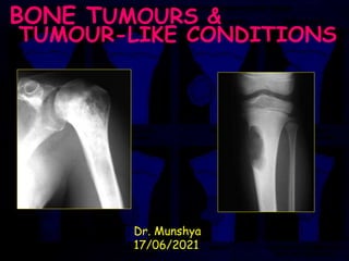
  • 1. JULY 2007 TUMOURS 1 BONE TUMOURS & TUMOUR-LIKE CONDITIONS Dr. Munshya 17/06/2021
  • 2. TUMOURS 2 Learning Outcomes By the end of this lecture you should be able to: 1. Classify bone tumours 2. Describe clinical, radiological and histological features of benign & malignant tumours & tumour-like conditions 3. Enumerate the common tumours in Zambia 4. Outline the principles of management of most common tumours
  • 3. TUMOURS 3 Introduction  Primary bone tumours are generally rare  Most common bone tumour is a secondary (metastatic) deposit  The most common malignant primary bone tumour in Zambia is Osteosarcoma
  • 4. TUMOURS 4 Nomenclature Primary bone tumours are of three varieties: 1. Benign 2. Malignant 3. Lesions that simulate bone tumours
  • 5. TUMOURS 5 Classification Histology Type Benign Malignant Haematopoietic Myeloma • Lymphoma Chondrogenic Osteochondroma Chondrosarcoma Chondroma Chondroblastoma Chondromyxoid Fibroma Osteogenic Osteiod Osteoma Osteosarcoma Osteoblastoma Fibrogenic Fibroma Fibrosarcoma Malignant fibrous Histiocytom Lipogenic Lipoma Liposarcoma Neurogenic Neurilemoma Notochordal Chordoma Vascular Haemangioma Haemangioendothelioma Unknown Origin Giant Cell Tumour (GCT) Ewings Tumour (Sarcoma) Histiocytoma Malignant Adamantinoma
  • 6. TUMOURS 6 Tumour-like Conditions (simulators) Young Patient Adult Elderly Patient Eosinophilic Granuloma Osteomyelitis Avulsion Fractures Aneurysmal Bone Cyst (ABC) Fibrous Dysplasia Osteofibrous Dysplasia Heterotopic osssification Unicameral Bone Cyst (UBC) Giant cell reparative granuloma Exuberant callus Synovial Chondromatosis Pigmented villonodular synovitis (PVNS) Stress fracture Heterotopic Ossification Ganglion cyst Metastatic bone disease Mastocytosis Hyperparathyroidism Paget’s disease Bone infarcts Ganglion cyst Cyst secondary to joint disease Epidermoid cyst
  • 7. TUMOURS 7 PRINCIPLES HISTORY & EXAMINATION STAGING WORKUP RADIOLOGY BIOPSY PROCEDURES CHEMOTHERAPY RADIOTHERAPY
  • 8. TUMOURS 8 HISTORY  Presenting complaint  Pain and / or swelling  Character / duration of symptoms (distinguish benign / malignant clinically)  Past and family history  Loss of weight  Other
  • 9. TUMOURS 9 AGE  Probability of OS in 12-25 yrs  Mets in > 50 yrs  Chondroblastoma vs GCT 10-20 yrs vs 17-30 yrs vs
  • 10. TUMOURS 10 CLINICAL EXAMINATION  General health  Anemia , wasting , spleen ?  Skin lesions  Precocious puberty  Exophthalmos ( EG , FD )
  • 11. TUMOURS 11 CLINICAL EXAMINATION (age,sex,site,past history)  Breasts  Thyroid  Chest  Liver  Kidney  Rectal
  • 12. TUMOURS 12 LOCAL EXAMINATION  Location ( epi , meta , diaphysis ) ( solitary or multiple )  Tumor size  Consistency ( bone or soft tissue )  Fixed or mobile  Solitary or multiple  N/V status  Lymph nodes ?
  • 13. TUMOURS 13 TUMOUR WORKUP  Bloods  Urinalysis  CXR  Plain X-rays  Bonescan  MRI  CT (lesion/chest)  Angiography  Biopsy
  • 14. TUMOURS 14 STAGING (ENNEKING) ENNEKING'S SURGICAL STAGES STAGE GRADE SITE METASTASES 1A 1B Low(G1) Low(G1) Intracompartmental(T1) Extracompartmental(T2) None(M0) None(M0) 2A 2B High(G2) High(G2) Intracompartmental(T1) Extracompartmental(T2) None(M0) None(M0) 3 Low(G1) or High(G2) Intracompartmental(T1) or Extracompartmental(T2) Yes(M1)
  • 15. TUMOURS 15 GRADING  G0= Histologically benign ( well differentiated and low cell to matrix ratio )  G1= Low grade malignant (few mitoses, moderate differentiation and local spread only )  G2= High grade malignant (frequent mitoses, poorly differentiated, high risk for metastases )
  • 16. TUMOURS 16 Features of aggressive tumours  Cellular atypia  Frequent mitoses  Extensive necrosis  Significant vascularity  Small amounts of immature matrix
  • 17. TUMOURS 17 Enneking’s 5 Questions 1. Age? 2. Site? 3. What is the tumour doing to the bone? 4. What is the bone doing to the tumour? 5. Any complications?
  • 18. TUMOURS 18 Osteoid Osteoma Incidence • Accounts for 10% of benign bone tumours • Male : Female 2:1 • Peak age 5 - 25 years (85% in this range) • Rare over 40 years Location:  Any bone, rarely multifocal tibia & femur in 50% spine - posterior elements Only occurs in bones formed by endochondral ossification May affect any part of a bone but is usually intracortical
  • 19. TUMOURS 19 Osteoid Osteoma Clinical Presentation  Pain is the commonest presentation  Pain often worse at night and relieved by aspirin (more likely NSAIDs)  10% occur in the spine and may -> scoliosis  Other sites may -> joint effusion, synovitis  Runs a self limiting course but usually -> surgery for pain relief  Pain usually decreases as the lesion matures lasting 18 - 30 months
  • 20. TUMOURS 20 Osteoid Osteoma X-Rays  Lytic nidus surrounded by sclerotic bone (which may mask the nidus)  Centre of nidus may be calcified  CT or tomograms -> diagnosis  Hot spot on bone scan
  • 21. TUMOURS 21 Osteoid Osteoma Differential Diagnosis • Brodie's abscess • Osteoblastoma • Stress fracture Treatment  NSAIDs  relieves symptoms  may take 3-4 years for symptoms to resolve  Surgical:  Nidus excision -> no recurrence (need only intact rim of reactive bone around the nidus to ensure complete excision)  Intraoperative localisation with: • Bone scan • Tetracycline (4mg tetracycline per kg qid 1-2 days pre operatively -> specimen excised under UV light) • CT • X-Ray excised tissue -> contains nidus
  • 22. TUMOURS 22 Osteochondroma (Exostosis)  Cartilage capped bony projection / exostosis  Commonest benign tumour of bone  Developmental abnormality of the metaphyseal area of any bone formed in cartilage (endochondral ossification)  Probably arise from aberrant cartilage from the perichondral ring of the growth plate
  • 23. TUMOURS 23 Osteochondroma (Exostosis) Incidence  Accounts for 45% of benign bone tumours  12% of all bone tumours  most become evident under 20 years  May be solitary or multiple (diaphyseal aclasis)  Any bone developing by endochondral ossification may be involved
  • 24. TUMOURS 24 Osteochondroma (Exostosis)  Present with lump or interference of tendon function  50% are distal femur, upper tibia or proximal humerus  May be sessile or pedunculated  Active growth during skeletal growth - >become latent  Move towards the diaphysis with growth and usually angle away from the growth plate  During growth period bone scan - >activity at the tip  Increased activity on bone scan after maturity suggests malignant change
  • 25. TUMOURS 25 Osteochondroma (Exostosis) Radiology  x-ray hallmark is blending of tumour into underlying metaphysis  flat, sessile lesion or a peduculated (stalk like) process  pedunculated osteochondromas tend to point away from the physis  look for a well defined metaphyseal excrescence of bone with a mottled density  Cartilaginous cap displays irregular areas of calcification
  • 26. TUMOURS 26 Osteochondroma (Exostosis) Pathology  Normal bone covered by a cap of normal cartilage  Cartilage cap resembles layers of the normal growth plate  The cartilage is more disorganized than normal  Binucleate chondrocytes in lacunae  Covered with a thin layer of periosteum
  • 27. TUMOURS 27 Osteochondroma (Exostosis) Treatment  Nil required unless symptomatic (persistent irritation (from bursitis or tendon) or neurovascular compromise)  Extra capsular marginal excision  Including the cartilaginous cap & overlying perichondrium  Deep bony base has minimal activity & may be removed piecemeal  Recurrence = < 5%  Decreased risk of recurrence if excised after maturity
  • 28. TUMOURS 28 Giant Cell Tumour (GCT)  Benign, usually solitary and locally aggressive  10% of benign bone lesions  Can undergo malignant transformation (5-10%)  Not seen until after the growth plate closes  Rarely metastasises (<1% to lungs)  Age 20 - 40 years  More common in females  Most commonly seen in the distal femur, proximal tibia and the distal radius  Nearly always located at the very end of a long bone (Subchondral)  Pathological fracture occurs in 10 - 15%  Neighbouring joint often irritated (effusion)
  • 29. TUMOURS 29 Giant Cell Tumour (GCT) Presentation  Pain, swelling  Pathological fracture
  • 30. TUMOURS 30 Giant Cell Tumour (GCT) X-Rays  Usually well defined lesion in the epiphysis extending up to the joint surface (subchondral) without marginal sclerosis, cortex thinned and sometimes ballooned  soap bubble appearance  Junction with normal bone poorly defined
  • 31. TUMOURS 31 Giant Cell Tumour (GCT) Pathology  Soft, friable tumour  Cut surface tan in colour, with areas of necrosis and haemorrhage  Numerous multinucleated giant cells. The stromal cells are homogenous mononuclear round/ovoid with large nuclei  The nuclei of the stromal cells are identical to the nuclei of the giant cells, a feature which distinguishes giant cell tumour from other conditions containing giant cells  Up to 50% have soft tissue extension but does not indicate malignancy
  • 32. TUMOURS 32 Giant Cell Tumour (GCT) Treatment  Intralesional excision by "extended" curettage  Curettage alone has a high local recurrence rate (50%) and the curettage is "extended" into the bone by a few millimetres by either using a burr, liquid nitrogen or phenol  The resulting cavity can be filled with bone graft or cement  En-bloc resection is possible if the bone is expendable e.g. proximal fibula, proximal radius  Amputation reserved for massive local recurrence, malignant change or infection  Radiotherapy reserved rare cases of unresectable tumours because of increased risk of secondary malignancy
  • 33. TUMOURS 33 Osteosarcoma  Primary malignant tumour arising from bone and producing bone Incidence  Male : Female 2:1  Bimodal age distribution: -  Peak 1: 10 - 20 years (age of rapid growth)  Peak 2 : 50 - 70 years (80% less than 30 and those more than 40 years usually secondary to Pagets)  Most common primary bone tumour in adolescents,  75% occur in the distal femur or around the knee  25% occur in the humerus  90% are metaphyseal in long bones  10% present with macroscopic metastatic disease
  • 34. TUMOURS 34 Osteosarcoma Types  Intramedullary (classical or ordinary) osteosarcoma  Surface osteosarcomas:  Parosteal osteosarcoma  Periosteal osteosarcoma  Secondary osteosarcomas:  Paget’s  Postradiation   Telangiectatic osteosarcoma
  • 36. TUMOURS 36 Osteosarcoma Clinical Presentation  Pain which is constant and worse at night  Pathological fracture is rare  May have a tender lump which may lack a definite edge and may be attached to muscle  If vascular may pulsate and feel warm
  • 37. TUMOURS 37 Osteosarcoma X-Rays  Variable with combination of bone destruction and bone formation  Sun ray spicules (Radial ossification) and  Codmans triangle (lifting of periosteum) may be evident  Cortical breach common  Adjacent soft tissue mass  Joint space rarely involved  25% Lytic  35% Sclerotic  40% Mixed
  • 38. TUMOURS 38 Osteosarcoma Pathology  Pleomorphic and anaplastic cell population with abundant fibrous and chondroid matrix  Stroma of spindle cells with numerous mitoses.  Most are high grade aggressive tumours usually about 10cm diameter at diagnosis (~ 10/12 growth)  50% -> osteoblastic  25% -> chondroid  25% -> fibroblastic
  • 39. TUMOURS 39 Osteosarcoma Differential Diagnosis  Post traumatic callus or myositis ossificans  Stress fracture - pathology may look similar  Osteomyelitis  Ewings
  • 40. TUMOURS 40 Osteosarcoma Consider osteosarcoma as a systemic disease therefore remove the primary site by en bloc excision and treat microscopic disease by chemotherapy Chemotherapy T10 regimen (methotrexate, vincristine, adriamycin)-> 60-75% survival Chemotherapy continues for 12 months in 4/52 cycles Radiotherapy -> palliation of local pain and to treat surgically inaccessible lesions and painful metastatic deposits radiotherapy may also be used pre-operatively to decrease the size and vascularity of the tumour. Surgery: Wide resection / Amputation
  • 41. TUMOURS 41 Chondrosarcoma  Primary malignant tumour whose cells produce cartilage matrix  May arise de novo or secondarily to an existing benign cartilaginous tumour
  • 42. TUMOURS 42 Chondrosarcoma Incidence  17% of primary malignant bone tumours  Peak incidence 30 - 60 years  Male : Female 2:1  Sites:  Pelvis 30%  Femur 20%  Femoral head 10%  Ribs 10%
  • 43. TUMOURS 43 Chondrosarcoma Clinical Presentation  Usually occurs in the metaphysis or diaphysis  Presents with constant ache or increased size of a pre-existing lump  Metastatic deposits are infrequent and usually go to lung
  • 44. TUMOURS 44 Chondrosarcoma X-Rays  Variable appearance with 60 - 70% have calcification and 50% have sub periosteal new bone  May be a large cystic lesion with cortical destruction and central calcification, endosteal scalloping and cortical expansion  Chondrosarcoma can also be classified as:  Intramedullary, which generally arise from enchondromas  Patients with Ollier's disease (multiple enchondromatosis) or Maffucci's syndrome (multiple enchondromas and hemangiomas) are at much higher risk of chondrosarcoma than the normal population  Surface which arise from osteochondromas/ exostoses.  Malignant change in an osteochondroma: increased size, fuzzy outline, cartilage cap more than 1cm thick, base more than 6cm diameter
  • 46. TUMOURS 46 Chondrosarcoma Pathology  Cellular pleomorphism and increased cellularity with focally calcified matrix
  • 47. TUMOURS 47 Chondrosarcoma Treatment  These tumours tend to metastasise late therefore attempt wide local excision initially  Radiotherapy useful for the treatment of surgically inaccessible sites however are relatively resistant to chemotherapy and radiotherapy Prognosis  Dependant on grade:  Low grade -> 65 - 85% 5 year survival  High grade -> 15 - 25% 5 year survival
  • 48. TUMOURS 48 Multiple Myeloma (MM)  Malignant tumor of plasma cells that causes widespread osteolytic bone damage  May affect any bone with haematopoietic red marrow (spine, skull, ribs, sternum and pelvis)  Age 50-80 year  M:F 2:1
  • 49. TUMOURS 49 Multiple Myeloma (MM) Presentation  Bone pain related to the deposits  Pathological fractures  Constitutional symptoms related to anaemia, thombocytopenia and renal failure  Other symptoms may include cachexia, spinal cord compression  Amyloidosis in 20%  Bacterial infections are common because of a lack of normal immunoglobulin production.
  • 50. TUMOURS 50 Multiple Myeloma (MM) Investigations  FBC normochromic, normocytic anaemia  ESR raised ++ (Often >100mm/hour)  Hypercalcaemia (20-40%)  Monoclonal immunoglobulin found on serum electrophoresis (90%)  Bence Jones proteins (light chain subunits of immunoglobulin) prese
  • 51. TUMOURS 51 Multiple Myeloma (MM) Radiology  The radiological appearance of multiple myeloma is characterised by irregular lytic defects of different sizes, often described as "punched out" and have no periosteal reaction  Skeletal survey is the most sensitive investigation as a bone scan can fail to have increased uptake in 25%  MRI is useful for delineating spinal lesions
  • 52. TUMOURS 52 Multiple Myeloma (MM) Histology  Biopsy reveals sheets of densely packed plasma cells  The degree of cytological atypia of these cells has no prognostic value  The osteolytic lesions are caused by increased osteoclastic resorption that is stimulated by cytokines released by the plasma cells
  • 53. TUMOURS 53 Multiple Myeloma (MM) Treatment  Radiotherapy  MM is sensitive to Radiotherapy, and reossification of tumour defects may occur within several months  Radiotherapy is recommended for intractable bone pain, it can be dramatically effective in relieving symptoms  Chemotherapy  Palliative only  Bisphosphonates useful in the treatment of hypercalcaemia.  Surgery  Prophylactic IM nails for femoral, humeral deposits  ORIF of other pathological fractures  15% may need spinal decompression due to deposits or fracture
  • 54. TUMOURS 54 Multiple Myeloma (MM) Prognosis  Untreated, a patient with bony lesions will only survive an average of 6-12 months with the cause of death usually by infection or haemorrhage  Improved survival following chemotherapy  With treatment a survival time of 3-5 years is not uncommon  Solitary lesions - 60% 5 year survival  Multiple lesions - 5% 5 year survival
  • 55. TUMOURS 55 Unicameral (Simple) Bone Cyst (UBC)  Benign lesion which occurs during growth  20% of benign bone lesions  Age 5-15 years  Not found in adults  Sex male to female is 3:1  The most common location is the proximal humerus (67%) followed by the proximal femur (15%)  UBC's may be found in unusual sites (e.g. calcaneum, pelvis) in patients >17 years  Cysts may be Active or Latent: Active cysts are located near the growth plate, but they move further away as the child grows and become inactive (latent)
  • 57. TUMOURS 57 Unicameral (Simple) Bone Cyst (UBC) Presentation  Asymptomatic  Usually presents as a pathological fracture (~ 65%) Aetiology  Unknown  Venous obstruction leading to a transudate of fluid  Fluid contains high levels of IL-1 & IL-6, which stimulate osteoclasts
  • 58. TUMOURS 58 Unicameral (Simple) Bone Cyst (UBC) Radiology  Well defined, central osteolytic area with a thin sclerotic margin (‘Fallen Leaf’ appearance)  Metaphyseal in young - moves towards diaphysis with growth  It fills and slightly expands the juxta epiphyseal metaphysis   CT not helpful unless the UBC is in the pelvis
  • 59. TUMOURS 59 Unicameral (Simple) Bone Cyst (UBC) Pathology  Histologically UBC's are thin walled cavities filled with blood tinged fluid.  The lining cells are cuboidal, but are not an endothelium  There is endosteal osteoclastic activity and periosteal new bone formation
  • 60. TUMOURS 60 Unicameral (Simple) Bone Cyst (UBC) Treatment  Treatment goal is to minimise fracture risk until the cyst heals (but this can take years)  Steroid injection 1-3 percutaneous injections repeated at 2 monthly intervals 60-80% success rate  Curettage and bone graft - 50% recurrence rate and possibility of damage to the growth plate  Bone marrow aspirate has recently been used  Intramedullary fixation / stabilization with flexible nails may help stabilize the lesion and may also promote resolution
  • 61. TUMOURS 61 Aneurysmal Bone Cyst (ABC)  Benign solitary, expansile and erosive lesion of bone  1% of benign bone lesions  Age most frequent in children (85% cases <20 years old)  Sex female to male is 2:1  ABC's can be found in any bone in the body  The most common location is the metaphysis of the lower extremity long bones, more so than the upper extremity  The vertebral bodies or arches of the spine may be involved  Approximately one-half of lesions in flat bones occur in the pelvis
  • 62. TUMOURS 62 Aneurysmal Bone Cyst (ABC) Presentation  Swelling, tenderness and pain  Occasionally there is limited range of motion due to joint obstruction  Spinal lesions can cause neurological symptoms secondary to cord compression  Pathological fractures are rare due to the eccentric location of the lesion
  • 63. TUMOURS 63 Aneurysmal Bone Cyst (ABC) Differential diagnosis Depending on the location, the differential includes  UBC,  chondromyxoid fibroma,  giant cell tumor,  osteoblastoma  highly malignant telangiectatic osteosarcoma.
  • 64. TUMOURS 64 Aneurysmal Bone Cyst (ABC) Radiographic features  On plain film, an ABC is normally placed eccentrically in the metaphysis and appears osteolytic  The periosteum is elevated and the cortex is eroded to a thin margin  The expansile nature of the lesion is often reflected by a "blow-out" or "soap bubble" appearance  CT scan can help delineate lesions in the pelvis or spine where plain film imaging may be inadequate  CT scan can narrow the differential diagnosis of ABC by demonstrating multiple fluid-fluid levels within the cystic spaces  MRI can also confirm the multiple fluid- fluid levels
  • 65. TUMOURS 65 Aneurysmal Bone Cyst (ABC) Pathology  Macroscopically, an ABC is like a blood filled sponge with a thin periosteal membrane. Soft, fibrous walls separate spaces filled with friable blood clot.  Microscopically, the ABC has cystic spaces filled with blood. The fibrous septa have immature woven bone trabeculae as well as macrophages filled with haemosiderin, fibroblasts, capillaries and giant cells.
  • 66. TUMOURS 66 Aneurysmal Bone Cyst (ABC) Treatment  The treatment approach will vary depending of the location and aggressiveness of the lesion  A slow growing, indolent ABC has been observed to regress spontaneously  Most lesions can be treated with curettage and application of a high- speed burr  Local recurrence rates vary widely,  Recurrence statistically related to young age and open growth plates, and may be less likely following wide excision than following curettage
  • 67. TUMOURS 67 Fibrous Dysplasia (FD) Normal medullary bone is replaced by variable amounts of structurally weak fibrous & osseous tissue
  • 68. TUMOURS 68 Fibrous Dysplasia (FD) Aetiology  ? developmental hamartoma Incidence  5 - 20% benign bone lesions  Relatively common and usually monostotic  Affects children and adolescents  Median age at onset 8 years  Male > Female ( Albrights - Female > Male)
  • 69. TUMOURS 69 Fibrous Dysplasia (FD) Sites  Ribs commonest (40%)  Lower limbs more than upper limbs  Craniofacial -> skull deformity  Epiphyses usually spared  Polyostotic -> pain, fracture (85%), deformity and skin pigmentation
  • 70. TUMOURS 70 Fibrous Dysplasia (FD) X-Rays  Lucent lesion in medullary space  Sclerotic margin with no discernable matrix.  Ground glass appearance typical  No periosteal reaction  Shepherds crook deformity of proximal femur caused by repeated # through proximal femur lesions
  • 71. TUMOURS 71 Fibrous Dysplasia (FD) Pathology  Bone replaced by firm, whitish tissue of gritty consistency  Vascular tumour with poorly orientated bone trabeculae separated by fibrous tissue.  Bone is woven rather than lamellar  lack of osteoblastic rimming of trabeculae
  • 72. TUMOURS 72 Fibrous Dysplasia (FD) Differential Diagnosis  Hyperparathyroidism  osteoblastoma  osteosarcoma
  • 73. TUMOURS 73 Fibrous Dysplasia (FD) Treatment  Monostotic -> curettage and grafting if symptomatic  Polyostotic -> symptomatic treatment  May require osteotomy for deformity or lengthening / shortening procedures
  • 74. TUMOURS 74 Fibrous Dysplasia Prognosis  Monostotic lesions cease activity at puberty but may be reactivated by pregnancy  Polyostotic - 85% -> pathological fracture  malignant change occurs after radiotherapy
  • 75. TUMOURS 75 Summary  Primary bone tumours are relatively rare  Osteosarcoma is the Commonest malignant primary bone tumour  Exostoses are the most common benign bone tumours  Patients with bone tumours need a thorough work up.