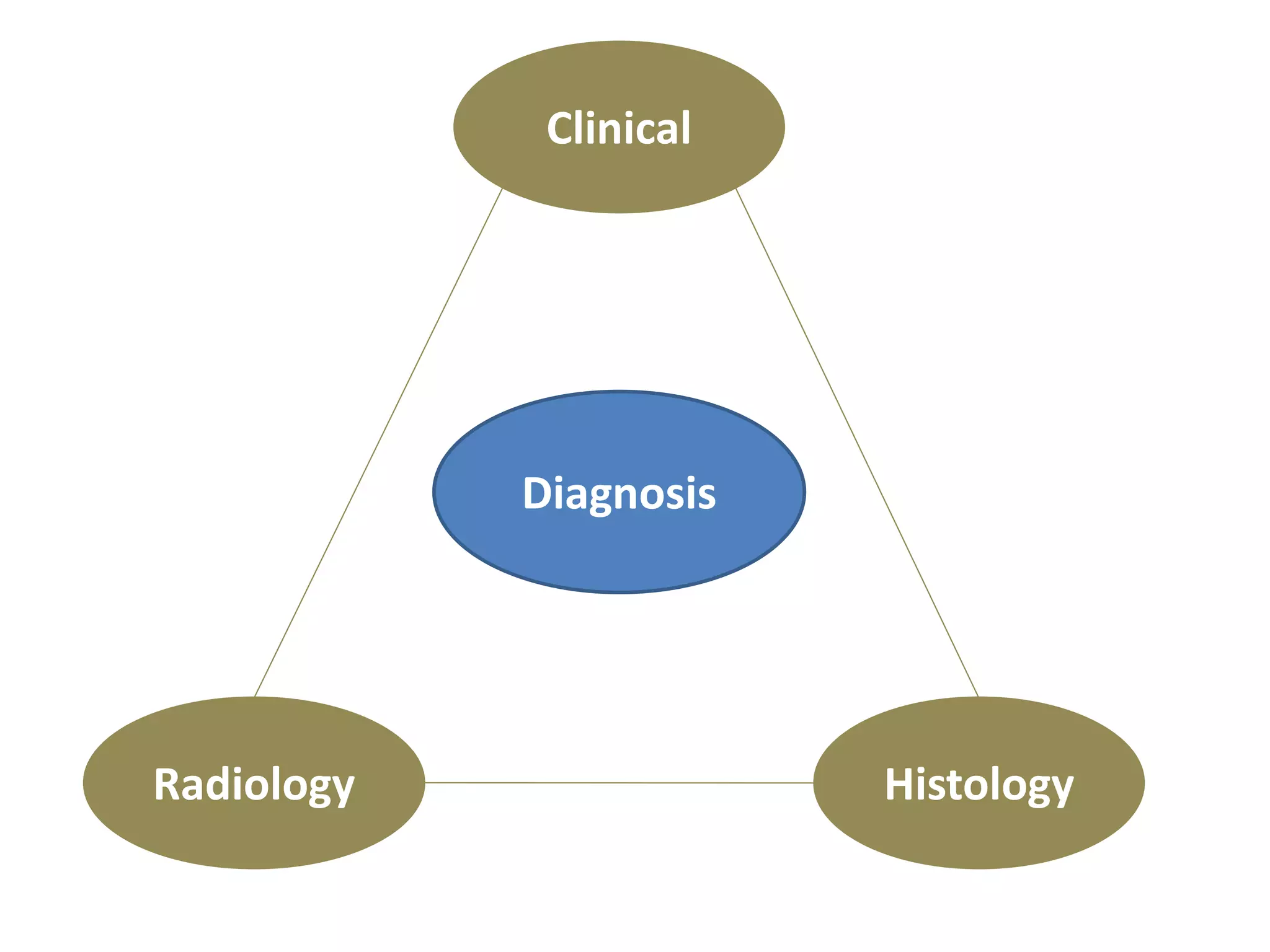
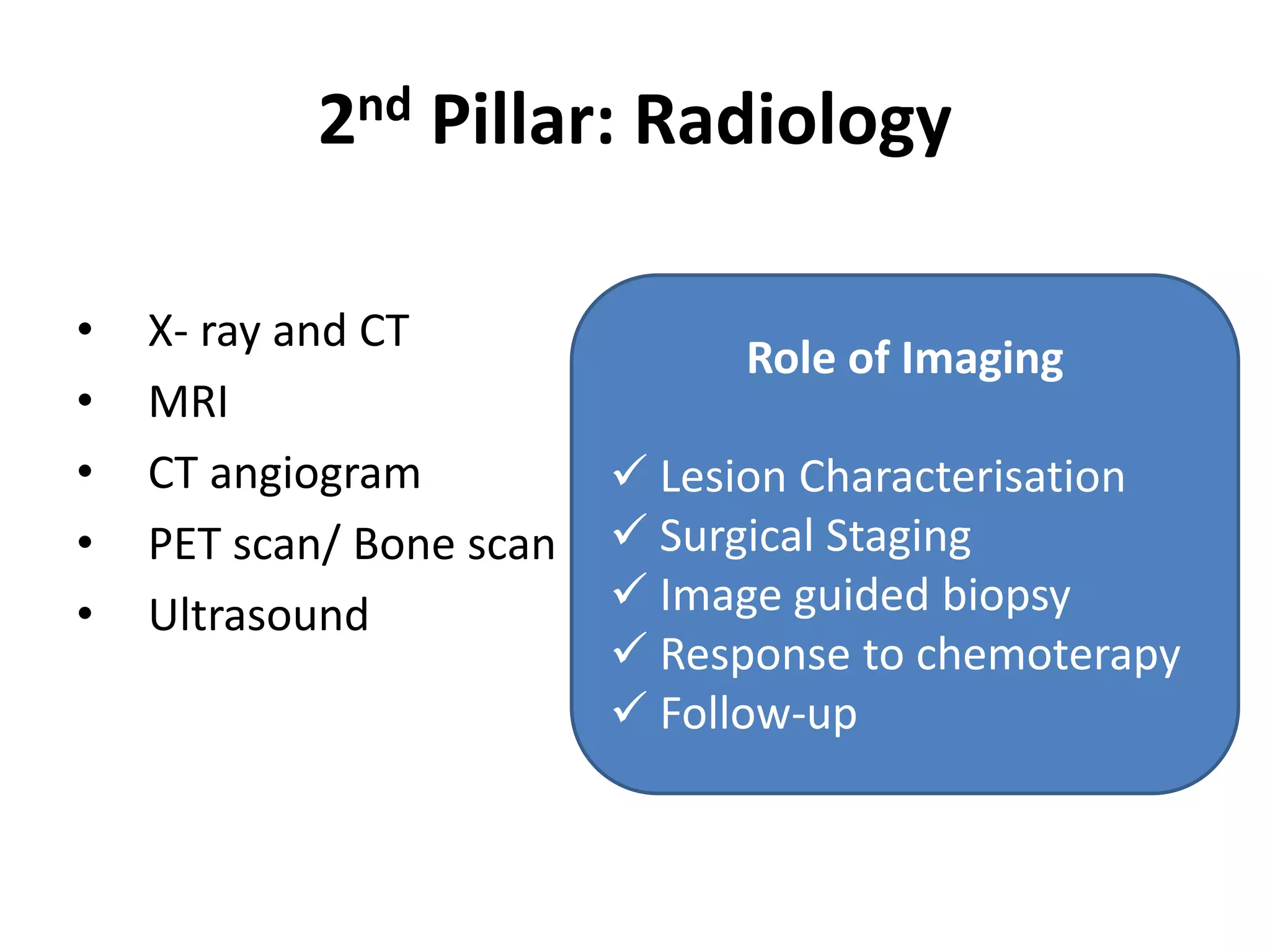
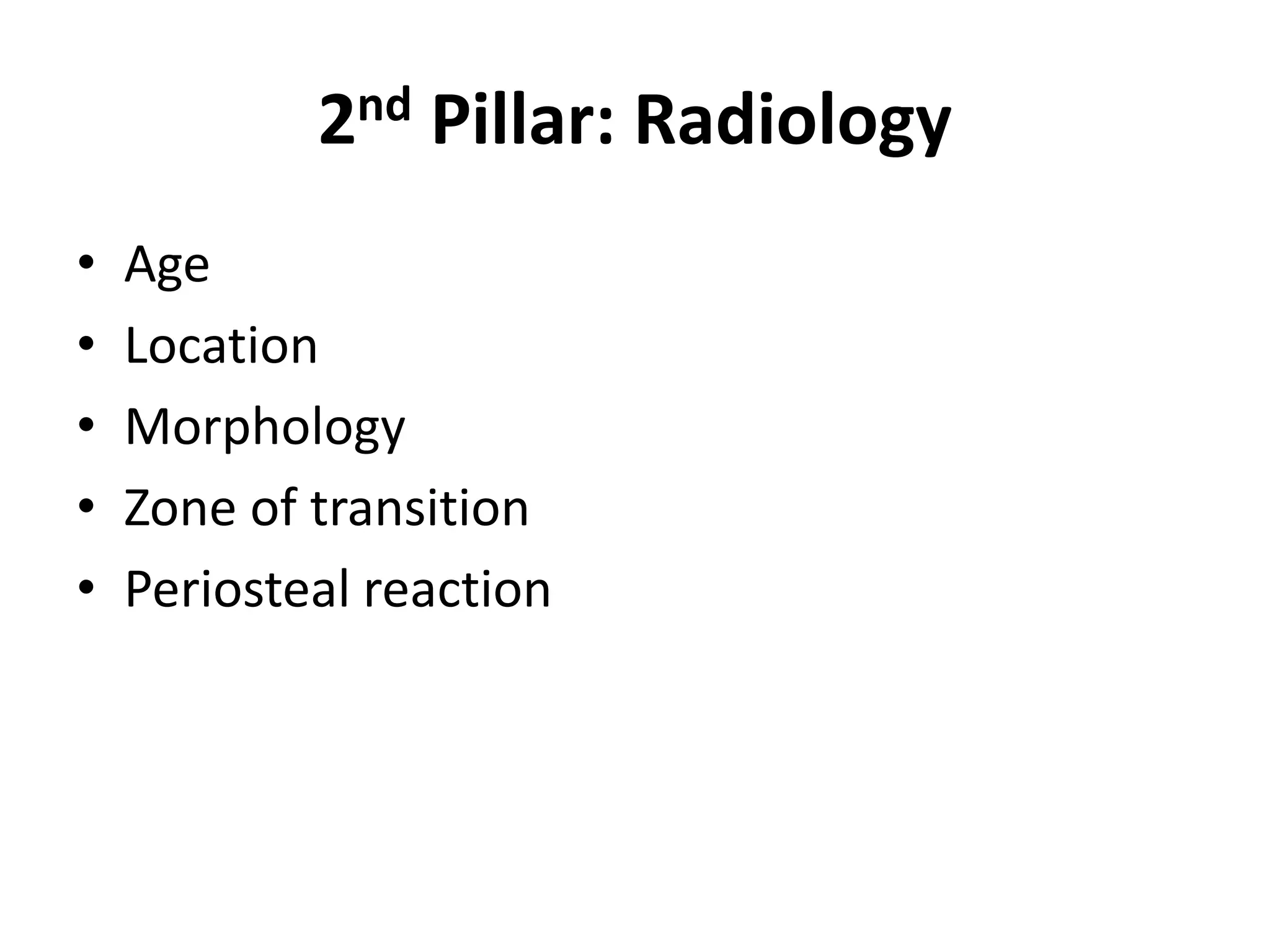
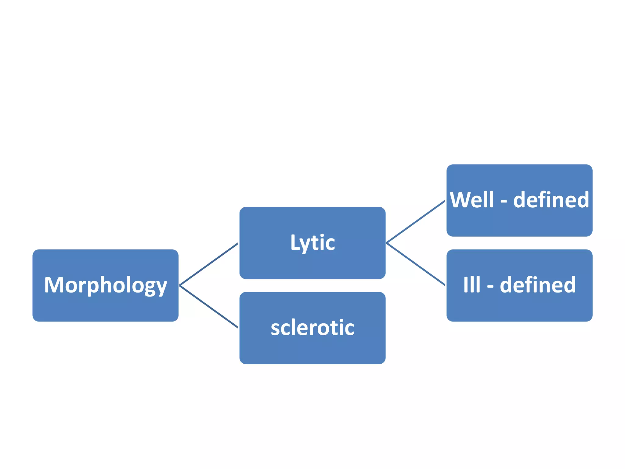
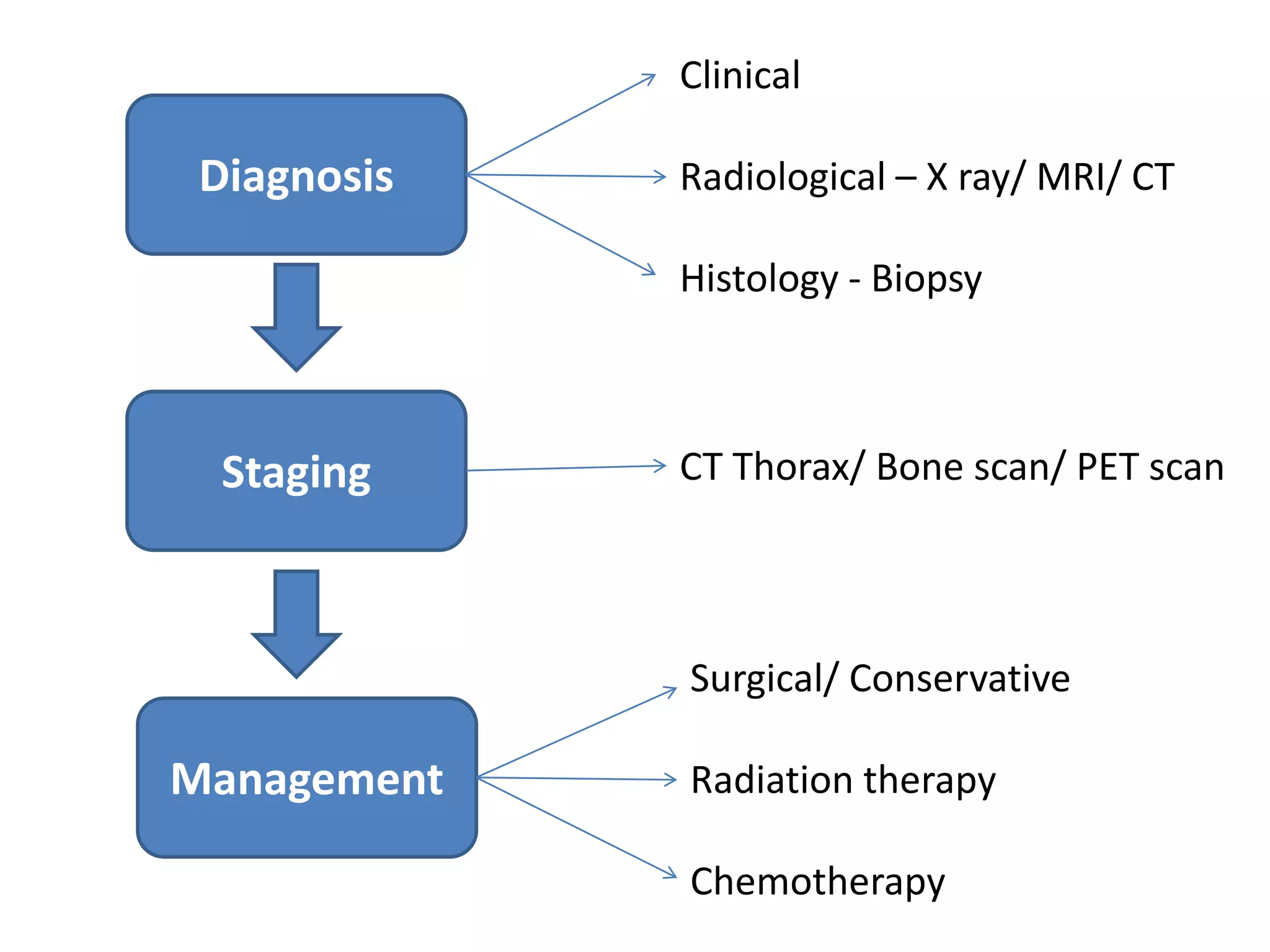
Approach to Bone Tumor
Dr GB sir
Department of Orthopaedic
Diagnosis
Staging
Management
Diagnosis
Clinical
Radiology Histology
1st Pillar - Clinical
History
• Pain and swelling
• Duration
• Onset and progression
• Remission
• Other joint/ organ involvement
1st Pillar - Clinical
Examination
Swelling:
• Numbers
• Shape – oval/ round/ diffuse
• Size
• Borders – well defined/ ill defined
• Surface – smooth/ irregular/ lobulated
• Consistency – firm/ cystic/ hard/ bony hard
• Plane – superficial/ deep
• Others special features
2nd Pillar: Radiology
• X- ray and CT
• MRI
• CT angiogram
• PET scan/ Bone scan
• Ultrasound
Role of Imaging
 Lesion Characterisation
 Surgical Staging
 Image guided biopsy
 Response to chemoterapy
 Follow-up
2nd Pillar: Radiology
• Age
• Location
• Morphology
• Zone of transition
• Periosteal reaction
Morphology
Lytic
Well - defined
Ill - defined
sclerotic
Zone of transition
• look at the zone of transition between the lesion and
the adjacent normal bone.
• The zone of transition is the most reliable indicator in
determining whether an osteolytic lesion is benign or
malignant.
• The zone of transition only applies to osteolytic
lesions since sclerotic lesions usually have a narrow
transition zone.
Zone of transition
Small zone of transition
• A small zone of transition results in a sharp, well-
defined border and is a sign of slow growth.
• A sclerotic border especially indicates poor biological
activity.
Zone of transition
Wide zone of transition
• An ill-defined border with a broad zone of transition
is a sign of aggressive growth (1).
• It is a feature of malignant bone tumors
Periosteal reaction
• A periosteal reaction is a non-specific reaction and
will occur whenever the periosteum is irritated by a
malignant tumor, benign tumor, infection or trauma.
• There are two patterns of periosteal reaction: a
benign and an aggressive type.
3rd pillar: HPE
• Performed only at the conculsion of imaging
work up.
 Helps determine the exact anatomic
approach to the tumor.
 Specifies the region of the tumor that
represents the underlying diseases.
 Biopsy can later the interpretation of the
imaging studies.
BIOPSY IS THE GOLD
STANDARD
Types of Biopsy
Biopsy
Bone Soft tissue
Types of Biopsy
Biopsy
Closed
Fine
needle
Core
needle
Open
Incisional Excisional Primary
wide
excisional
Diagnosis
Staging
Management
Clinical
Radiological – X ray/ MRI/ CT
Histology - Biopsy
CT Thorax/ Bone scan/ PET scan
Surgical/ Conservative
Radiation therapy
Chemotherapy

Approach to bone tumor

  • 1.
    Approach to BoneTumor Dr GB sir Department of Orthopaedic
  • 2.
  • 3.
  • 4.
    1st Pillar -Clinical History • Pain and swelling • Duration • Onset and progression • Remission • Other joint/ organ involvement
  • 5.
    1st Pillar -Clinical Examination Swelling: • Numbers • Shape – oval/ round/ diffuse • Size • Borders – well defined/ ill defined • Surface – smooth/ irregular/ lobulated • Consistency – firm/ cystic/ hard/ bony hard • Plane – superficial/ deep • Others special features
  • 6.
    2nd Pillar: Radiology •X- ray and CT • MRI • CT angiogram • PET scan/ Bone scan • Ultrasound Role of Imaging  Lesion Characterisation  Surgical Staging  Image guided biopsy  Response to chemoterapy  Follow-up
  • 7.
    2nd Pillar: Radiology •Age • Location • Morphology • Zone of transition • Periosteal reaction
  • 9.
  • 11.
    Zone of transition •look at the zone of transition between the lesion and the adjacent normal bone. • The zone of transition is the most reliable indicator in determining whether an osteolytic lesion is benign or malignant. • The zone of transition only applies to osteolytic lesions since sclerotic lesions usually have a narrow transition zone.
  • 12.
    Zone of transition Smallzone of transition • A small zone of transition results in a sharp, well- defined border and is a sign of slow growth. • A sclerotic border especially indicates poor biological activity.
  • 13.
    Zone of transition Widezone of transition • An ill-defined border with a broad zone of transition is a sign of aggressive growth (1). • It is a feature of malignant bone tumors
  • 14.
    Periosteal reaction • Aperiosteal reaction is a non-specific reaction and will occur whenever the periosteum is irritated by a malignant tumor, benign tumor, infection or trauma. • There are two patterns of periosteal reaction: a benign and an aggressive type.
  • 18.
    3rd pillar: HPE •Performed only at the conculsion of imaging work up.  Helps determine the exact anatomic approach to the tumor.  Specifies the region of the tumor that represents the underlying diseases.  Biopsy can later the interpretation of the imaging studies.
  • 19.
    BIOPSY IS THEGOLD STANDARD
  • 20.
  • 21.
  • 25.
    Diagnosis Staging Management Clinical Radiological – Xray/ MRI/ CT Histology - Biopsy CT Thorax/ Bone scan/ PET scan Surgical/ Conservative Radiation therapy Chemotherapy