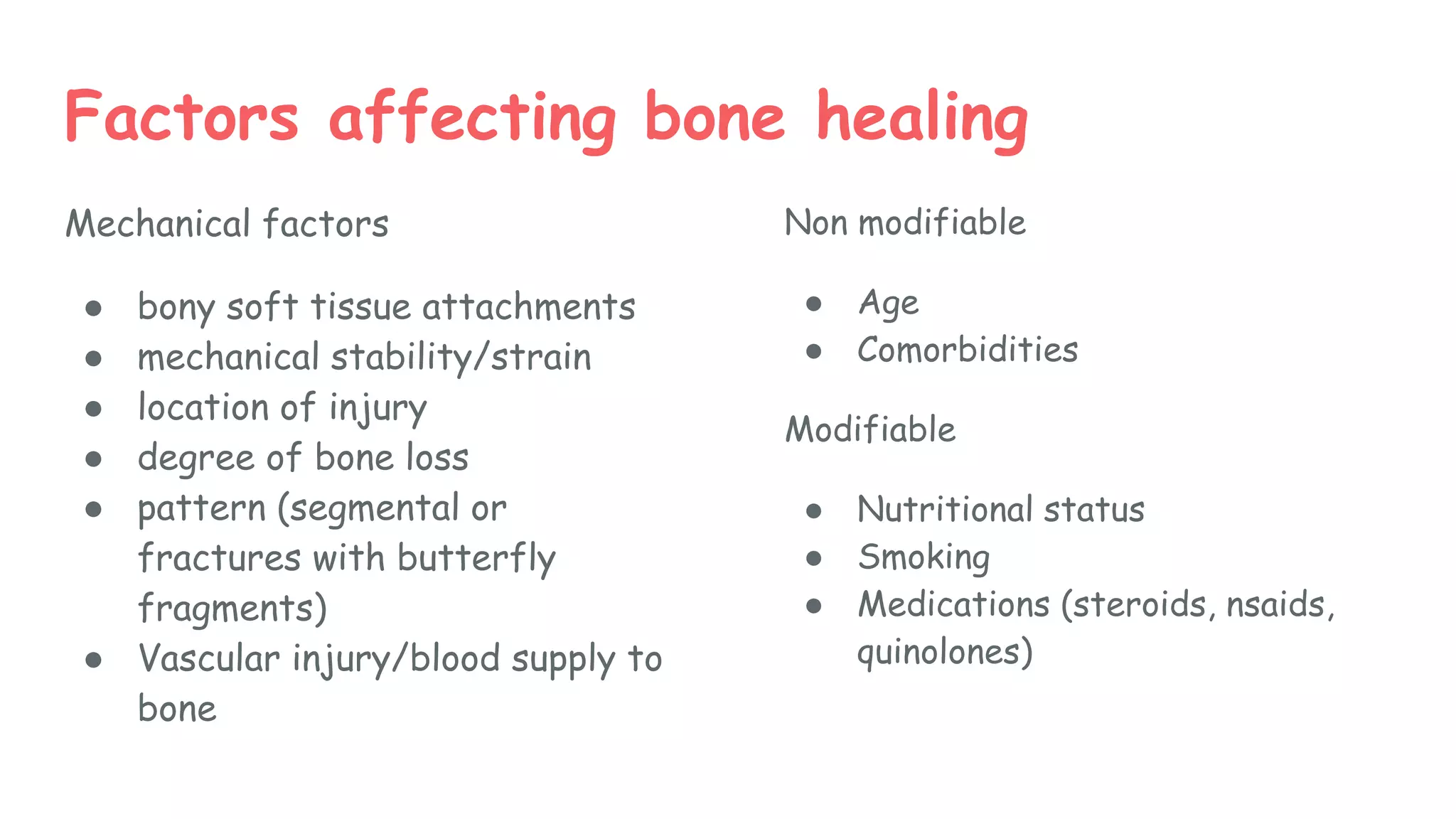
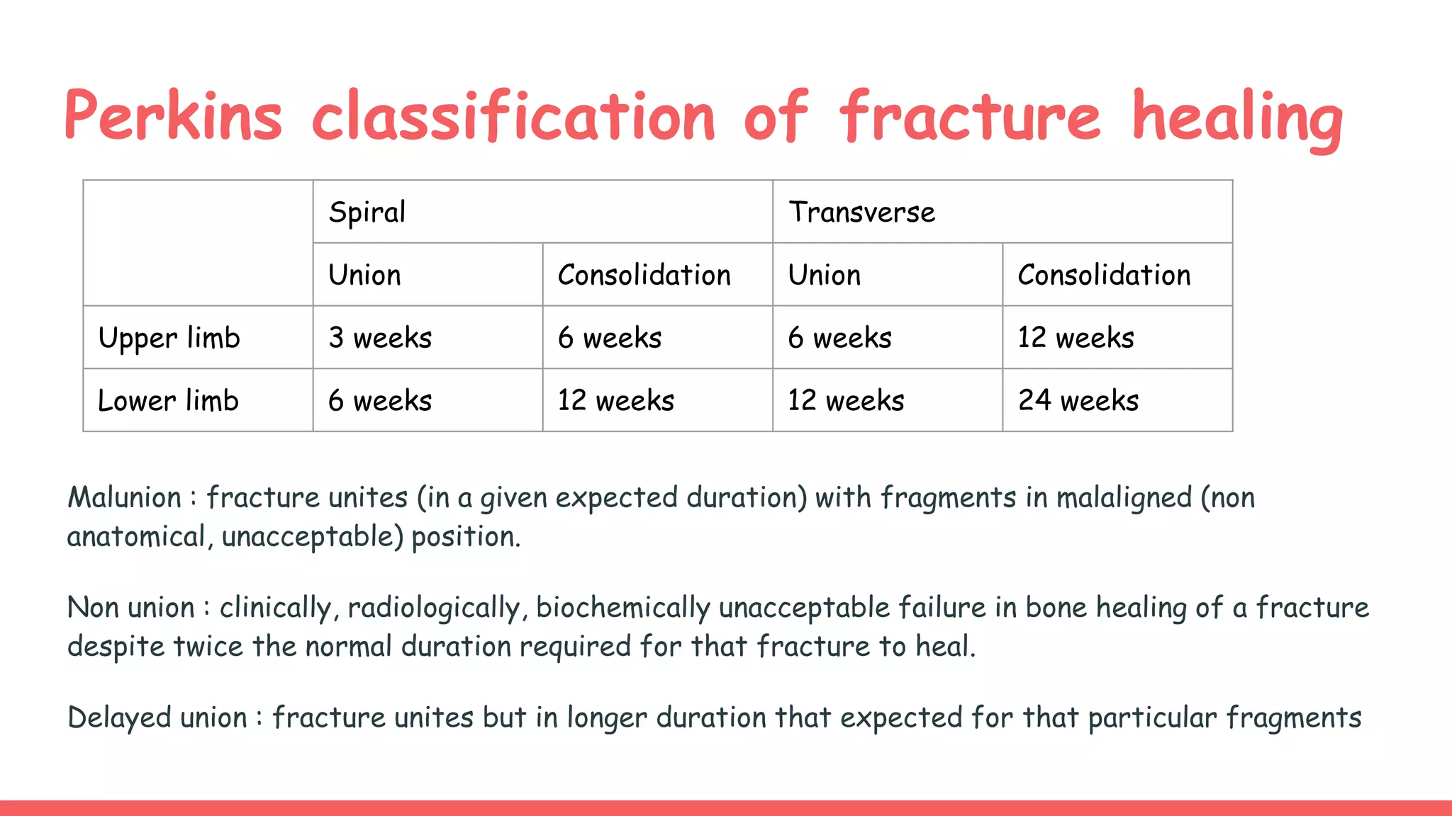
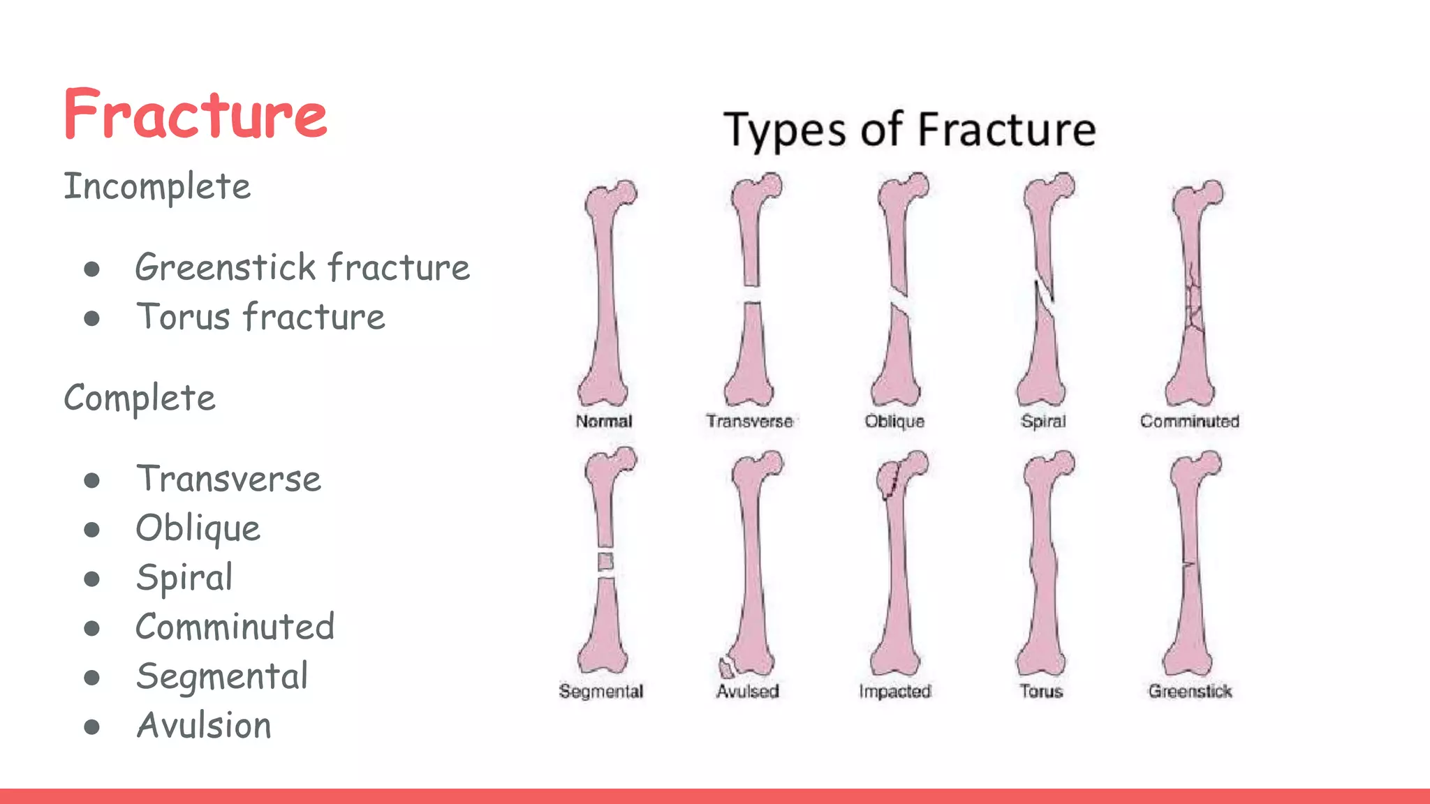
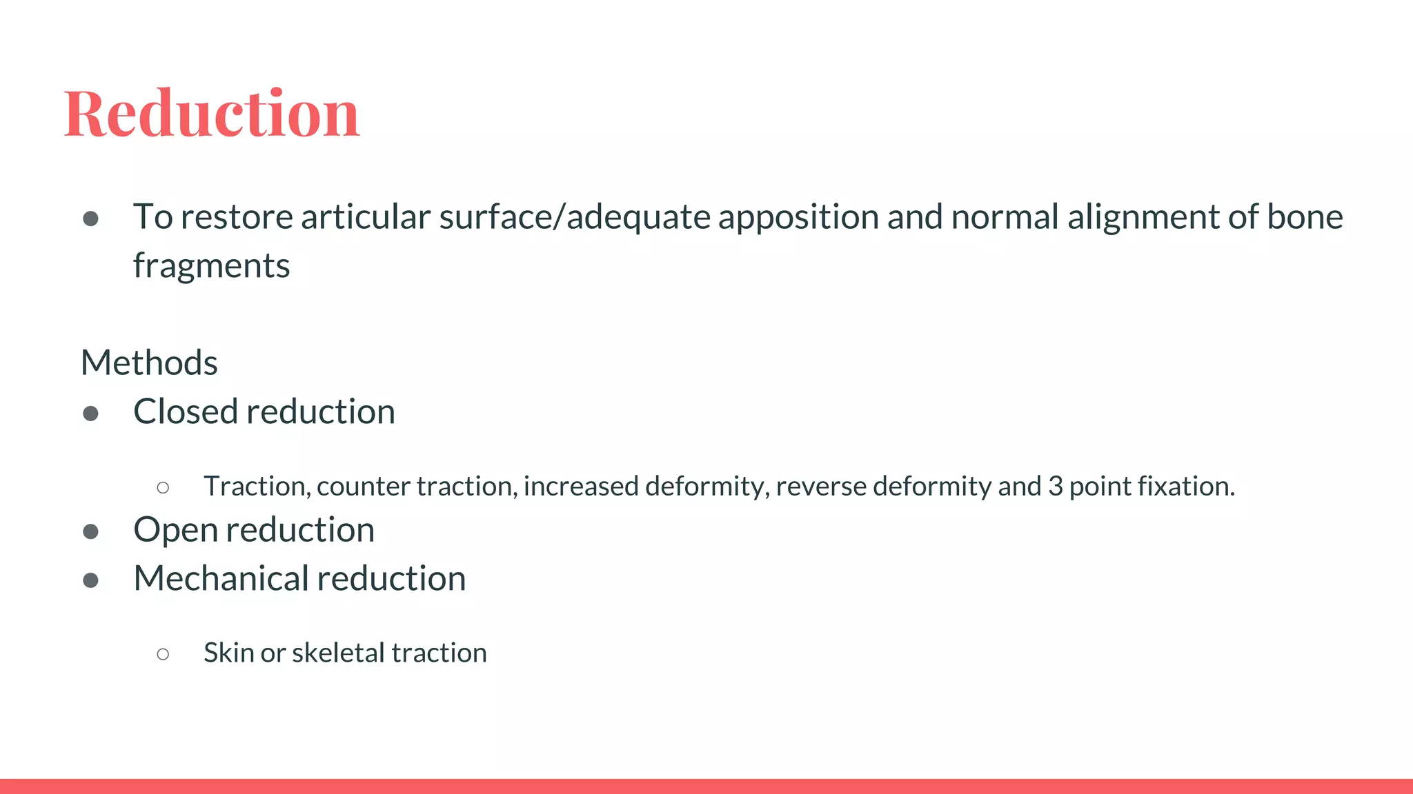
2. Outline
1. What is bone?
2. Bone metabolism
3. Bone healing
4. Fractures
○ Classification
○ Clinical feature
○ Investigations
○ Management
3. What is bone?
Bone is an organ which responsible
for
● Serves as attachment sites
for muscles
● Protection of internal organs
● Reservoir for minerals in the
body 99% of calcium in body
are stored as hydroxyapatite
crystals
● Haematopoiesis site
4. Bone composition
1. Organic phase 35%
Matrix
● Collagen : type 1 collagen gives to tensile
strength
● Non collagen protein : osteocalcin,
osteonectin, osteopontin
● Proteoglycan : compressive strength
Cells
● Osteoblast, osteoclast, osteocytes
2. Inorganic phase 60%
Calcium hydroxyapatite, calcium phosphate
3. Water 5%
6. Location
● bone (99%)
● blood and extracellular fluid (0.1%)
● Intracellular
Forms of calcium
● bone: majority is hydroxyapatite
● serum
Ca++ bound to protein (45%)
free-ionized Ca++ (45%)
bound to various anions, eg. citrate, bicarbonate (10%)
Dietary requirements
2000 mg/day for lactating women
1500 mg/day for pregnant women, postmenopausal woman, and patients with a healing bone fracture
1300 mg/day for adolescents and young adults
750 mg/day for adults
600 mg/day for children
Calcium
7. Phosphate
Location
● bone (86%)
● blood and extracelluar fluid (0.08%)
● intracellular 14%
Forms of phosphate
● bone :majority is hydroxyapatite
● serum
mostly inorganic phosphate (H2PO4-)
Regulation
● plasma phosphate is mostly unbound and reabsorbed by the kidney
● may be excreted in urine
● elevated serum phosphate can lead to increased release of PTH and bone resorption
Dietary intake
1000-1500 mg/day
11. Bone healing
1. Primary bone healing
● Bone healing by direct union
1. Secondary bone healing
● Bone healing by callus
12. Primary bone healing (strain is < 2%)
● When the fracture is immobilised by nails/plate and achieve absolute
stability
● occurs via Haversian remodeling
● Healing occur by intramembranous healing
○ Occurs without cartilage model or soft callus
○ undifferentiated mesenchymal cells will differentiate into osteoblast and
deposit organic matrix. New bone formation directly between fragments
13. Secondary bone healing -enchondral
healing (strain is between 2%-10%)
Inflammation
● Hematoma forms, macrophages, neutrophils and platelets release several
cytokines. Fibroblasts and mesenchymal cells migrate to fracture site and
forms granulation tissue
Repair
● Primary callus forms within two weeks. Enchondral ossification converts
soft callus to hard callus (woven bone). Type II collagen (cartilage) is
produced and then followed by type I collagen (bone) expression
Amount of callus is inversely proportional to extent of immobilization
14. Secondary bone healing
Remodelling
● cartilaginous calcification takes place.
● newly formed bone (woven bone) is remodeling via organized
osteoblastic/osteoclastic activity forming lamellar bone
● Shaped through
Wolff's law: bone remodels in response to mechanical stress
piezoelectic charges : bone remodels is response to electric charges:
compression side is electronegative and stimulates osteoblast formation,
tension side is electropostive and simulates osteoclasts
15.
16. Union of fractures
Blount’s law
1. Fractures in children unite faster than adults
2. Fractures closer to joints unite faster
3. Displacements at fracture site will remodel
4. Angulations in the plane of movement of joint (up to 20 degrees) will remodel
(acceptable)
5. Rotations will never remodel (unacceptable)
17. Factors affecting bone healing
Mechanical factors
● bony soft tissue attachments
● mechanical stability/strain
● location of injury
● degree of bone loss
● pattern (segmental or
fractures with butterfly
fragments)
● Vascular injury/blood supply to
bone
Non modifiable
● Age
● Comorbidities
Modifiable
● Nutritional status
● Smoking
● Medications (steroids, nsaids,
quinolones)
18. Perkins classification of fracture healing
Malunion : fracture unites (in a given expected duration) with fragments in malaligned (non
anatomical, unacceptable) position.
Non union : clinically, radiologically, biochemically unacceptable failure in bone healing of a fracture
despite twice the normal duration required for that fracture to heal.
Delayed union : fracture unites but in longer duration that expected for that particular fragments
Spiral Transverse
Union Consolidation Union Consolidation
Upper limb 3 weeks 6 weeks 6 weeks 12 weeks
Lower limb 6 weeks 12 weeks 12 weeks 24 weeks
20. What is fracture?
Define as break in structural/cortical continuity of bone
Classified into
1. Closed fracture : broken bone that overlying skin is intact with no
penetrating/protrusion of bone
2. Open fracture : broken bone that penetrate the skin or communicating
with external surrounding, no matter how small the rent is.
● Incomplete/complete
● Displaced/Undisplaced
● Trauma/Stress/Pathological
21. Fractures
Stress fracture
● Repetitive mechanical loading on
normal bone
● Prolonged stress cause
resorption of bone faster than
replacement thus more liable to
fracture
Common sites
● Metatarsal
● Calcaneum
● Femoral neck
● Tibia/fibula
Pathological fracture
● Occurs after trivial injury
Cause by
● Osteoporosis/osteoporosis
● Metabolic bone disease
● Infection
● Tumour
25. Open fracture classification - Gustillo Anderson
classification
Wound Size Soft tissue injury Contamination Impact force Risk of
amputation
Type I <1cm Minimal Minimal Low energy 5%
Type II 1-10cm Moderate Moderate Moderate
energy
10%
Type
IIIA
>10cm (extensive soft tissue injury and contamination, high energy force)
III A adequate soft tissue coverage
III B inadequate soft tissue coverage with periosteal stripping
III C neurovascular injury
20%
Type IIIB 30%
Type IIIC >40%
26.
27. Fractures
Clinical features
● Pain
● Swelling
● Deformity
● Loss of function
● Neurovascular compromise
Investigation
Plain x-rays
Rules of 2
● 2 views(AP, lateral)
● 2 joints (above and below fracture
site)
● 2 sides (in case of paediatric fracture
to compare epiphysis)
● 2 times (before and after reduction)
● 2 opinions
CT scan/MRI/Biolochemical test
28. Displacement
• Translation (shift) – The fragments may be shifted sideways, backward or forward in relation
to each other, such that the fracture surfaces lose contact. The fracture will usually unite as
long as sufficient contact between surfaces is achieved.
• Angulation (tilt) – The fragments may be tilted or angulated in relation to each other.
• Rotation (twist) – One of the fragments may be twisted on its longitudinal axis; the bone
looks straight but the limb ends up with a rotational deformity.
• Length – The fragments may be distracted and separated, or they may overlap, due to muscle
spasm, causing shortening of the bone.
29. Fracture
Principle of management
● First aid
○ Which consist of primary and secondary survey
● Reduction
● Immobilization
● Active rehabilitation
30. Reduction
● To restore articular surface/adequate apposition and normal alignment of bone
fragments
Methods
● Closed reduction
○ Traction, counter traction, increased deformity, reverse deformity and 3 point fixation.
● Open reduction
● Mechanical reduction
○ Skin or skeletal traction
31. Closed manual reduction Indications :
• Minimally displaced fracture/dislocations
• Fracture that are stable after reduction and held in cast and splint
• Done under sedation
Contraindication
• Unstable fracture
• Multiple fracture
• Uncooperative patient
Open reduction Indications
• Non union
• Open fracture
• Failed closed reduction
• Neurovascular compromised
• Intra articular fracture
• Salter Harris 3,4,5 or depending on site
• Polytrauma
Mechanical reduction • Difficult manipulation due to strong muscle pull, usually in femoral of
tibial shaft fracture
• Reduce fracture through ligament pull
• Allows soft tissue tension to decrease and better alignment
32.
33. Immobilization
External Internal
Non rigid fixator Rigid fixator
1. Splints
2. Casts
• applied involving one joint
above and one joint below
Complications
• Stiffness
• Pressure sores
• Compartment syndrome
• Non compliant
1. Traction
• Skin and skeletal traction
Complication
• Vascular and nerve injury
• Pin site infection
• Deep vein thrombosis
2. External fixation
Indication
• Open fracture
• Comminuted fracture
• Temporary stabilization prior
to definite management
• Lengthening of bone
Complication
• Neurovascular injury
• Pin site infection
• Stiffness of muscle
Indication
• Unstable fracture that prone
to re-displace after reduction
• Fracture that unite poorly
• Pathological fracture
• Non reducible fracture except
by open
Complication
• Bleeding
• Infection
• Implant failure
• Non union
• Re-fracture
Types
• Percutaneous pinning(Kirschner
wire)
• Extramedullary fixation
(Screw,plate,nails)
• Intramedullary fixation
34.
35.
36.
37.
38.
39. Open fracture
Types Management
I >3L of irrigation
2nd generation cephalosporin (cefuroxime, cefaclor)
II >6L of irrigation
2nd generation cephalosporin (cefuroxime, cefaclor)
III >9L of irrigation
2nd generation cephalosporin (cefuroxime, cefaclor) + gentamycin + C
penicillin to cover for clostridium
1. Tetanus toxoid
2. Antibiotics
3. Wound irrigation and debridement (golden hours :6 hours)
4. Wound coverage
41. Complication of fracture
Local Immediate Pain
Disability
Early Compartment syndrome
Neurovascular injury
Late Delayed union
Malunion
Nonunion
Avascular necrosis
Myositis ossificans
Secondary osteoarthritis
Osteomyelitis
Systemic DVT/PE
Shock
Sepsis
Fat embolism syndrome
42. Summary
● Bone composed by organic phase, inorganic phase and water.
● Bone metabolism involved parathyroid hormone, calcitonin, and vitamin D
which act on skin, gastrointestinal tract and kidney
● Bone healing can be divided into primary and secondary healing which affected
by various factors.
● Several laws eg wolff’s law, piezoelectic charges, and blounts law involve in
bone healing.
● Fracture can be divided into several classifications.
● Management of fracture involved multidisciplinary, systemic and long term
approach