
Recommended
More Related Content
Similar to Body fluids and circulation
Similar to Body fluids and circulation (20)
More from SwastikPattnayak1
Recently uploaded
Recently uploaded (20)
Body fluids and circulation
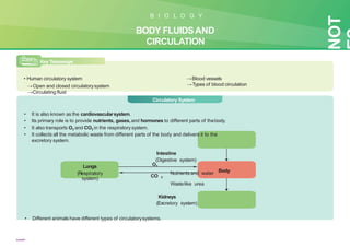
- 1. Key Takeaways • Human circulatory system →Open and closed circulatorysystem →Circulating fluid →Heart →Blood vessels →Types of blood circulation NOT B I O L O G Y BODY FLUIDSAND CIRCULATION CIRCULA TORY SYSTEM Lungs (Respiratory system) Body O2 CO 2 Circulatory System • It is also known as the cardiovascularsystem. • Its primary role is to provide nutrients, gases,and hormones to different parts of thebody. • It also transports O2 and CO2 in the respiratorysystem. • It collects all the metabolic waste from different parts of the body and delivers it to the excretory system. Intestine (Digestive system) Nutrients and water Wastelike urea Kidneys (Excretory system) • Different animals have different types of circulatorysystems. swastik
- 2. Open Closed • Example:All arthropods, molluscs • The blood pumped by the heart is always circulated through aclosed network of blood vessels. • The flow of fluid through this systemcan be regulated. T ypes of circulatorysystems seen in animals 02 Heart Valve • The heart pumps the circulatoryfluid known as hemolymph into open spaces or body cavities. • The hemolymph returns to theheart via small pores known asostia. Ostia Heart Body cells Body cells • Example:Annelids, chordates Blood vessels Heart Heart Heart Heart Heart Ostia Blood Annelids Arthropods Molluscs Chordates Blood vessels Blood vessels Blood vessels Heart Hemolymph in sinuses surrounding organs swastik
- 3. 03 Human CirculatorySystem Circulating fluid Heart Blood vessels Blood Lymph • It is a fluid connective tissue. • It transports oxygen, nutrients, antibodies, etc. • It is a colourless fluid. • It carries immune cells, nutrients, etc. Human heart • It is a muscularorgan. • It is derived from mesodermalcells. • It is located between the lungs in the thoracic cavity, slightly towards the left of thesternum (breastbone). • It is roughly the size of a clenched fist and 12cmlong. Size 12cm Human heart Circulating fluids
- 4. 04 • It has three layers. Layers in the heart Pericardium Pericardium Myocardium Pericardial Fluid Pericardium (Around heart) It is a protective covering around the heart,and the space between pericardium and myocardium is filled with pericardialfluid. Myocardium (Muscle heart) It is a muscular wall of heart. It contracts to pump blood out of the heart andthen relaxes as the heartrefills with returning blood. Endocardium (Inner heart) Pericardium Myocardium Endocardium It remains in contact with blood. Structure of a human heart • Human heart consists of four chambers: →Right and left atria. →Right and left ventricles. • Atria →These are the small upper chambers. Debananda Sahoo, VGYAN CLASSES
- 5. 05 →The walls of atria are thin. →The atria are named based on their position →Larger right atrium and the left atrium. →They are separated by a thin muscular interatrialseptum. • Ventricles →These are the large lower chambers. →They are separated from atria by thick fibrous tissue called atrioventricularseptum. →The ventricles are named based on their position →The right ventricles and the left ventricle. →The left and right ventricles are separated by the thick muscular tissuecalled interventricular septum Superior vena cava Carries oxygen poor- blood from the upper parts of the body Pulmonaryveins Blood enters the left atrium through the four pulmonary vein openings. Right atrium Receives blood from the body through vena cava Aorta Pumps blood to the rest of the body Pulmonaryartery Pumps blood to thelungs Pulmonarysemilunar valve Guards the opening of the right ventricle to the pulmonaryartery Aortic semilunarvalve Guards the opening ofthe left ventricle to the aorta Left atrium Receives oxygenated blood from the lungs Inferior vena cava Carries oxygen-poor blood from the lower parts of the body Right ventricle Receives blood from the right atrium and then pumps blood into the pulmonary artery or pulmonary trunk Left ventricle Receives blood from the left atrium and thenpumps blood into the aorta which then supplies to the whole body Debananda Sahoo, VGYAN CLASSES
- 6. 06 • Comparison of left and right ventricles Left ventricle The left ventricle is larger and three times thicker than the right ventricle. →As the left ventriclehas to pump blood to the whole body throughaorta. Right ventricle Ventricles Rightventricle Left ventricle Comprises of thinwall Comprises of thick wall Receives deoxygenated blood fromthe right atrium Receives oxygenated blood from the left atrium Pumps blood to lungs through the pulmonarytrunk Pumps blood to the whole bodythrough the aorta • Septum →The septum’s primary function in the heart is to isolate the two sidesof the heart. Atria Interatrial septum Separates left and right atrium Interventricular septum Separates left and right ventricles Atrioventricular septum Atria and ventricles on the same sideare separated by atrioventricular septum. Debananda Sahoo, VGYAN CLASSES
- 7. 07 • External structure of the heart • Valve →Valves are flaps of fibrous tissues located in the cardiac chambers. →They ensure that blood flows in a single direction(unidirectional). →Flaps also prevent blood from flowing backwards. Tricuspid valve Bicuspid or mitral valve • It has three muscular flaps. • It guards the opening between right atrium and right ventricle. • It has two flaps. • It guards the passage from left atrium to left ventricle. Chordae tendinae Papillarymuscles They anchor the bicuspid and tricuspid valves to theventricular papillary muscles and keep the valves in closed position during ventricular systole. Grooves (Sulci) Different divisions in the heart create depressions known as grooves. Atrioventricular sulcus The thick atrioventricular septum formsgroove on the surface of heart and runs between the atria and ventricles and the surface of the heart. Interatrial groove The thick interatrial septum formsgroove on the surface of the heart. Anterior view Posteriorview Interventricular sulcus The thick interventricular septum formsgroove on the surface of the heart and runs between the two ventricles and the surface of theheart. Debananda Sahoo, VGYAN CLASSES
- 8. 08 Blood vessels • Bluish or deep red in colour as theycarry deoxygenated blood. Arteries • Pinkish or bright red in colour as theycarry oxygenated blood • Blood flows with a high pressure andspeed. Veins Capillaries • Arteries branch out into arteriolesand veins into venules. • These arterioles and venules branch out into smaller, thinner blood vessels known as capillaries. • Allow exchange of gases and other materials with the cells of theorgans. • They act as a connection between arteries and veins. Layers of arteries and veins Heart Heart Arteries Carry blood Veins Carry blood Different parts of body Different parts of body Tunicaintima Tunicamedia Tunicaexterna Squamous endoithelium Smooth muscle and elastic fibres Fibrous connective tissue with collagen fibres Debananda Sahoo, VGYAN CLASSES
- 9. 09 The total length of blood vessels in an average human adult is twice of Earth’s circumference at the equator! Did you know? T ypes of blood circulation Single circulation • Seen in 2-chambered heart • Contains one atrium and one ventricle • Example: Fish • The deoxygenated blood is pumped from ventricles to gills. Incomplete double circulation • Seen in 3-chambered heart • Contains two atria and one ventricle • Examples: Reptiles and amphibians except crocodile • The left atrium receives oxygenated blood from gills/skin/lungs and the right atrium receives deoxygenated blood from thebody. • They inter-mix in a single ventricle, which pumps it out to gills/skin/ lungs and to different parts of body, thus it is known as incomplete circulation. • Blood passes through the heart twice, hence known as double circulation. • It is very inefficient. Complete double circulation • Seen in 4-chambered heart • Contains two atria and two ventricles • Examples: Birds and mammals Debananda Sahoo, VGYAN CLASSES
- 10. 10 SummarySheet Open Closed • The heart pumps the circulatoryfluid known as hemolymph into open spaces or body cavities. • The hemolymph returns to theheart via small pores known asostia. • The blood pumped by the heart is always circulated through aclosed network of blood vessels. • The flow of fluid through this systemcan be regulated. Types of circulatorysystems seen in animals Arteries Veins Involved in carrying oxygenated blood Involved in carrying deoxygenated blood Carry blood away from the heart tovarious parts of the body Carry blood towards the heart fromvarious parts of the body Valves are absent Valves are present Gills Lungs Various body Right atrium Ventricle Right ventricle Various body parts Left ventricle Right atrium Left atrium Left atrium Incomplete double circulation Complete double circulation Heart Single circulation parts Debananda Sahoo, VGYAN CLASSES
- 11. 1 1 Human CirculatorySystem Circulating fluid Heart Myocardium (Muscle heart) Blood vessels Blood Veins Transports nutrients, oxygen, antibodies, etc. Endocardium (Inner heart) Arteries Carry blood from the heart to differentparts of the body Carry blood from different body parts tothe heart Capillaries Connecting link between arteries andveins Lymph Carries immune cells, nutrients, etc. Pericardium (Around heart) Debananda Sahoo, VGYAN CLASSES
- 12. Atrium Ventricle Thin-walled Thick-walled Forms the upper chamber of theheart Forms the lower chamber of theheart Divided into right and leftatria Divided into left and right ventricles Collects blood from the body andsupplies to ventricles Supplies blood to different parts ofthe body 12 Single circulation Incomplete double circulation Complete double circulation • Seen in 2-chambered heart • Contains one atrium and one ventricle • Example: Fish • 3-chambered heart • Contains two atriaand one ventricle • Examples: Reptilesand amphibians except crocodile • Blood passesthrough the heart twice • Mixing of bloodoccurs • Inefficient • 4-chambered heart • Contains two atriaand two ventricles • Examples: Birds and mammals • No mixing of blood • Two separate circulatory pathways are present T ypes of blood circulation Debananda Sahoo, VGYAN CLASSES