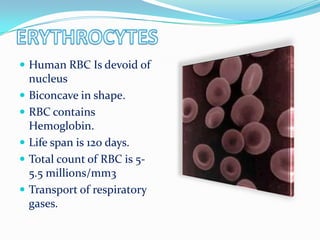
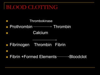
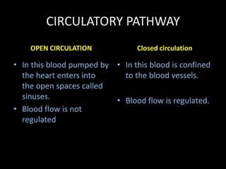
 Living organisms are made up of cells .
 This cells need oxygen and nutrients for their growth.
 These are supplied through circulatory system.
 In lower organisms like sponges and hydra circulation
is through water but in higher organisms it through
blood and lymph.
 Plasma
 Formed Elements;
(a) Erythrocytes
(b) Leucocytes
(c) Thrombocytes
 It has 90_92% of water,6_8% of proteins.
There are 3 types of proteins.
 Fibrinogen –Helps in coagulation of blood.
 Globulin-Involved in defense mechanism.
 Albumin- Helps in osmotic balance.
 It has minerals.
 Plasma without fibrinogen is called serum.
BLOOD PLASMA
 Human RBC Is devoid of
nucleus
 Biconcave in shape.
 RBC contains
Hemoglobin.
 Life span is 120 days.
 Total count of RBC is 5-
5.5 millions/mm3
 Transport of respiratory
gases.
 Colorless.
 Nucleated.
 Total count 6000 to
8000mm3.
 Short lived.
TYPES OF LEUCOCYTES
 Granulocytes
 Agranulocytes
• 2-3% of Leucocytes.
• Resist infections.
• Associated with allergic reactions.
• NEUTROPHILS
• 60-65% of Leucocytes.
• Phagocytes in nature.
• It secretes histomine, serotonin& heparin.
• Involved in inflammatory reactions.
EOSINOPHILS
LYMPHOCYTES
• It forms 20-25 % of formed element.
• It is of 2 types
• B-Lymphocytes &T-Lymphocytes.
• Both are responsible for immune responses of the body.
• MONOCYTES
• These are phagocyte in nature .It forms 6 –8% of formed
element.
BLOOD CLOTTING
 Thrombokinase
 Prothrombin Thrombin
 Calcium
 Fibrinogen Thrombin Fibrin

 Fibrin +Formed Elements Bloodclot
Blood group Antigens Antibody DONATE
BLOOD TO
A A anti-B A,AB
B B anti-A B,AB
AB A,B -------- AB
O ------- Anti-
A,B
ALL
RH GROUPING
 Another surface antigen is also present in
RBC.
 This surface antigen was first reported in
Rhesus Monkey. So it is called Rhesus
factor or RH factor.
 80% of people are Rh+ve ,they have Rh
factor.
RH INCOMPATIBILIITY
 FEMALE * MALE
 Rh-ve Rh+ve
FOETUS (Rh+ve)
safe
At child birth
Anti Rh
antibody
Second Foetus
Destroy the 2nd
foetus
 As the blood passes through the capillaries in tissues, some
water along with many small water soluble substances move
out into the spaces between the cells of tissues leaving the
larger proteins and most of the formed elements in the blood
vessels.
 This fluid released out is called the interstitial fluid or tissue
fluid.
 This fluid present in the lymphatic system is called the lymph.
 It has lymphocytes.
 It is colorless.
 Fats are absorbed through lymph.
CIRCULATORY PATHWAY
OPEN CIRCULATION
• In this blood pumped by
the heart enters into
the open spaces called
sinuses.
• Blood flow is not
regulated
Closed circulation
• In this blood is confined
to the blood vessels.
• Blood flow is regulated.
Structure of Human heart
 It is located in thoracic
cavity in between 2
lungs.
 It is protected by
pericardium.
 It has 4 chambers (2
Atria& 2 ventricles)
 Heart is made up of
cardiac muscle.
 Right auricle is separated
from Right ventricle by
means of a septum with 3
flaps called tricuspid
valve.
 Left auricle is separated
from left ventricle by
means of a septum with
2flaps called bicuspid
valve/mitral valve.
 Cardiac cycle initiates from SA node(
Pace maker). It also maintains the
rhythm of heart beat.
 It is located in the right upper corner of
right atrium.
 From SA node the electrical impulses
reach the AV node.
 AV node is located in the lower left
corner of the right atrium.
 From AV node it reaches the Bundle of
His.
 Finally it reaches the ventricles
through Purkinjee fibers.
 Human heart is myogenic in nature.
Cardiac cycle
CARDIAC CYCLE
CARDIAC OUTPUT
• It is the amount of blood pumped by heart
per minute is called heart output or
cardiac output.
• Cardiac output =72x70 ml
=5040 ml.
TYPES OF BLOOD VESSELS
 Carry blood from heart
to different parts of the
body.
 Carry oxygenated
blood except
pulmonary artery.
 Deeply seated.
 Valves are absent.
 Have thick walls
 Carry blood from
different parts of the
body to heart.
 Carry deoxygenated
blood except
pulmonary vein.
 Superficially seated.
 Valves are present.
 Have thin walls.
ARTERIES VEINS
DOUBLE CIRCULATION
 Pulmonary circulation
 Systemic circulation
Right Atria Right ventricle Pulmonary artery
Systemic arota Left ventricle Left atria
Pulmonary vein
Body parts
Lungs
Deoxygenated blood
Deoxygenated blood
Deoxygenated blood
oxygenated blood
oxygenated blood
oxygenated bloodoxygenated blood
oxygenated blood
Deoxygenated blood
PULMONARY CIRCULATION
SYSTEMIC CIRCULATION
ECG; It is the graphical
representation of electrical activity
of cardiac chambers.
P wave-Depolarization of atria
QRS _Depolarization of ventricles.
TWAVE Repolarisation of cardiac
chambers.
DISORDER OF
CIRCULATORY SYSTEM
 High Blood Pressure
(Hypertension)
 Coronary Artery Disease (CAD)
 Angina Pectoris
 Heart Failure
 A persistent rise in diastolic pressure
above 90 mmHg and or systolic
pressure above 140mmHg is termed as
hypertension.
 It is due to the deposition of cholesterol on the walls of
arteries leading to narrowing of arteries.
 This also causes hypertension.
 Blood supply to the cardiac muscle is reduced.
ARTERIOSCLEROSIS
 Loss of elasticity of the walls of arteries
due to ageing.
MYOCARDIAL INFRACTION
 Very low blood flow to the cardiac muscle. As a result
cardiac muscle cannot sustain its function.
• Acute pain in the chest due to very less
supply of oxygen to the heart muscle.
MD. SARWAR AZAD
TIRTHA MAITRA
RAZIUR RAHMAN
S.V.S HEMANT KUMAR
ANUBHUTI
CHAURASIA

Body fluid and circulation

  • 2.
     Living organismsare made up of cells .  This cells need oxygen and nutrients for their growth.  These are supplied through circulatory system.  In lower organisms like sponges and hydra circulation is through water but in higher organisms it through blood and lymph.
  • 3.
     Plasma  FormedElements; (a) Erythrocytes (b) Leucocytes (c) Thrombocytes
  • 4.
     It has90_92% of water,6_8% of proteins. There are 3 types of proteins.  Fibrinogen –Helps in coagulation of blood.  Globulin-Involved in defense mechanism.  Albumin- Helps in osmotic balance.  It has minerals.  Plasma without fibrinogen is called serum. BLOOD PLASMA
  • 5.
     Human RBCIs devoid of nucleus  Biconcave in shape.  RBC contains Hemoglobin.  Life span is 120 days.  Total count of RBC is 5- 5.5 millions/mm3  Transport of respiratory gases.
  • 6.
     Colorless.  Nucleated. Total count 6000 to 8000mm3.  Short lived.
  • 7.
    TYPES OF LEUCOCYTES Granulocytes  Agranulocytes
  • 8.
    • 2-3% ofLeucocytes. • Resist infections. • Associated with allergic reactions. • NEUTROPHILS • 60-65% of Leucocytes. • Phagocytes in nature. • It secretes histomine, serotonin& heparin. • Involved in inflammatory reactions. EOSINOPHILS
  • 9.
    LYMPHOCYTES • It forms20-25 % of formed element. • It is of 2 types • B-Lymphocytes &T-Lymphocytes. • Both are responsible for immune responses of the body. • MONOCYTES • These are phagocyte in nature .It forms 6 –8% of formed element.
  • 10.
    BLOOD CLOTTING  Thrombokinase Prothrombin Thrombin  Calcium  Fibrinogen Thrombin Fibrin   Fibrin +Formed Elements Bloodclot
  • 11.
    Blood group AntigensAntibody DONATE BLOOD TO A A anti-B A,AB B B anti-A B,AB AB A,B -------- AB O ------- Anti- A,B ALL
  • 12.
    RH GROUPING  Anothersurface antigen is also present in RBC.  This surface antigen was first reported in Rhesus Monkey. So it is called Rhesus factor or RH factor.  80% of people are Rh+ve ,they have Rh factor.
  • 13.
    RH INCOMPATIBILIITY  FEMALE* MALE  Rh-ve Rh+ve FOETUS (Rh+ve) safe At child birth Anti Rh antibody Second Foetus Destroy the 2nd foetus
  • 14.
     As theblood passes through the capillaries in tissues, some water along with many small water soluble substances move out into the spaces between the cells of tissues leaving the larger proteins and most of the formed elements in the blood vessels.  This fluid released out is called the interstitial fluid or tissue fluid.  This fluid present in the lymphatic system is called the lymph.  It has lymphocytes.  It is colorless.  Fats are absorbed through lymph.
  • 15.
    CIRCULATORY PATHWAY OPEN CIRCULATION •In this blood pumped by the heart enters into the open spaces called sinuses. • Blood flow is not regulated Closed circulation • In this blood is confined to the blood vessels. • Blood flow is regulated.
  • 16.
    Structure of Humanheart  It is located in thoracic cavity in between 2 lungs.  It is protected by pericardium.  It has 4 chambers (2 Atria& 2 ventricles)
  • 17.
     Heart ismade up of cardiac muscle.  Right auricle is separated from Right ventricle by means of a septum with 3 flaps called tricuspid valve.  Left auricle is separated from left ventricle by means of a septum with 2flaps called bicuspid valve/mitral valve.
  • 18.
     Cardiac cycleinitiates from SA node( Pace maker). It also maintains the rhythm of heart beat.  It is located in the right upper corner of right atrium.  From SA node the electrical impulses reach the AV node.  AV node is located in the lower left corner of the right atrium.  From AV node it reaches the Bundle of His.  Finally it reaches the ventricles through Purkinjee fibers.  Human heart is myogenic in nature.
  • 19.
  • 20.
  • 21.
    CARDIAC OUTPUT • Itis the amount of blood pumped by heart per minute is called heart output or cardiac output. • Cardiac output =72x70 ml =5040 ml.
  • 22.
    TYPES OF BLOODVESSELS  Carry blood from heart to different parts of the body.  Carry oxygenated blood except pulmonary artery.  Deeply seated.  Valves are absent.  Have thick walls  Carry blood from different parts of the body to heart.  Carry deoxygenated blood except pulmonary vein.  Superficially seated.  Valves are present.  Have thin walls. ARTERIES VEINS
  • 24.
    DOUBLE CIRCULATION  Pulmonarycirculation  Systemic circulation
  • 25.
    Right Atria Rightventricle Pulmonary artery Systemic arota Left ventricle Left atria Pulmonary vein Body parts Lungs Deoxygenated blood Deoxygenated blood Deoxygenated blood oxygenated blood oxygenated blood oxygenated bloodoxygenated blood oxygenated blood Deoxygenated blood PULMONARY CIRCULATION SYSTEMIC CIRCULATION
  • 26.
    ECG; It isthe graphical representation of electrical activity of cardiac chambers. P wave-Depolarization of atria QRS _Depolarization of ventricles. TWAVE Repolarisation of cardiac chambers.
  • 27.
    DISORDER OF CIRCULATORY SYSTEM High Blood Pressure (Hypertension)  Coronary Artery Disease (CAD)  Angina Pectoris  Heart Failure
  • 28.
     A persistentrise in diastolic pressure above 90 mmHg and or systolic pressure above 140mmHg is termed as hypertension.
  • 29.
     It isdue to the deposition of cholesterol on the walls of arteries leading to narrowing of arteries.  This also causes hypertension.  Blood supply to the cardiac muscle is reduced.
  • 30.
    ARTERIOSCLEROSIS  Loss ofelasticity of the walls of arteries due to ageing.
  • 31.
    MYOCARDIAL INFRACTION  Verylow blood flow to the cardiac muscle. As a result cardiac muscle cannot sustain its function.
  • 32.
    • Acute painin the chest due to very less supply of oxygen to the heart muscle.
  • 33.
    MD. SARWAR AZAD TIRTHAMAITRA RAZIUR RAHMAN S.V.S HEMANT KUMAR ANUBHUTI CHAURASIA