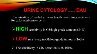
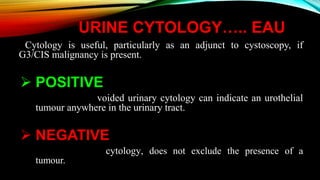
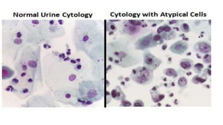
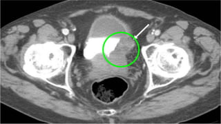
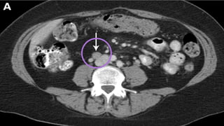
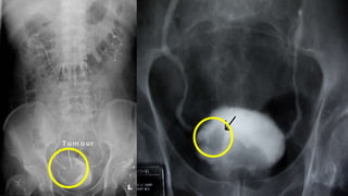
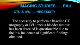
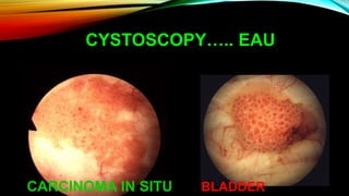
CARCINOMA URINARY BLADDER
DIAGNOSITIC MODALITIES
By
Dr. Muhammad Saifullah
Post-Graduate Resident (FINAL YEAR)
Department of Urology & Renal Transplantation
Allied Hospital, Faisalabad.
URINE CYTOLOGY….. EAU
Examination of voided urine or bladder-washing specimens
for exfoliated cancer cells.
HIGH sensitivity in G3/high-grade tumours (84%)
LOW sensitivity in G1/low-grade tumours (16%)
 The sensitivity in CIS detection is 28-100%.
URINE CYTOLOGY….. EAU
Cytology is useful, particularly as an adjunct to cystoscopy, if
G3/CIS malignancy is present.
 POSITIVE
voided urinary cytology can indicate an urothelial
tumour anywhere in the urinary tract.
 NEGATIVE
cytology, does not exclude the presence of a
tumour.
URINE CYTOLOGY….. EAU
LIMITATIONS…..
• User-dependent.
• Evaluation can be hampered by
i. Low cellular yield,
ii. Urinary tract infections
iii.Stones
iv.Intravesical instillations
URINE CYTOLOGY….. EAU
NOTE:
• In experienced hands specificity exceeds
90%.
• In patients with suspect cytology it is
reasonable to repeat the investigation.
IMAGING STUDIES….. EAU
TRANSABDOMINAL
ULTRASOUND….. ADVANTAGES
• Characterisation of Renal masses
• Detection of hydronephrosis
• Visualisation of intraluminal masses in the bladder.
Ultrasound is therefore a useful tool for detection of
obstruction in patients with haematuria.
IMAGING STUDIES….. EAU
TRANSABDOMINAL
ULTRASOUND….. LIMITATIONS
•Cannot exclude the presence of Upper Tract
Urothelial Carcinoma and therefore, cannot replace
CT urography.
•The diagnosis of CIS cannot be made with
Ultrasound.
IMAGING STUDIES….. EAU
COMPUTED TOMOGRAPHY
UROGRAPHY ….. ADVANTAGES
Computed tomography (CT) urography is used
to:
 Detect papillary tumours in the urinary tract,
which can be seen as filling defects
 Hydronephrosis.
 Status of lymph nodes.
 Neighbouring organs.
IMAGING STUDIES….. EAU
INTRAVENOUS UROGRAPHY …..
ADVANTAGES
Intravenous urography (IVU) can be an
alternative if CT is not available.
In muscle-invasive tumours of the bladder and in
Upper Tract Urothelial Carcinomas, CT Urography
gives more information than IVU (including status
of lymph nodes and neighbouring organs).
IMAGING STUDIES….. EAU
CTU & IVU….. NECESSITY
The necessity to perform a baseline CT
urography or IVU once a bladder tumour
has been detected is questionable due to
the low incidence of significant findings
obtained.
URINARY MOLECULAR
MARKER TESTS….. EAU
None of these markers have been
accepted for diagnosis or follow-up in
routine practice or clinical guidelines.
URINARY MOLECULAR
MARKER TESTS….. EAU
i. UroVysion (FISH)
ii. Microsatellite analysis
iii. Immunocyt/uCyt +
iv. Nuclear matrix Protein 22
URINARY MOLECULAR
MARKER TESTS….. EAU
v. BTA stat
vi. BTA TRAK
vii. Cytokeratins
BTA: Bladder Tumour Antigen
UROVYSION (FISH)…..
 Sensitive and specific test
used to diagnose urothelial
carcinoma in urine.
 It detects aneuploidy of
chromosomes 3, 7 and 17,
and loss of both 9p21 loci
in malignant urothelial
cells.
UROVYSION (FISH)…..
There is a strong correlation
between the morphologic features of
the nuclei and polysomy. Polysomic
cells tend to have large and irregular
nuclei and to have a mottled
chromatin staining pattern.
UROVYSION (FISH)…..
For this reason, the microscopic
analysis portion of the UroVysion
assay uses a scanning technique to
assess the slides for cells that are most
likely to have chromosomal
abnormalities.
UROVYSION (FISH)…..
 UroVysion probe set:
i. CEP3 (RED)
ii. CEP7 (GREEN)
iii. CEP17 (AQUA)
iv. 9p21 (GOLD)
CEP indicates chromosome enumeration probe.
UROVYSION (FISH)…..
EAU
NORMAL (‘‘DISOMIC’’) CELL TRISOMY 7 CELLS SHOWING 3 COPIES OF
CEP7 (GREEN) BUT 2 COPIES OF THE OTHER
3 PROBES
UROVYSION (FISH)…..
EAU
TETRASOMIC CELL SHOWING 4 COPIES
OF ALL 4 PROBES
POLYSOMIC CELL WITH GAINS (3 OR MORE
COPIES) FOR 2 OR MORE OF THE 4 PROBES
IMMUNOCYT/UCYT+ …..
 An immunocytochemical test
 Developed by Fradet and Lockhard in 1997
 Uses fluorescent-labeled antibodies to 3 markers
that are commonly found on malignant
exfoliated urothelial cells.
IMMUNOCYT/UCYT+ …..
 One antibody is directed against a
highmolecular-weight form of glycosylated
carcinoembryonic antigen, 19A211 and is
labeled red.
 The other two antibodies, LDQ10 and M344, are
directed against mucins, which are cytoplasmic
antigens specific for bladder cancer and are
labeled with fluorescein.
IMMUNOCYT/UCYT+ …..
Positive Immunocyt/uCyt+
test result demonstrating
GREEN fluorescence.
Negative immunocyt/uCyt+
test result
Positive Immunocyt/uCyt+ test
result demonstrating
RED fluorescence.
BTA STAT & BTA TRAK …..
 Immunoassays that detect human complement
factor H related protein in urine, employing the
same antibody pair.
 The BTA stat….. qualitative test which can be
performed in a consultation setting
 The BTA TRAK….. quantitative test that is
performed in the laboratory.
BTA STAT & BTA TRAK …..
CYTOKERATIN…..
 Cytokeratin 7 (CK7) and
cytokeratin 20 (CK20) are
2 types of intermediate
filament protein.
 Expression of CK7 is seen
in the majority of primary
urinary bladder carcinomas.
CYTOKERATIN…..
 CK20 is restricted to
superficial and occasional
intermediate cells of the
normal urothelium of the
bladder. Aberrant CK20
expression has been
documented in urothelial
carcinoma.
URINARY MOLECULAR
MARKER TESTS
(LIMITATIONS)….. EAU
 Benign conditions and BCG influence many
urinary marker tests
 False-positive results of UroVysion and
microsatellite analysis can be attributed to occult
disease
 Sensitivity is usually higher at the cost of lower
specificity, compared to urine cytology.
URINARY MOLECULAR MARKER
TESTS (POTENTIAL
APPLICATIONS)….. EAU
 SCREENING of the population at risk of
bladder cancer.
 Haematuria dipstick, NMP22 or UroVysion
in BC screening.
 Routine application of screening is not
recommended.
URINARY MOLECULAR MARKER
TESTS (POTENTIAL
APPLICATIONS)….. EAU
PRIMARY DETECTION….. Exploration of
patients after haematuria or other symptoms
suggestive of bladder cancer.
 Adjunct to cystoscopy to detect invisible
tumours, particularly CIS.
URINARY MOLECULAR MARKER
TESTS (POTENTIAL
APPLICATIONS)….. EAU
PRIMARY DETECTION
 Urinary cytology is highly specific, but
urinary markers lack this high
specificity and are not recommended
for primary detection.
URINARY MOLECULAR MARKER
TESTS (POTENTIAL
APPLICATIONS)….. EAU
SURVEILLANCE of NMIBC
 Follow-up of low/intermediate-risk NMIBC
 Reduce the number of cystoscopy procedures,
urinary markers should be able to detect
recurrence before the tumours are large and
numerous.
URINARY MOLECULAR MARKER
TESTS (POTENTIAL
APPLICATIONS)….. EAU
SURVEILLANCE of NMIBC
 The limitation of urinary cytology is its low
sensitivity for low-grade recurrences.
 Several urinary markers are better, but still
do not detect half of the low-grade tumours
identified by cystoscopy.
ACCORDING TO CURRENT KNOWLEDGE,
NO URINARY MARKER
CAN REPLACE
CYSTOSCOPY
DURING FOLLOW UP OR HELP TO LOWER
CYSTOSCOPIC FREQUENCY IN A ROUTINE
FASHION
CYSTOSCOPY….. EAU
 The diagnosis of papillary BC ultimately
depends on cystoscopic examination of the
bladder and histological evaluation of the
resected tissue.
 CIS is diagnosed by a combination of
cystoscopy, urine cytology, and histological
evaluation of multiple bladder biopsies.
CYSTOSCOPY….. EAU
 A flexible instrument with
topical intra-urethral
anaesthetic lubricant
instillation results in better
compliance compared to a
rigid instrument, especially
in men
CYSTOSCOPY….. EAU
PAPILLARY GROWTH SESSILE
CYSTOSCOPY….. EAU
CARCINOMA IN SITU BLADDER
Bladder Cancer Diagnostic Modalities

Bladder Cancer Diagnostic Modalities

  • 1.
    CARCINOMA URINARY BLADDER DIAGNOSITICMODALITIES By Dr. Muhammad Saifullah Post-Graduate Resident (FINAL YEAR) Department of Urology & Renal Transplantation Allied Hospital, Faisalabad.
  • 3.
    URINE CYTOLOGY….. EAU Examinationof voided urine or bladder-washing specimens for exfoliated cancer cells. HIGH sensitivity in G3/high-grade tumours (84%) LOW sensitivity in G1/low-grade tumours (16%)  The sensitivity in CIS detection is 28-100%.
  • 4.
    URINE CYTOLOGY….. EAU Cytologyis useful, particularly as an adjunct to cystoscopy, if G3/CIS malignancy is present.  POSITIVE voided urinary cytology can indicate an urothelial tumour anywhere in the urinary tract.  NEGATIVE cytology, does not exclude the presence of a tumour.
  • 6.
    URINE CYTOLOGY….. EAU LIMITATIONS….. •User-dependent. • Evaluation can be hampered by i. Low cellular yield, ii. Urinary tract infections iii.Stones iv.Intravesical instillations
  • 7.
    URINE CYTOLOGY….. EAU NOTE: •In experienced hands specificity exceeds 90%. • In patients with suspect cytology it is reasonable to repeat the investigation.
  • 8.
    IMAGING STUDIES….. EAU TRANSABDOMINAL ULTRASOUND…..ADVANTAGES • Characterisation of Renal masses • Detection of hydronephrosis • Visualisation of intraluminal masses in the bladder. Ultrasound is therefore a useful tool for detection of obstruction in patients with haematuria.
  • 10.
    IMAGING STUDIES….. EAU TRANSABDOMINAL ULTRASOUND…..LIMITATIONS •Cannot exclude the presence of Upper Tract Urothelial Carcinoma and therefore, cannot replace CT urography. •The diagnosis of CIS cannot be made with Ultrasound.
  • 11.
    IMAGING STUDIES….. EAU COMPUTEDTOMOGRAPHY UROGRAPHY ….. ADVANTAGES Computed tomography (CT) urography is used to:  Detect papillary tumours in the urinary tract, which can be seen as filling defects  Hydronephrosis.  Status of lymph nodes.  Neighbouring organs.
  • 14.
    IMAGING STUDIES….. EAU INTRAVENOUSUROGRAPHY ….. ADVANTAGES Intravenous urography (IVU) can be an alternative if CT is not available. In muscle-invasive tumours of the bladder and in Upper Tract Urothelial Carcinomas, CT Urography gives more information than IVU (including status of lymph nodes and neighbouring organs).
  • 16.
    IMAGING STUDIES….. EAU CTU& IVU….. NECESSITY The necessity to perform a baseline CT urography or IVU once a bladder tumour has been detected is questionable due to the low incidence of significant findings obtained.
  • 17.
    URINARY MOLECULAR MARKER TESTS…..EAU None of these markers have been accepted for diagnosis or follow-up in routine practice or clinical guidelines.
  • 18.
    URINARY MOLECULAR MARKER TESTS…..EAU i. UroVysion (FISH) ii. Microsatellite analysis iii. Immunocyt/uCyt + iv. Nuclear matrix Protein 22
  • 19.
    URINARY MOLECULAR MARKER TESTS…..EAU v. BTA stat vi. BTA TRAK vii. Cytokeratins BTA: Bladder Tumour Antigen
  • 20.
    UROVYSION (FISH)…..  Sensitiveand specific test used to diagnose urothelial carcinoma in urine.  It detects aneuploidy of chromosomes 3, 7 and 17, and loss of both 9p21 loci in malignant urothelial cells.
  • 21.
    UROVYSION (FISH)….. There isa strong correlation between the morphologic features of the nuclei and polysomy. Polysomic cells tend to have large and irregular nuclei and to have a mottled chromatin staining pattern.
  • 22.
    UROVYSION (FISH)….. For thisreason, the microscopic analysis portion of the UroVysion assay uses a scanning technique to assess the slides for cells that are most likely to have chromosomal abnormalities.
  • 23.
    UROVYSION (FISH)…..  UroVysionprobe set: i. CEP3 (RED) ii. CEP7 (GREEN) iii. CEP17 (AQUA) iv. 9p21 (GOLD) CEP indicates chromosome enumeration probe.
  • 24.
    UROVYSION (FISH)….. EAU NORMAL (‘‘DISOMIC’’)CELL TRISOMY 7 CELLS SHOWING 3 COPIES OF CEP7 (GREEN) BUT 2 COPIES OF THE OTHER 3 PROBES
  • 25.
    UROVYSION (FISH)….. EAU TETRASOMIC CELLSHOWING 4 COPIES OF ALL 4 PROBES POLYSOMIC CELL WITH GAINS (3 OR MORE COPIES) FOR 2 OR MORE OF THE 4 PROBES
  • 26.
    IMMUNOCYT/UCYT+ …..  Animmunocytochemical test  Developed by Fradet and Lockhard in 1997  Uses fluorescent-labeled antibodies to 3 markers that are commonly found on malignant exfoliated urothelial cells.
  • 27.
    IMMUNOCYT/UCYT+ …..  Oneantibody is directed against a highmolecular-weight form of glycosylated carcinoembryonic antigen, 19A211 and is labeled red.  The other two antibodies, LDQ10 and M344, are directed against mucins, which are cytoplasmic antigens specific for bladder cancer and are labeled with fluorescein.
  • 28.
    IMMUNOCYT/UCYT+ ….. Positive Immunocyt/uCyt+ testresult demonstrating GREEN fluorescence. Negative immunocyt/uCyt+ test result Positive Immunocyt/uCyt+ test result demonstrating RED fluorescence.
  • 29.
    BTA STAT &BTA TRAK …..  Immunoassays that detect human complement factor H related protein in urine, employing the same antibody pair.  The BTA stat….. qualitative test which can be performed in a consultation setting  The BTA TRAK….. quantitative test that is performed in the laboratory.
  • 31.
    BTA STAT &BTA TRAK …..
  • 32.
    CYTOKERATIN…..  Cytokeratin 7(CK7) and cytokeratin 20 (CK20) are 2 types of intermediate filament protein.  Expression of CK7 is seen in the majority of primary urinary bladder carcinomas.
  • 33.
    CYTOKERATIN…..  CK20 isrestricted to superficial and occasional intermediate cells of the normal urothelium of the bladder. Aberrant CK20 expression has been documented in urothelial carcinoma.
  • 34.
    URINARY MOLECULAR MARKER TESTS (LIMITATIONS)…..EAU  Benign conditions and BCG influence many urinary marker tests  False-positive results of UroVysion and microsatellite analysis can be attributed to occult disease  Sensitivity is usually higher at the cost of lower specificity, compared to urine cytology.
  • 35.
    URINARY MOLECULAR MARKER TESTS(POTENTIAL APPLICATIONS)….. EAU  SCREENING of the population at risk of bladder cancer.  Haematuria dipstick, NMP22 or UroVysion in BC screening.  Routine application of screening is not recommended.
  • 36.
    URINARY MOLECULAR MARKER TESTS(POTENTIAL APPLICATIONS)….. EAU PRIMARY DETECTION….. Exploration of patients after haematuria or other symptoms suggestive of bladder cancer.  Adjunct to cystoscopy to detect invisible tumours, particularly CIS.
  • 37.
    URINARY MOLECULAR MARKER TESTS(POTENTIAL APPLICATIONS)….. EAU PRIMARY DETECTION  Urinary cytology is highly specific, but urinary markers lack this high specificity and are not recommended for primary detection.
  • 38.
    URINARY MOLECULAR MARKER TESTS(POTENTIAL APPLICATIONS)….. EAU SURVEILLANCE of NMIBC  Follow-up of low/intermediate-risk NMIBC  Reduce the number of cystoscopy procedures, urinary markers should be able to detect recurrence before the tumours are large and numerous.
  • 39.
    URINARY MOLECULAR MARKER TESTS(POTENTIAL APPLICATIONS)….. EAU SURVEILLANCE of NMIBC  The limitation of urinary cytology is its low sensitivity for low-grade recurrences.  Several urinary markers are better, but still do not detect half of the low-grade tumours identified by cystoscopy.
  • 40.
    ACCORDING TO CURRENTKNOWLEDGE, NO URINARY MARKER CAN REPLACE CYSTOSCOPY DURING FOLLOW UP OR HELP TO LOWER CYSTOSCOPIC FREQUENCY IN A ROUTINE FASHION
  • 41.
    CYSTOSCOPY….. EAU  Thediagnosis of papillary BC ultimately depends on cystoscopic examination of the bladder and histological evaluation of the resected tissue.  CIS is diagnosed by a combination of cystoscopy, urine cytology, and histological evaluation of multiple bladder biopsies.
  • 42.
    CYSTOSCOPY….. EAU  Aflexible instrument with topical intra-urethral anaesthetic lubricant instillation results in better compliance compared to a rigid instrument, especially in men
  • 44.
  • 45.