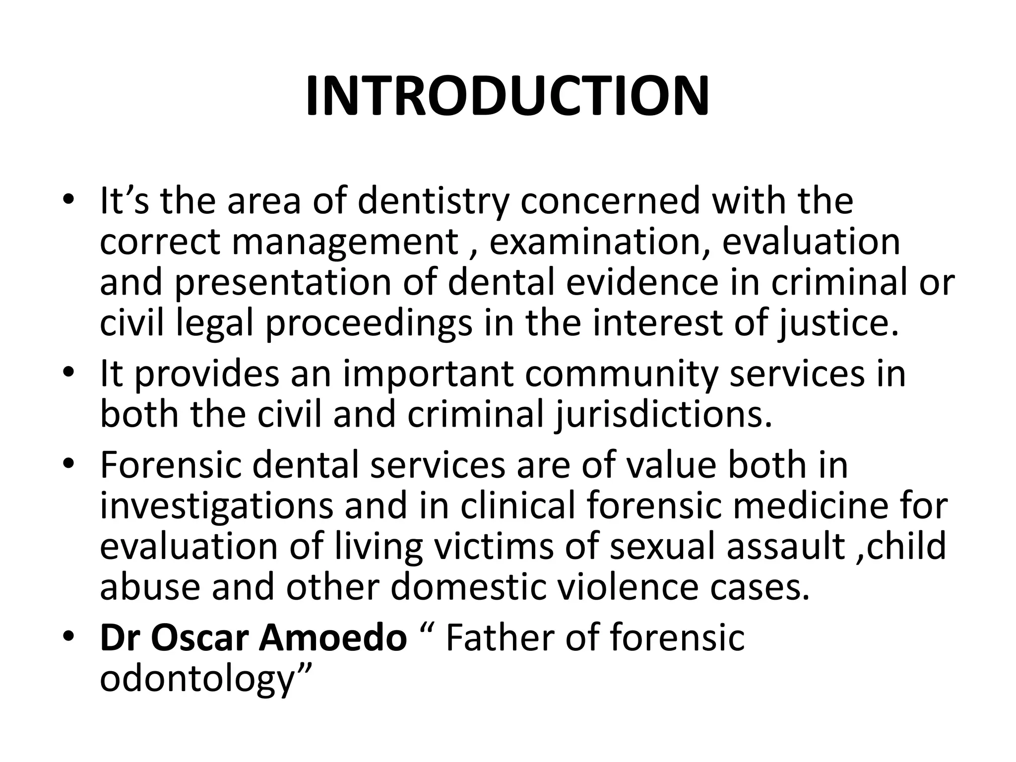
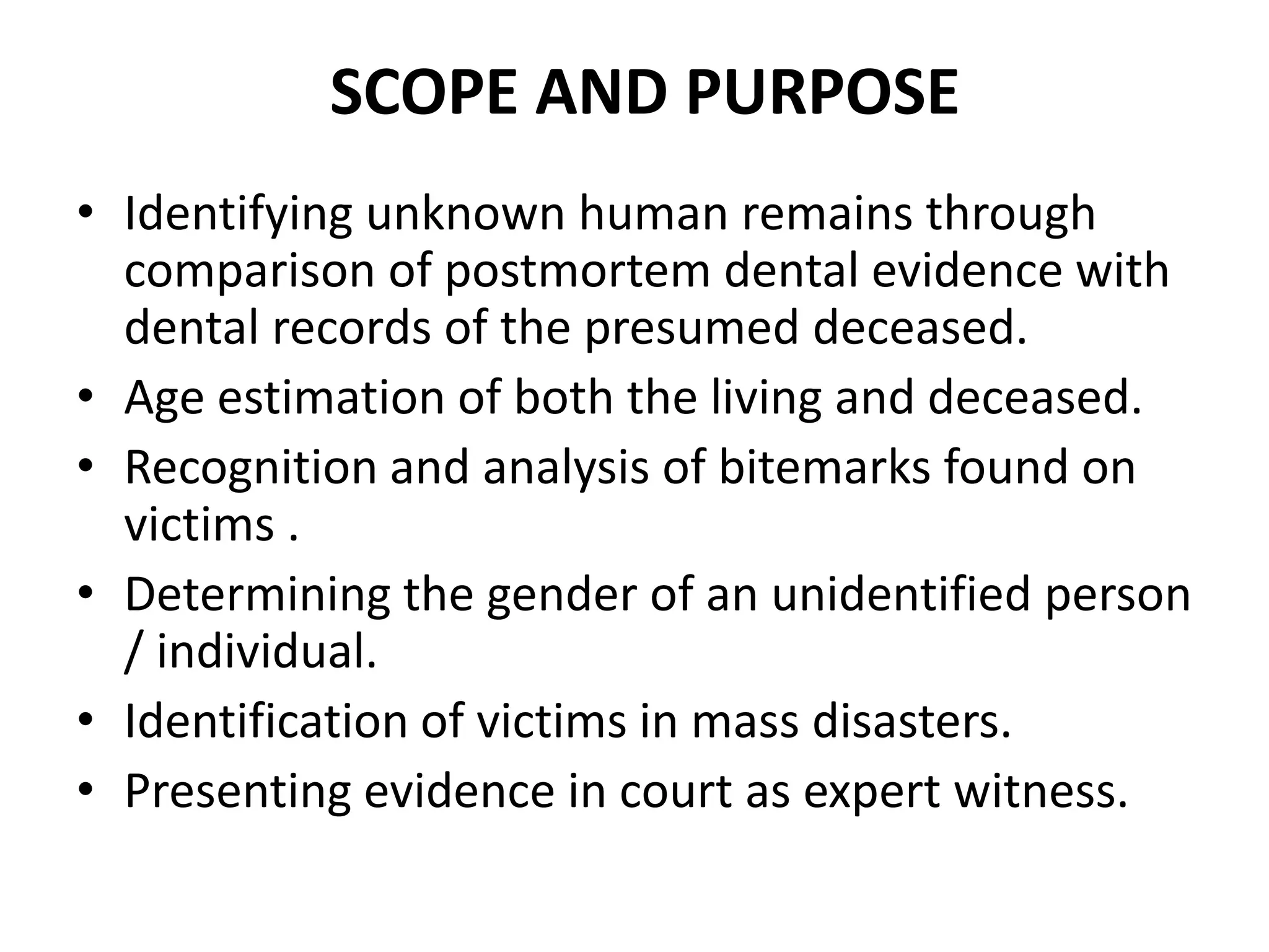
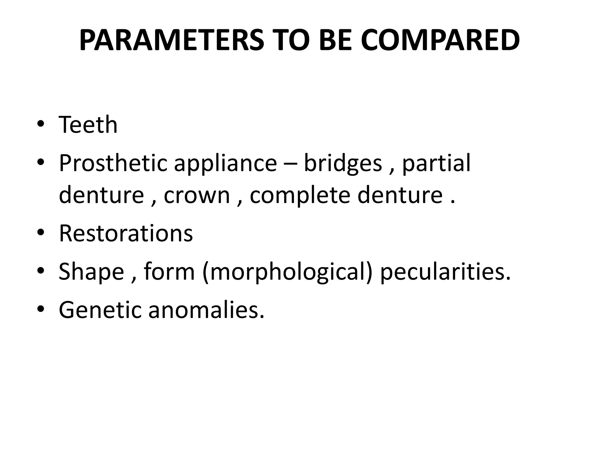
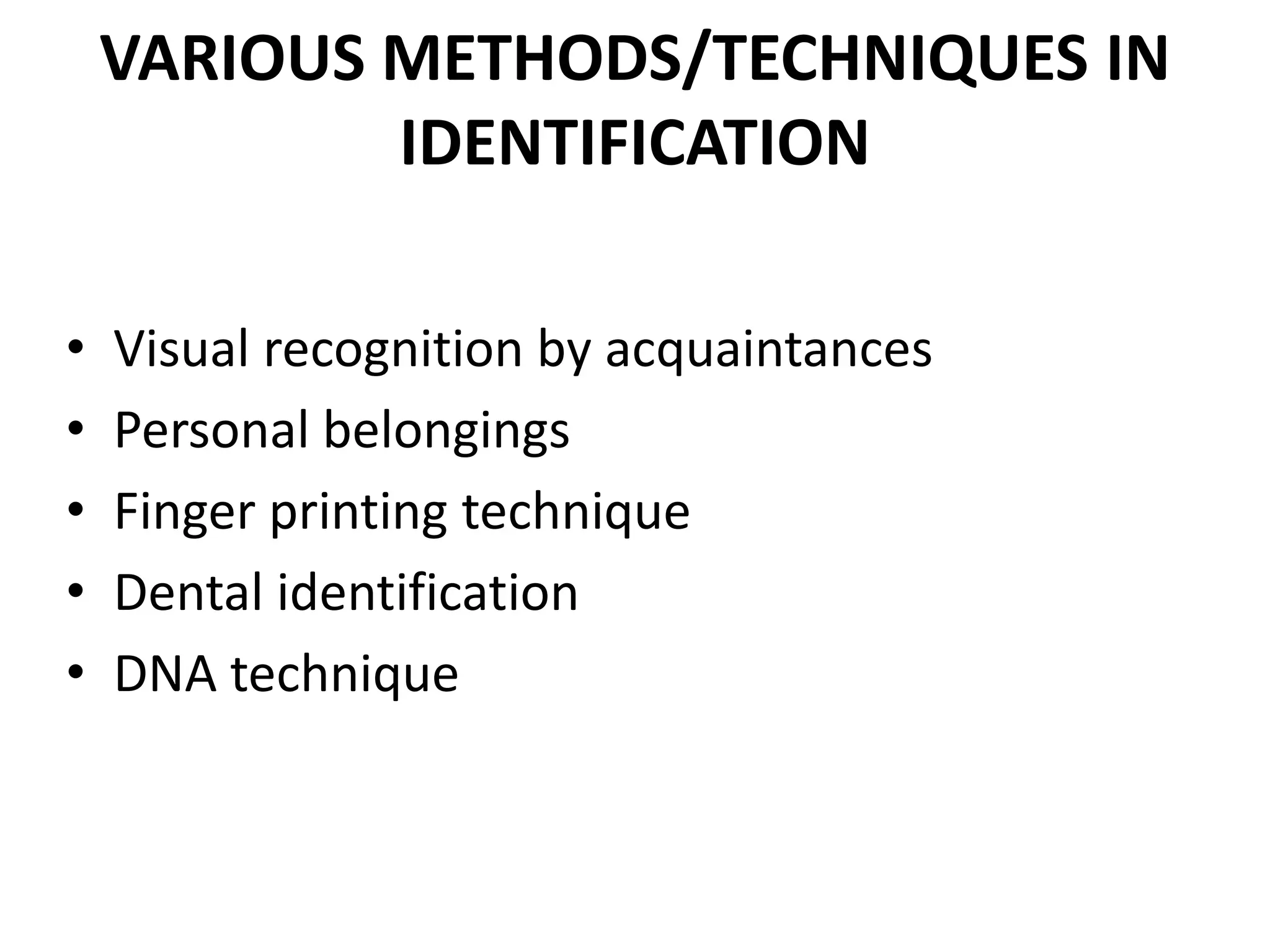
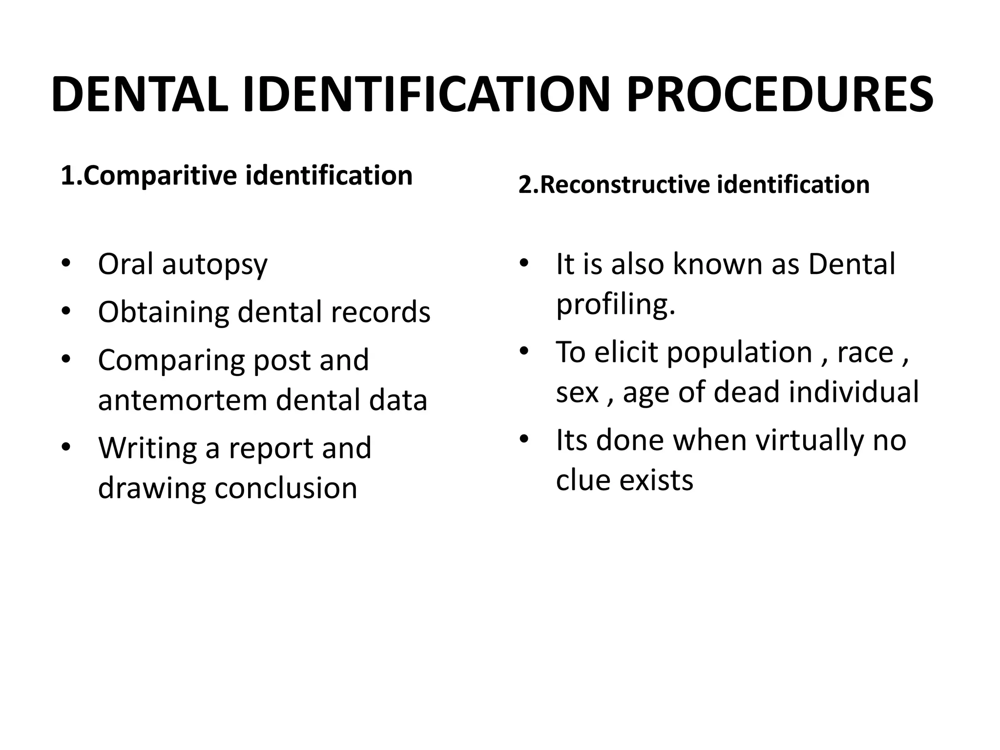
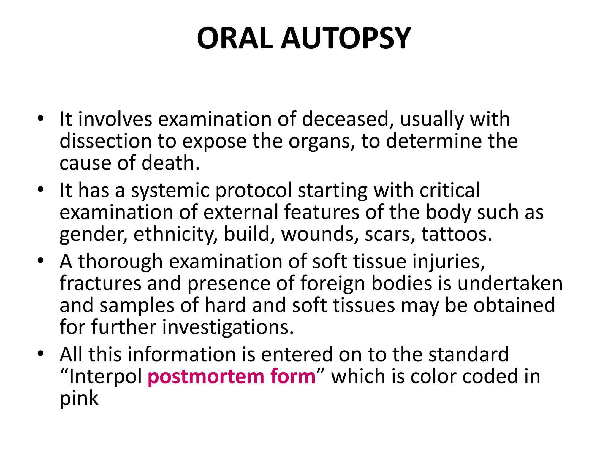
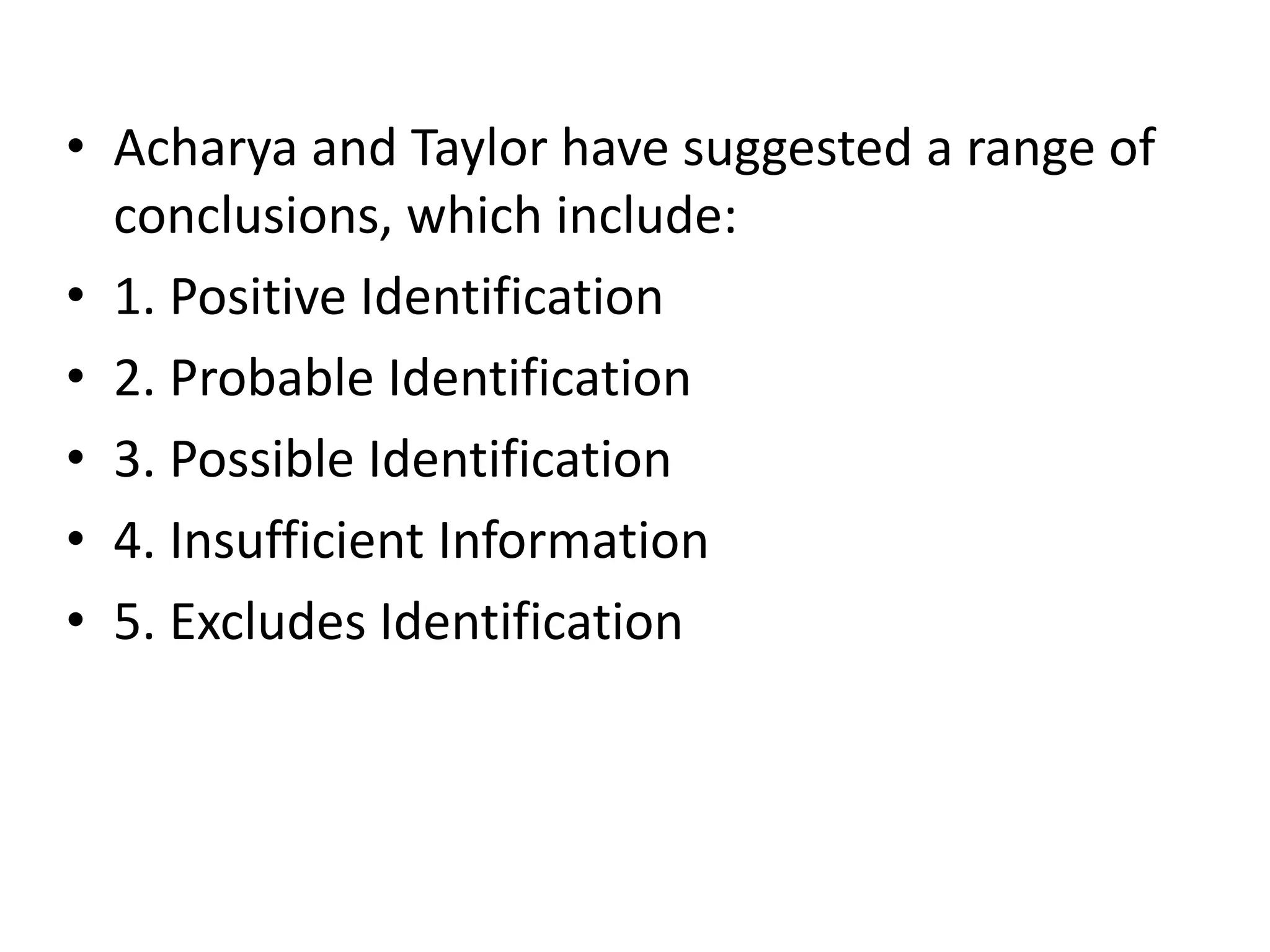
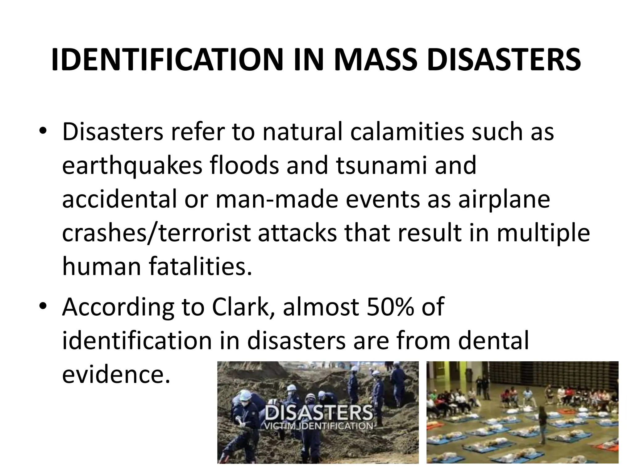
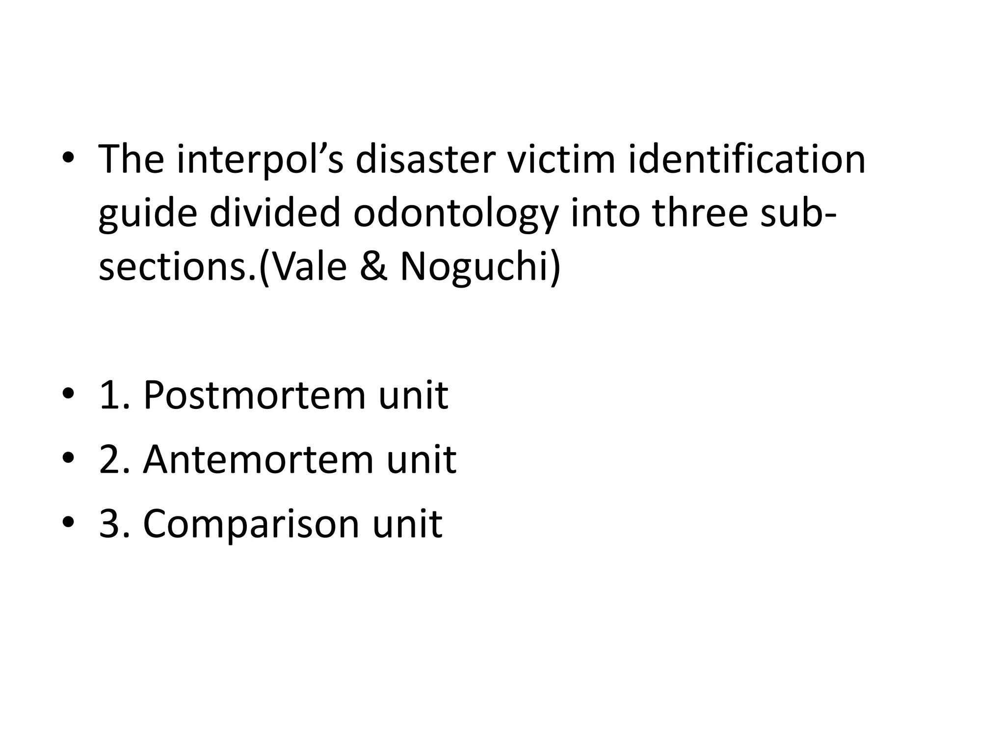
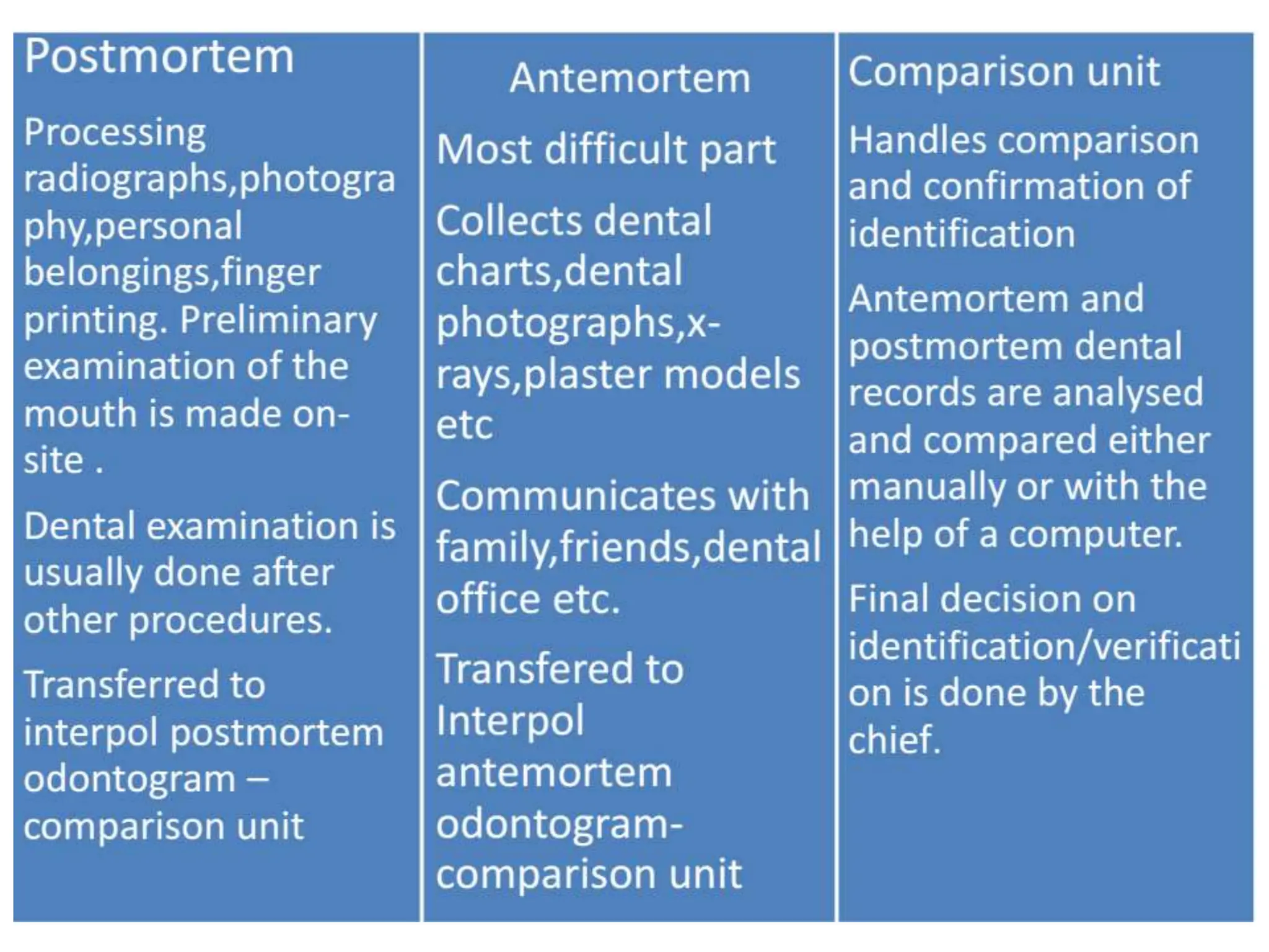
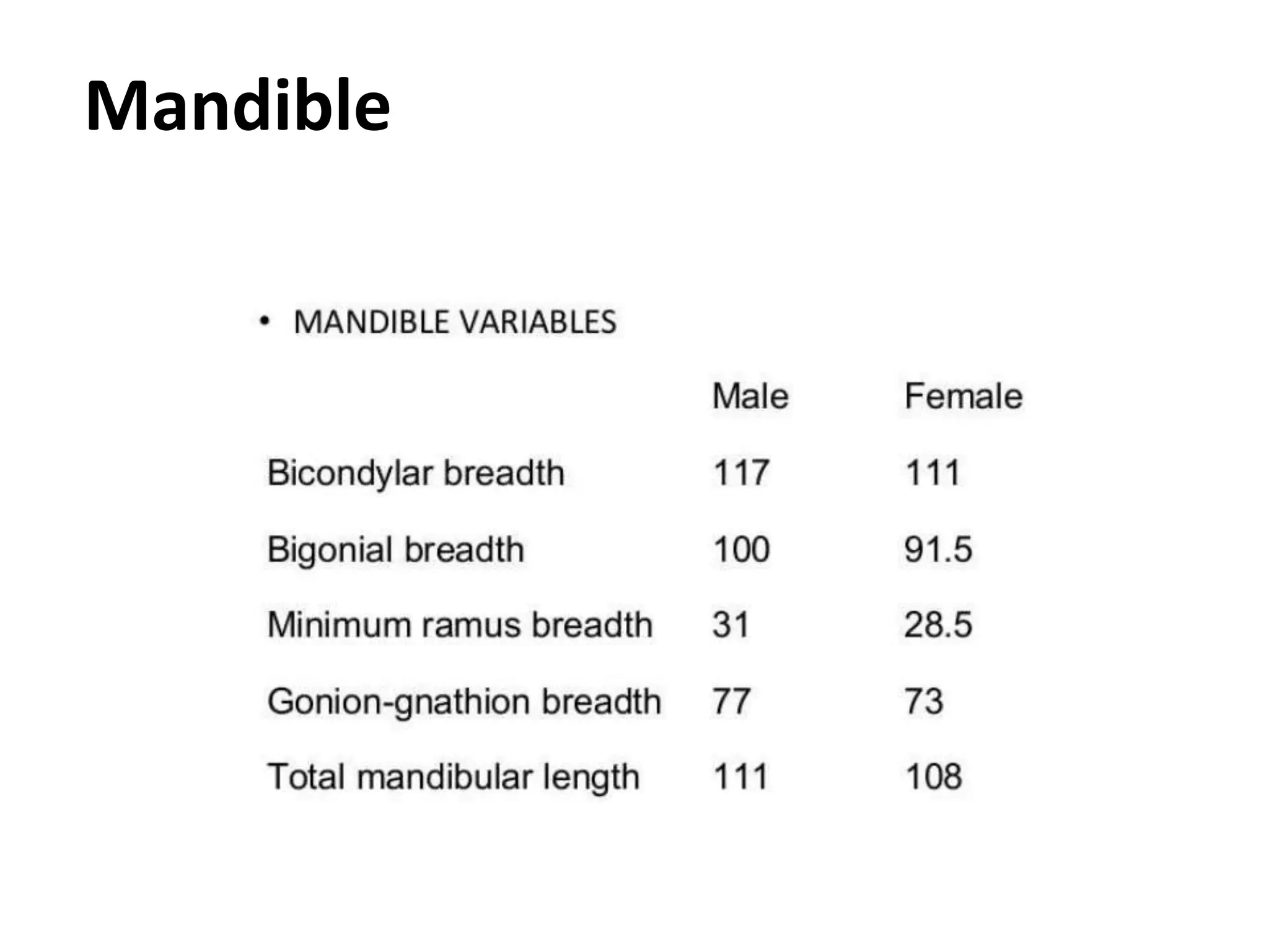
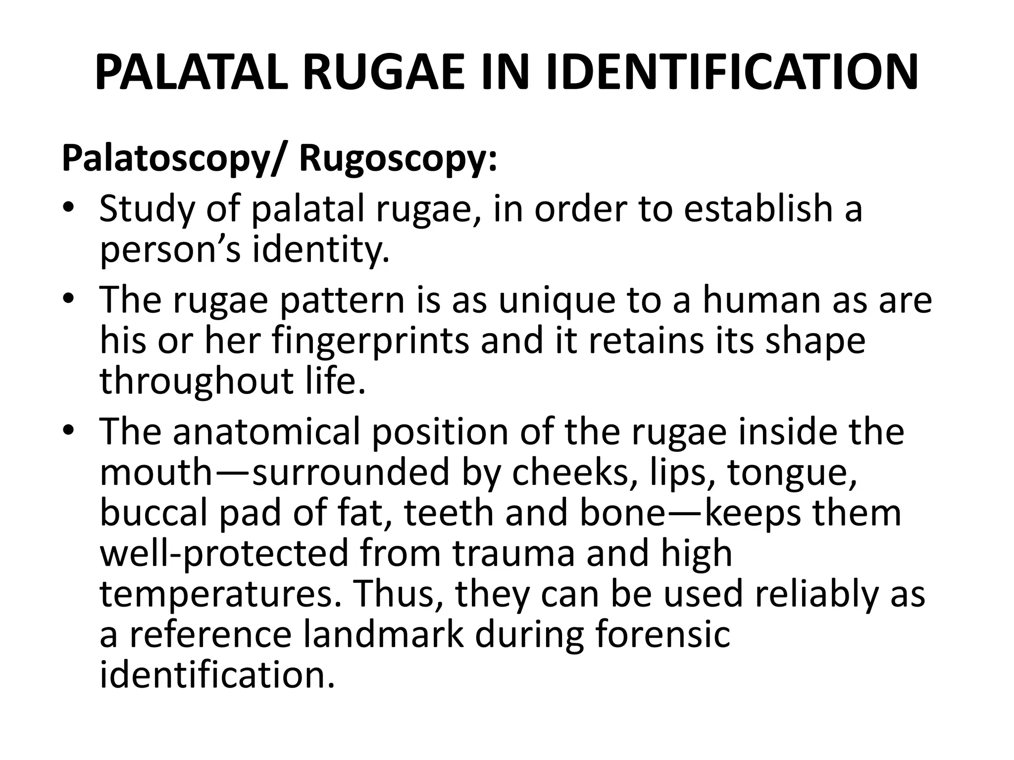
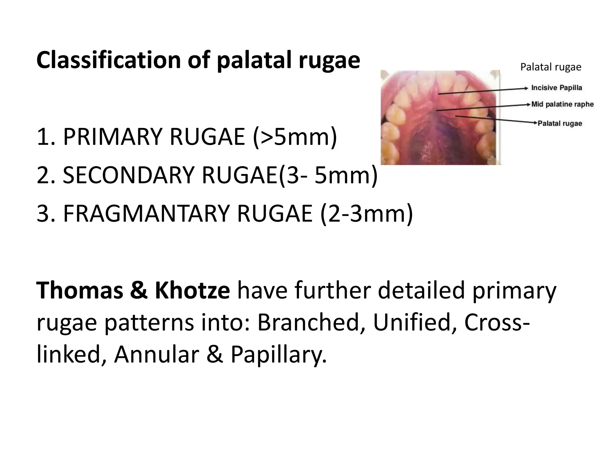
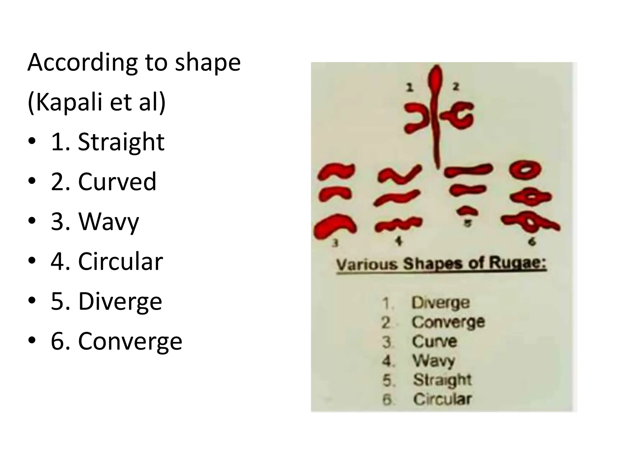
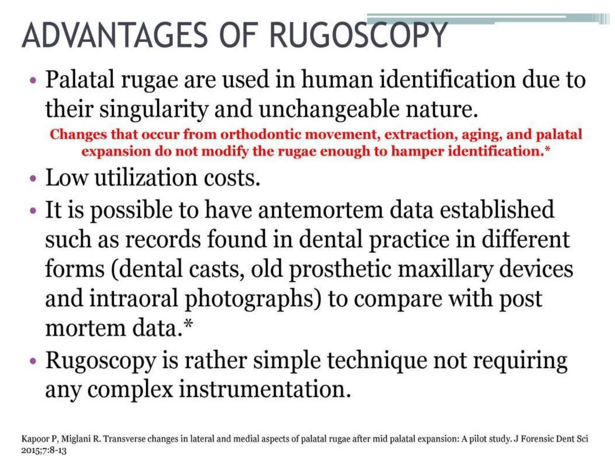
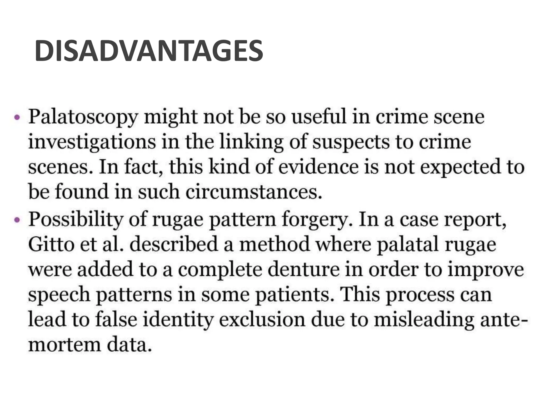
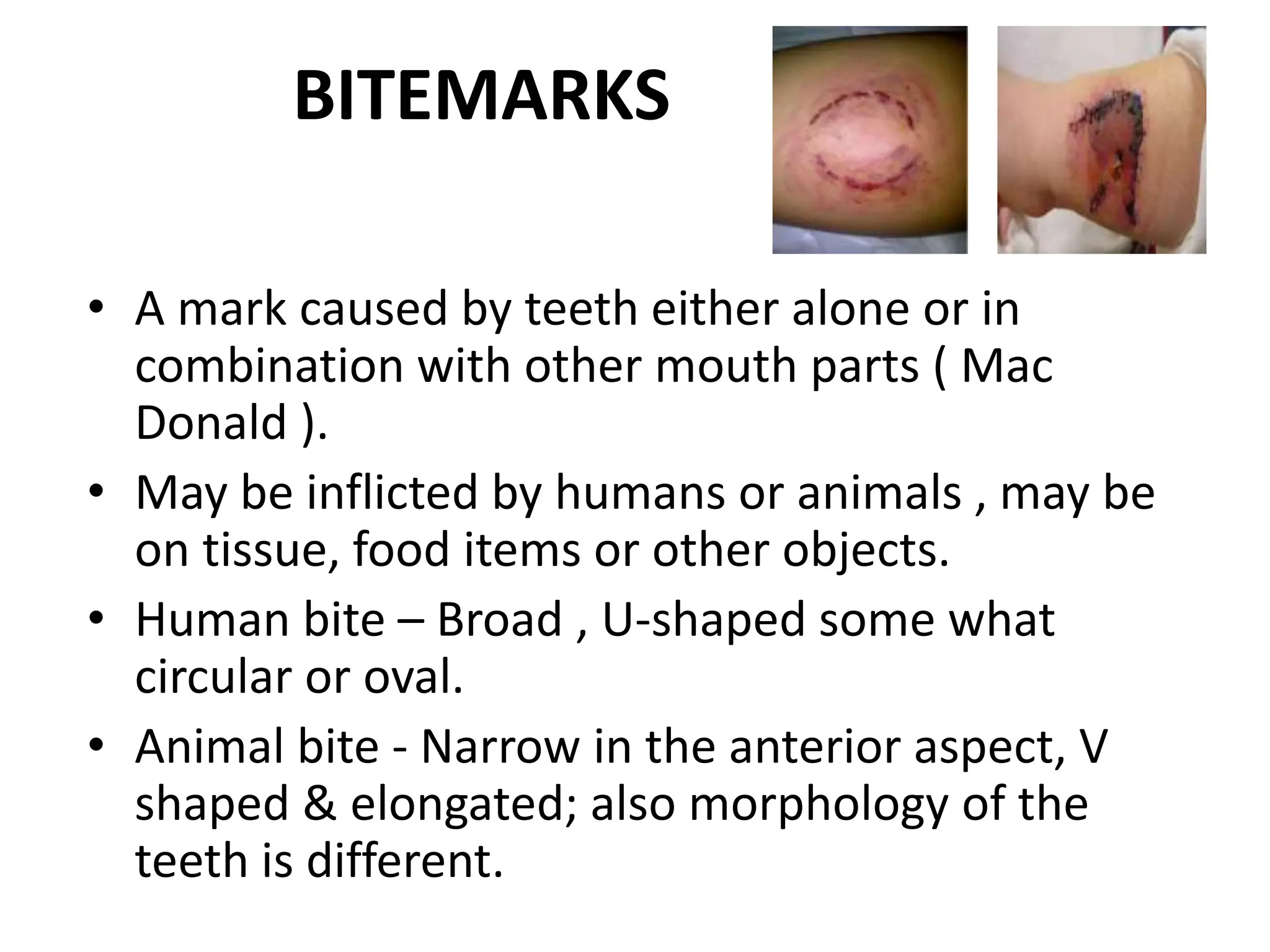
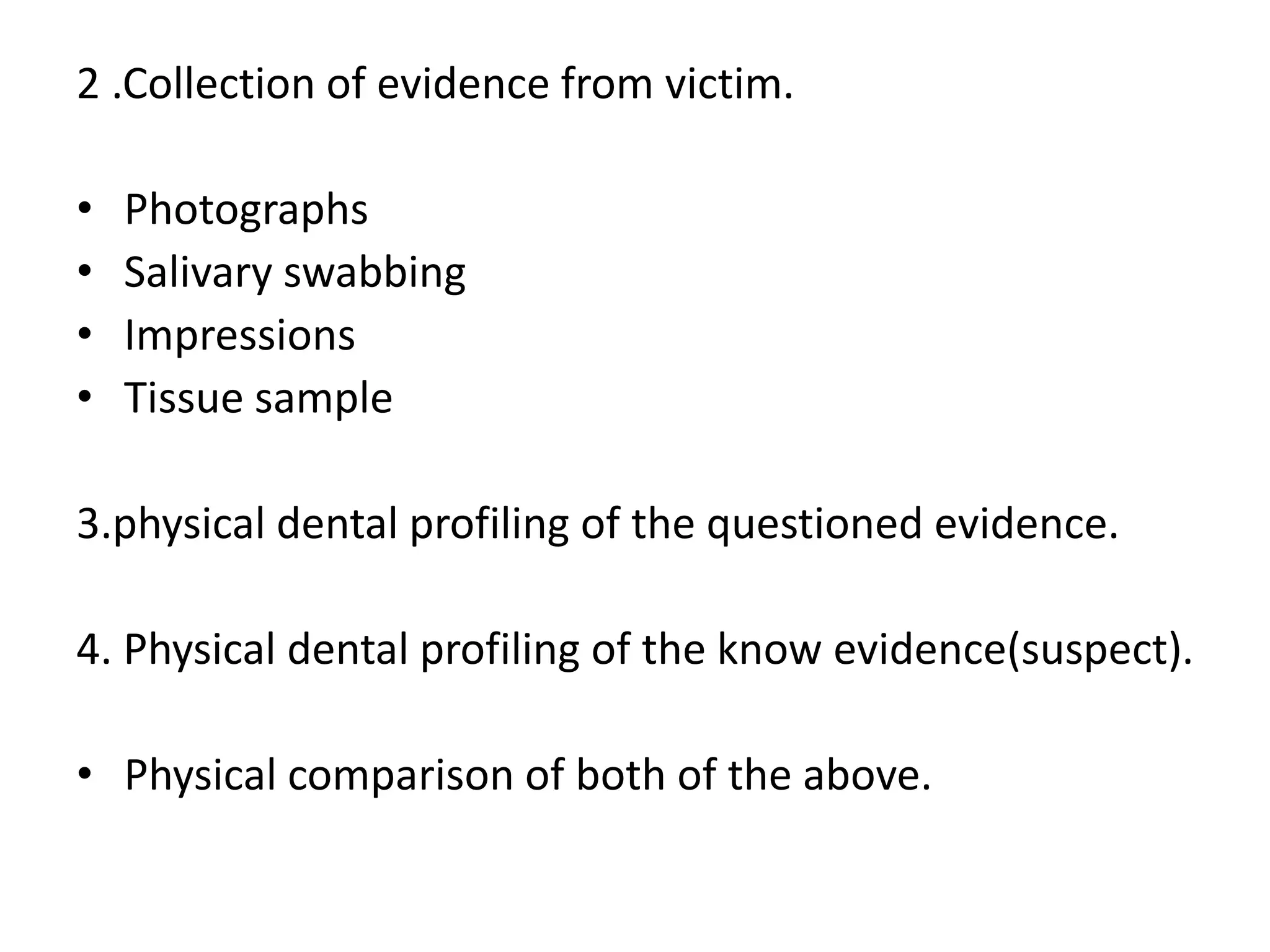
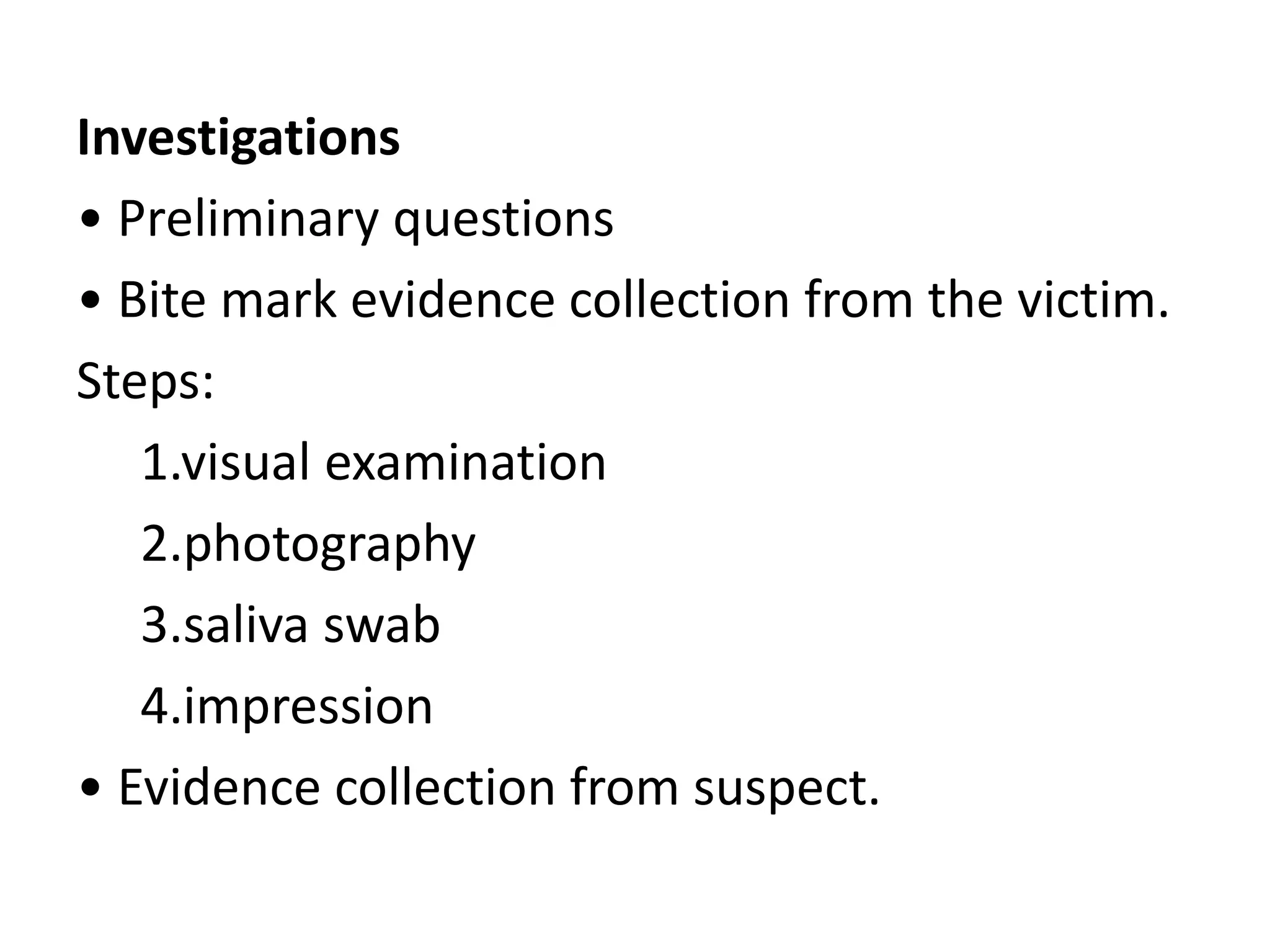
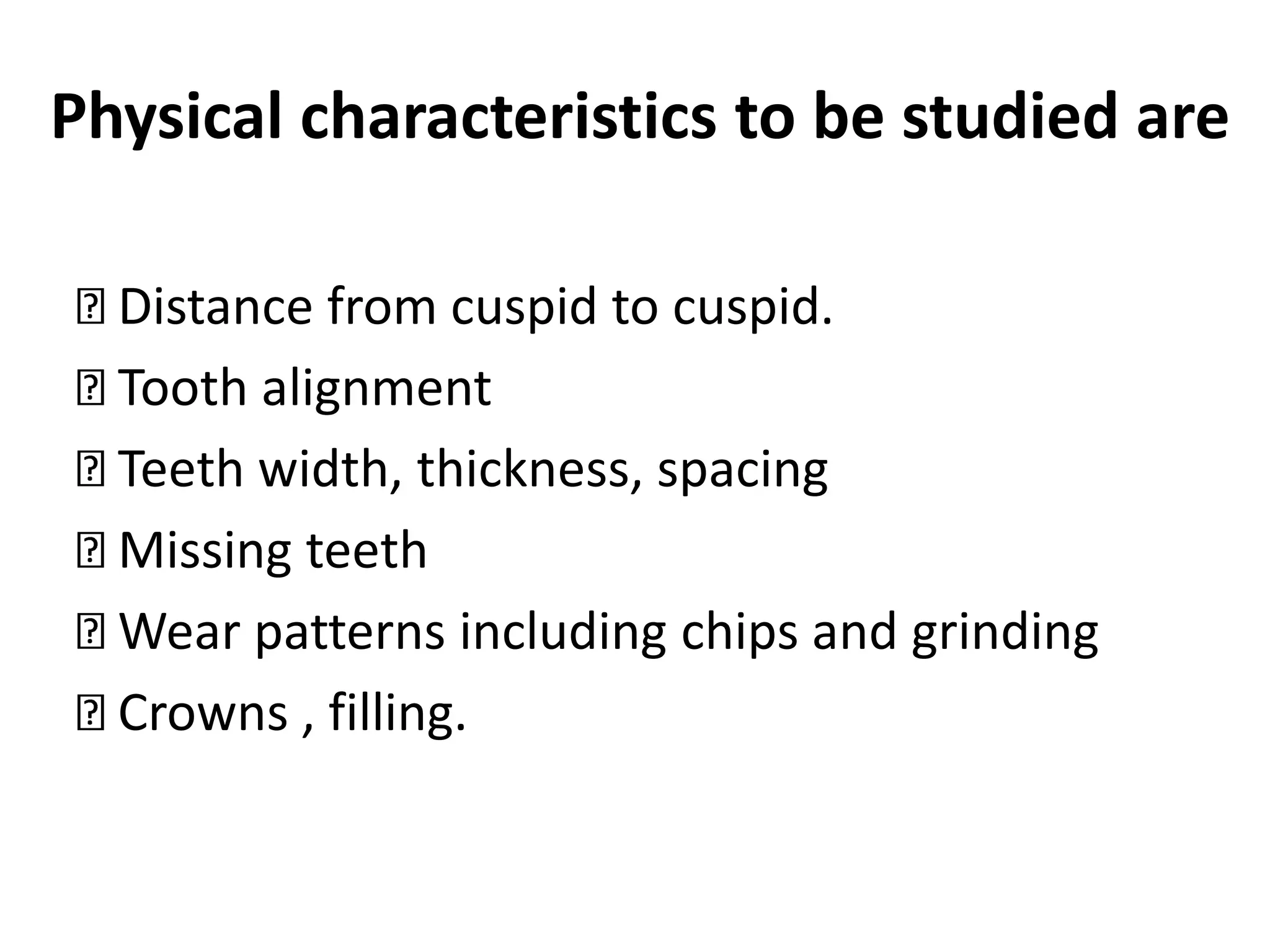
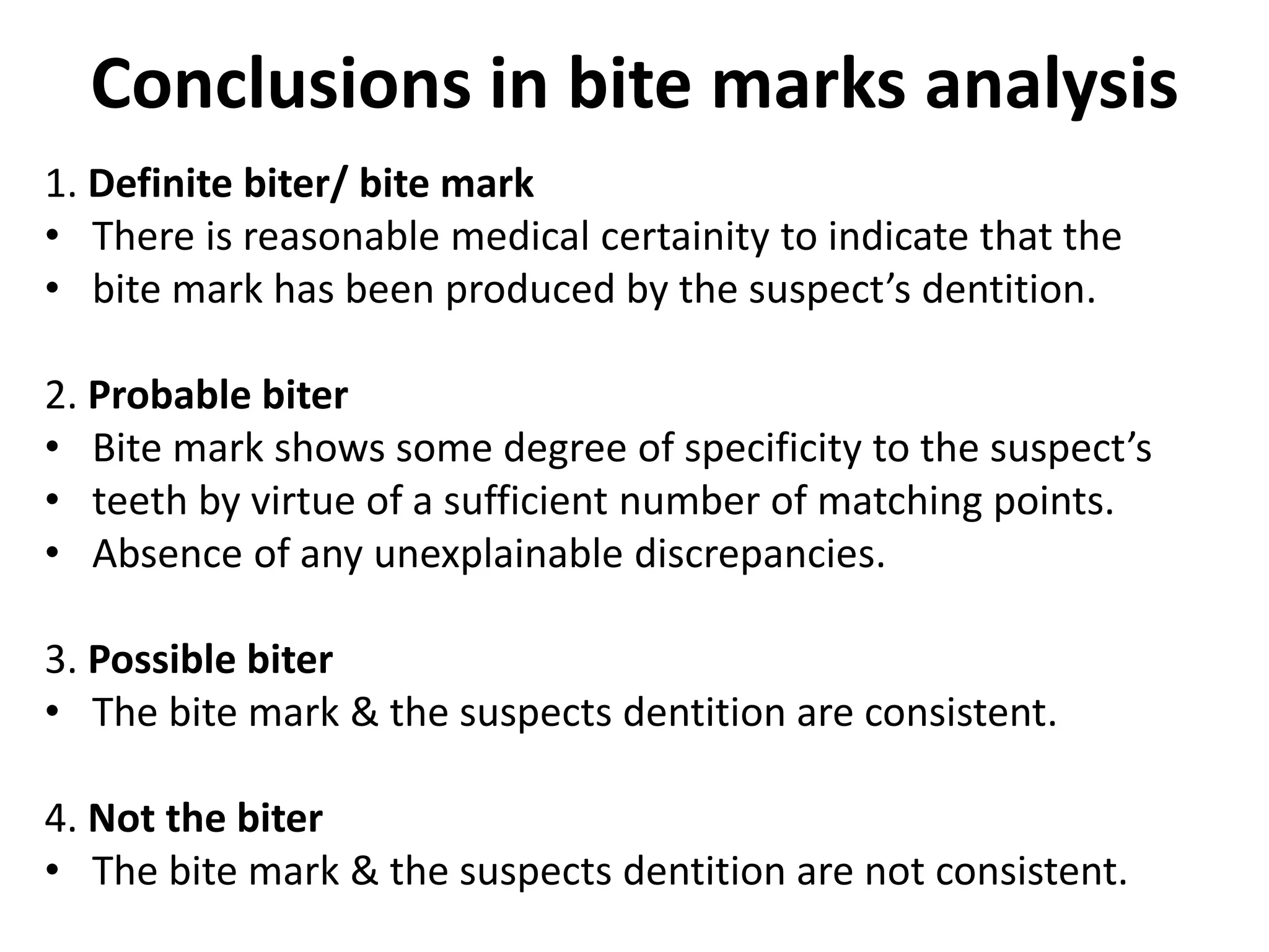
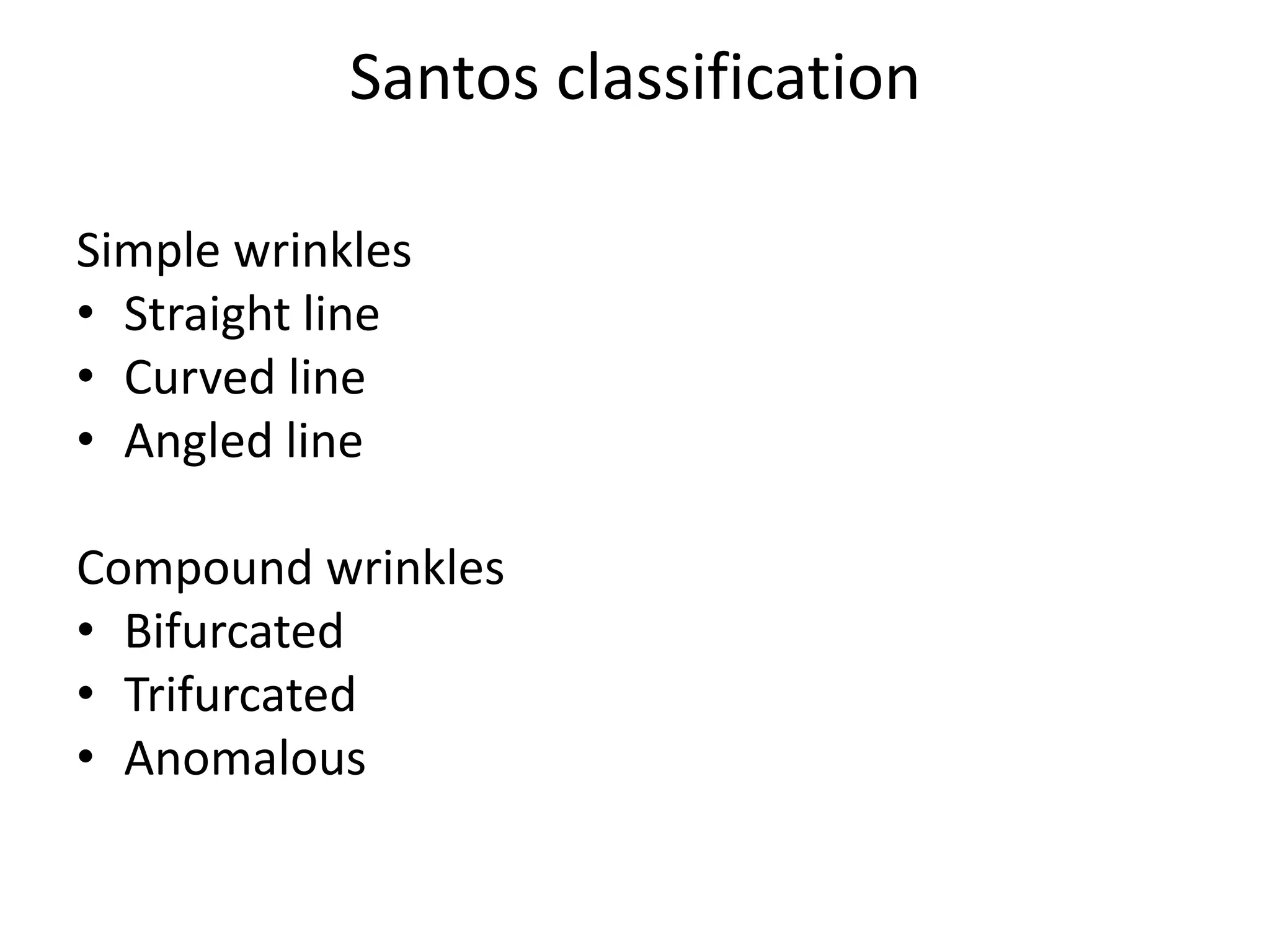
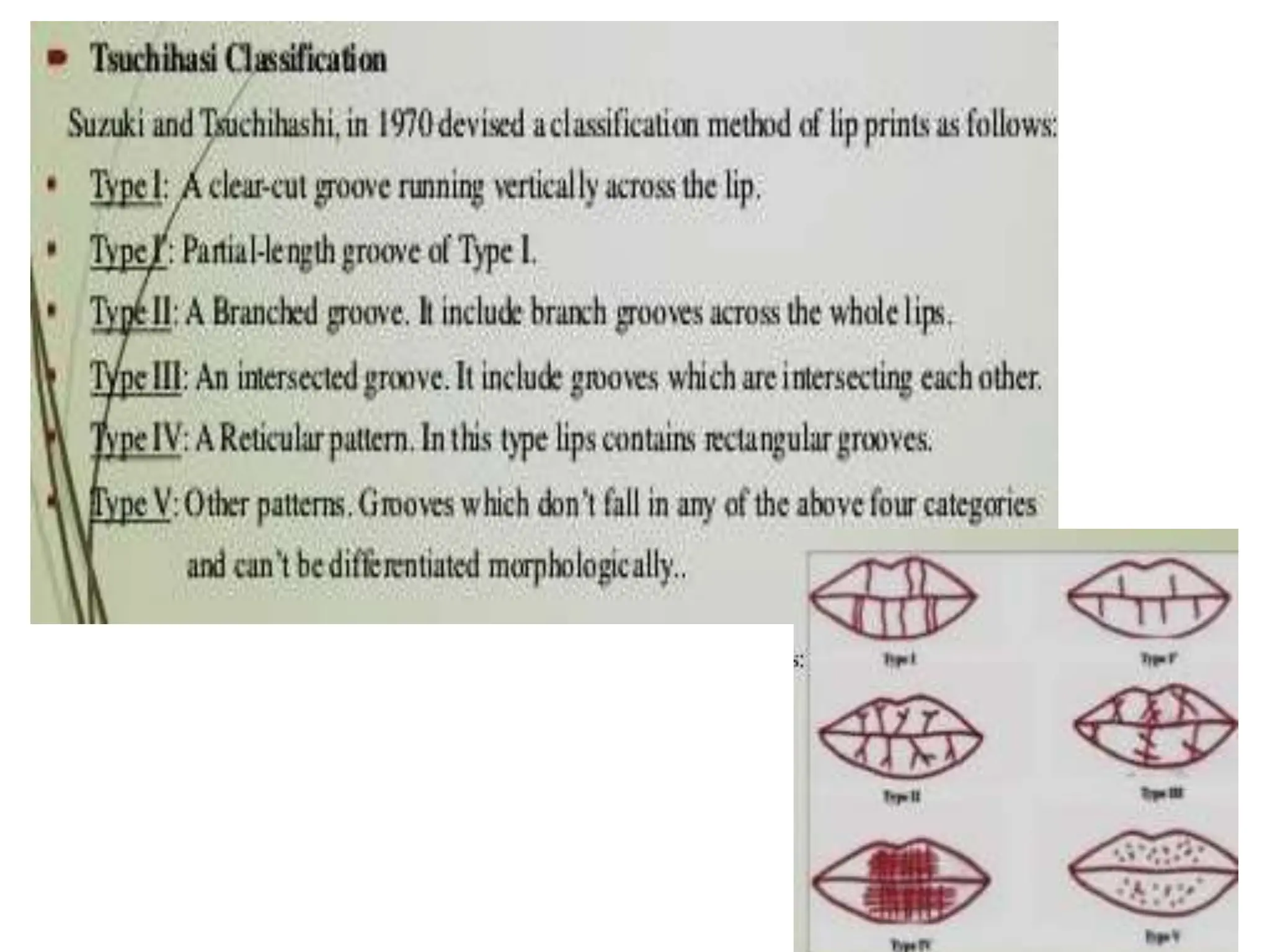
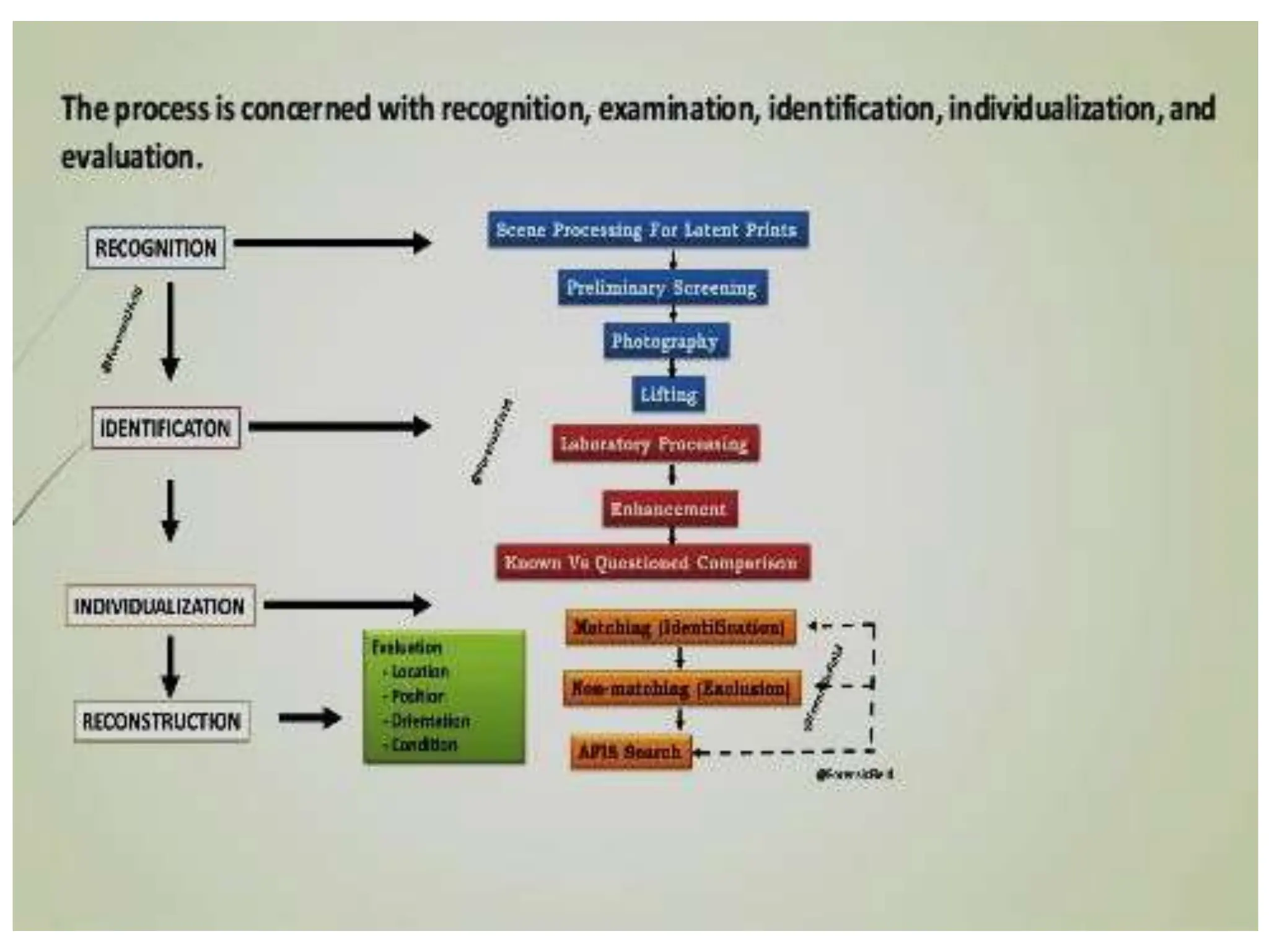
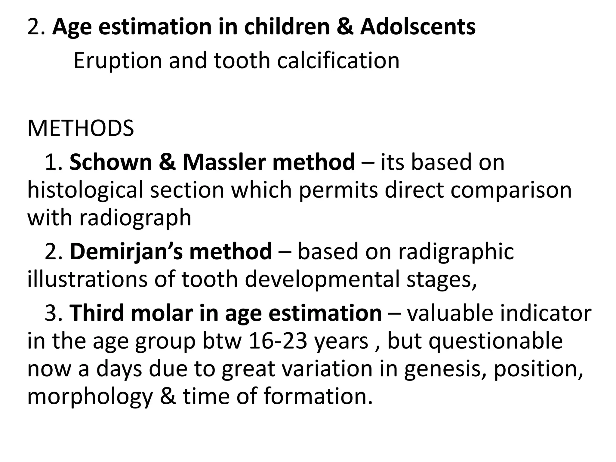
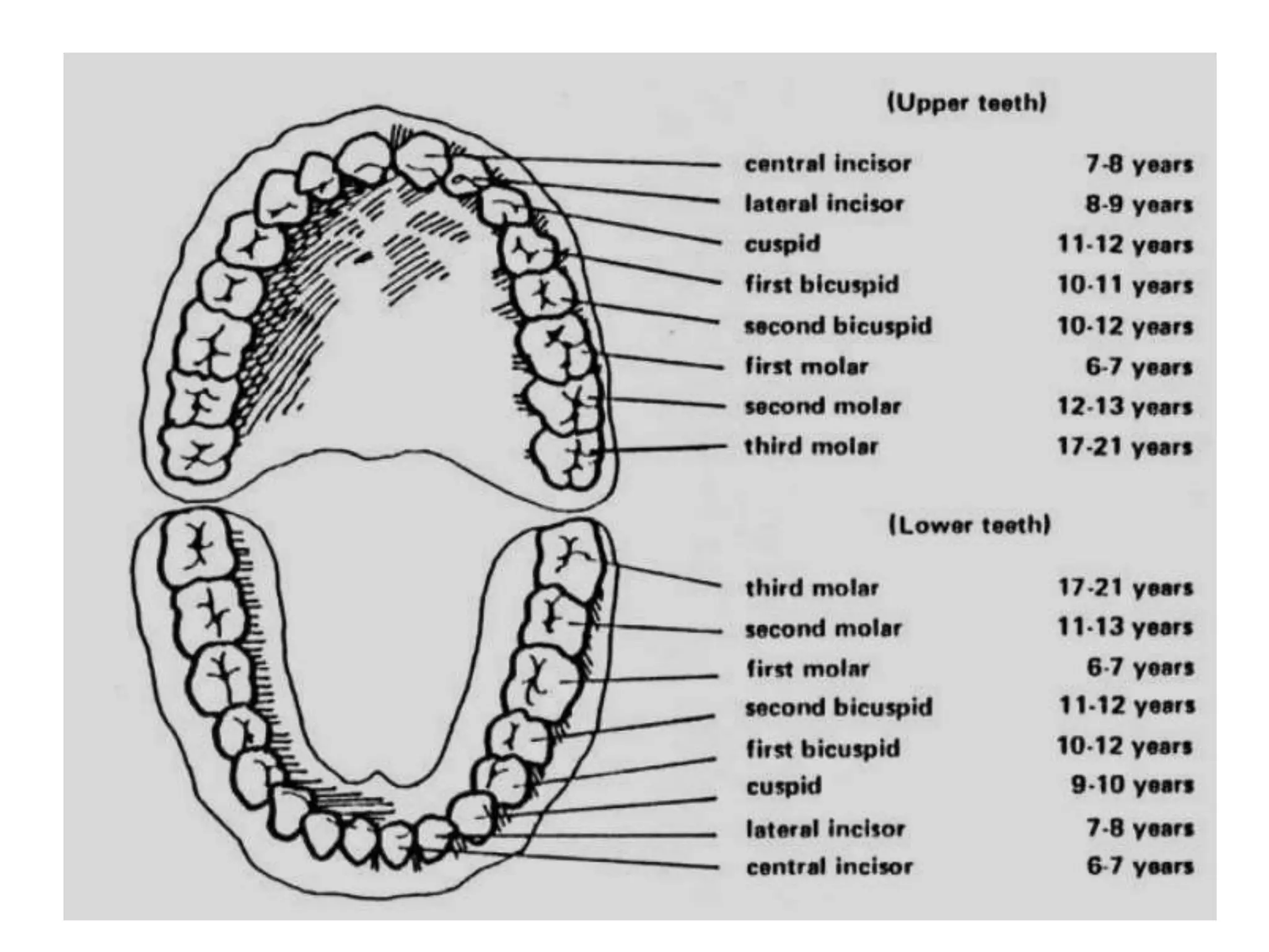
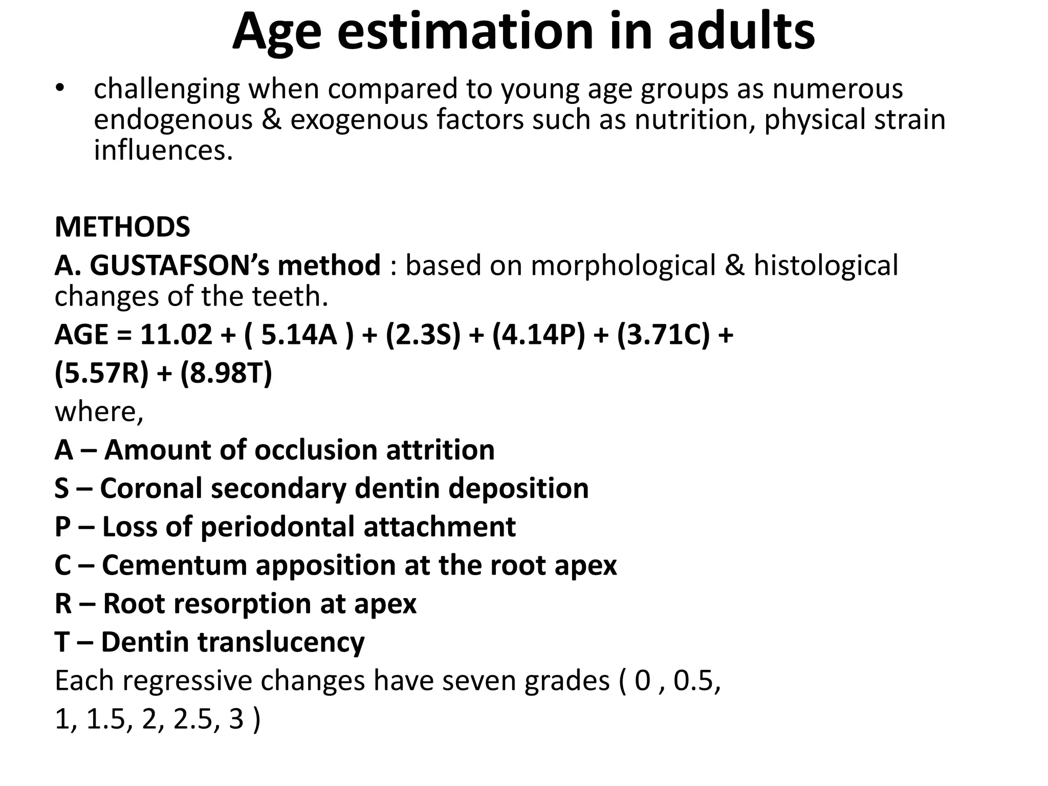
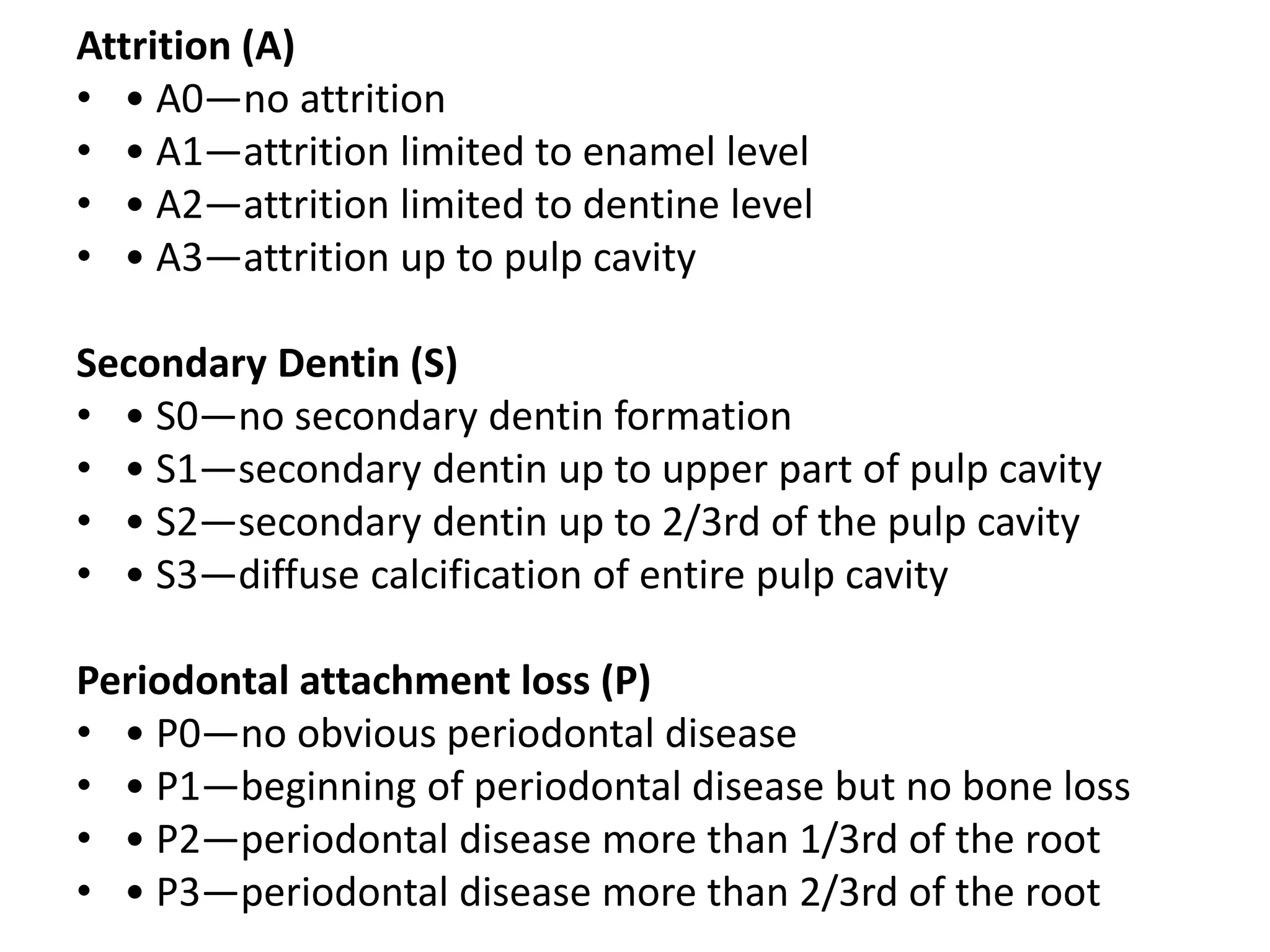
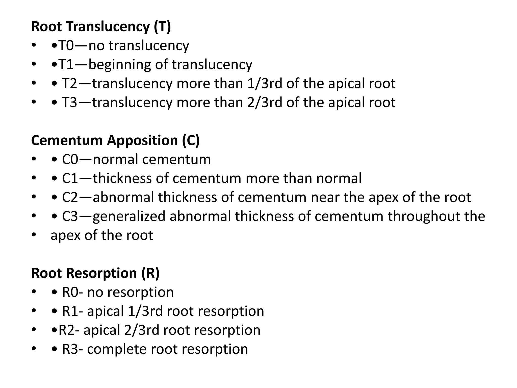
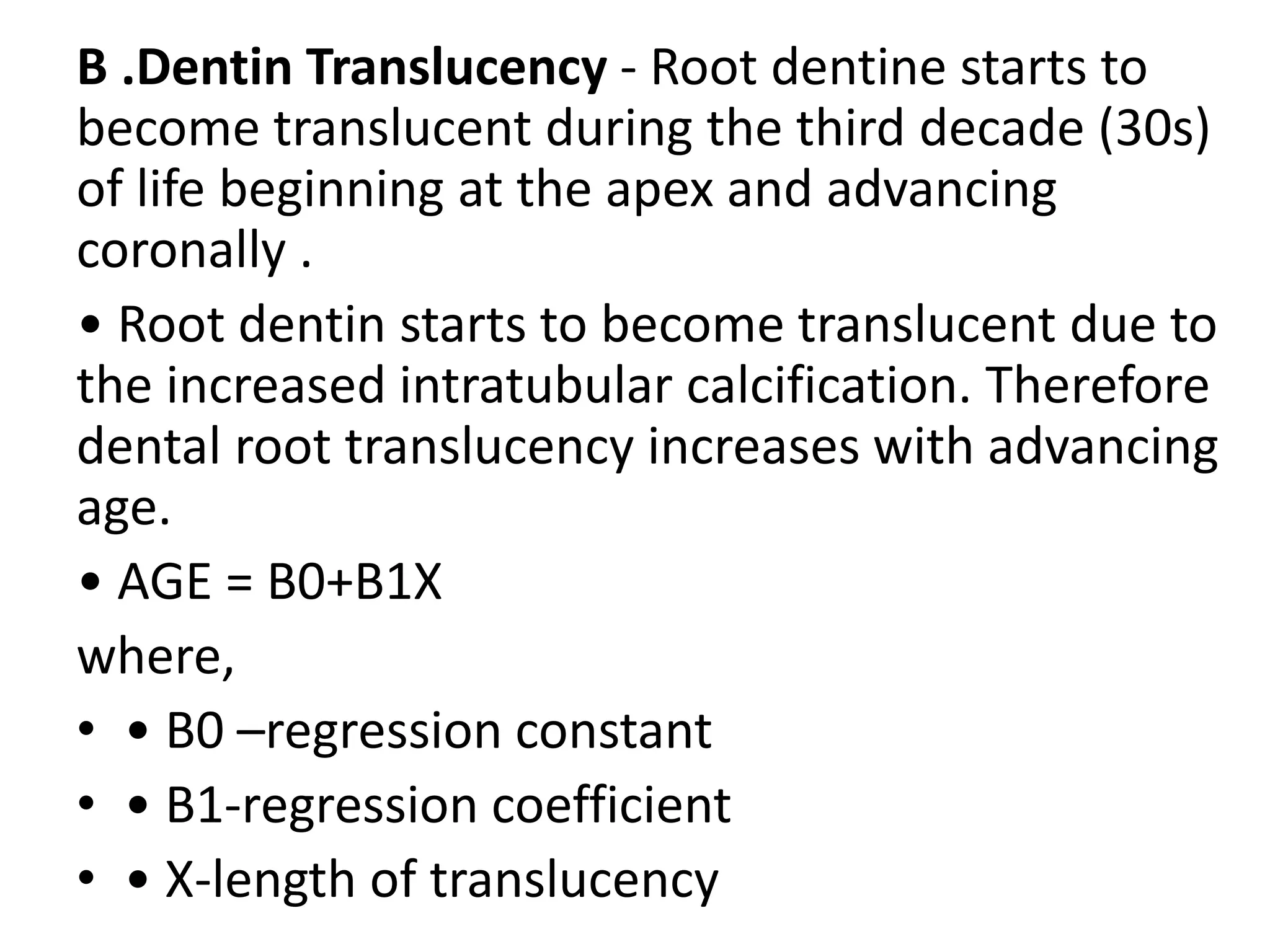
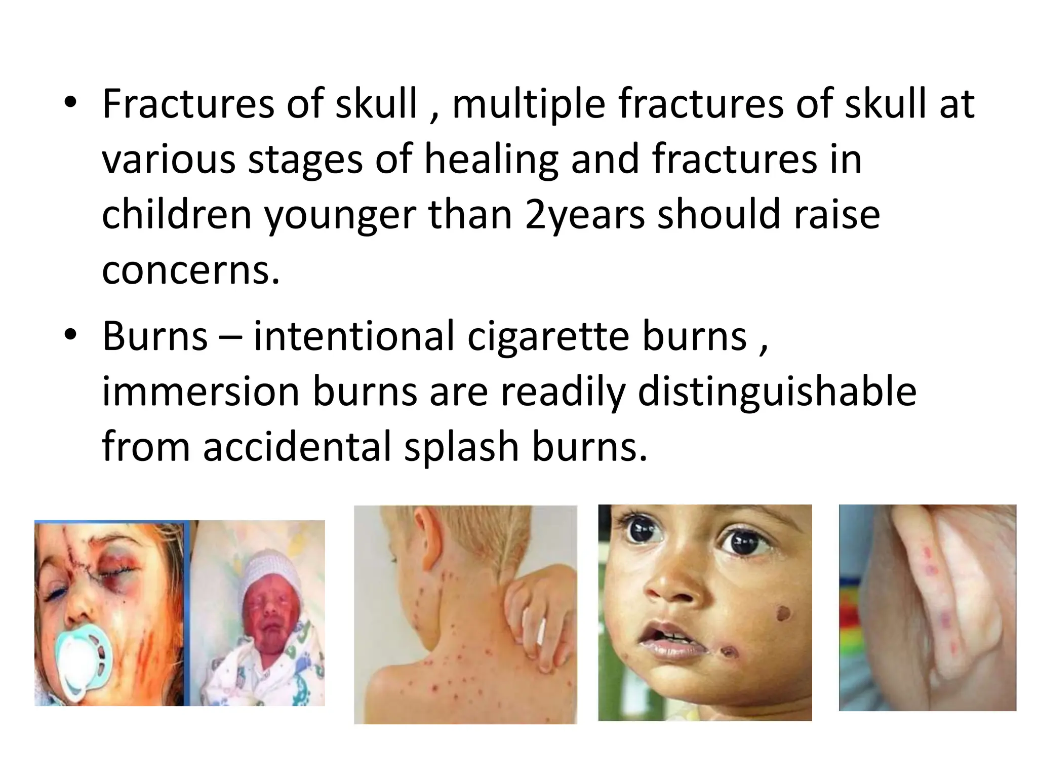
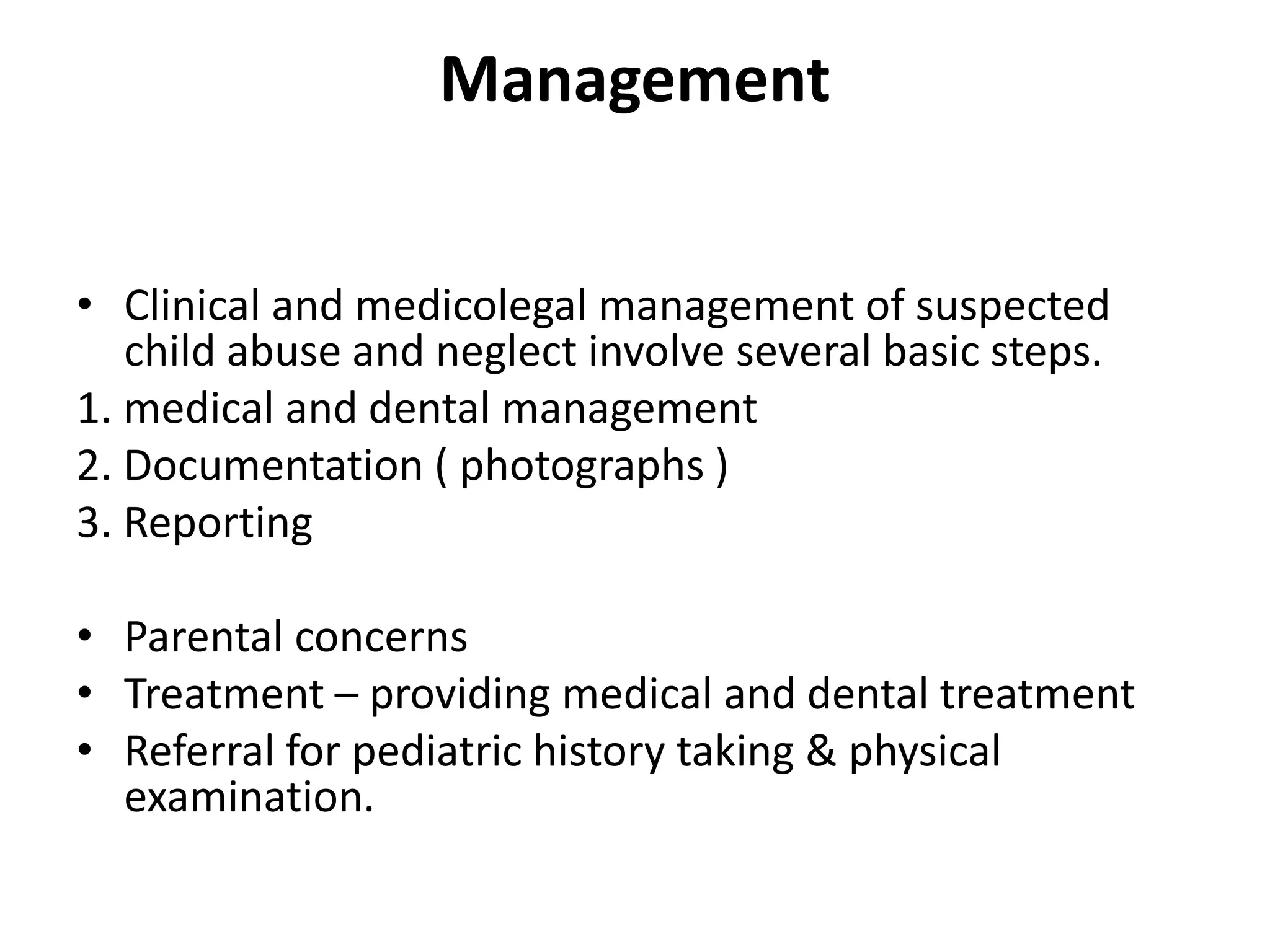
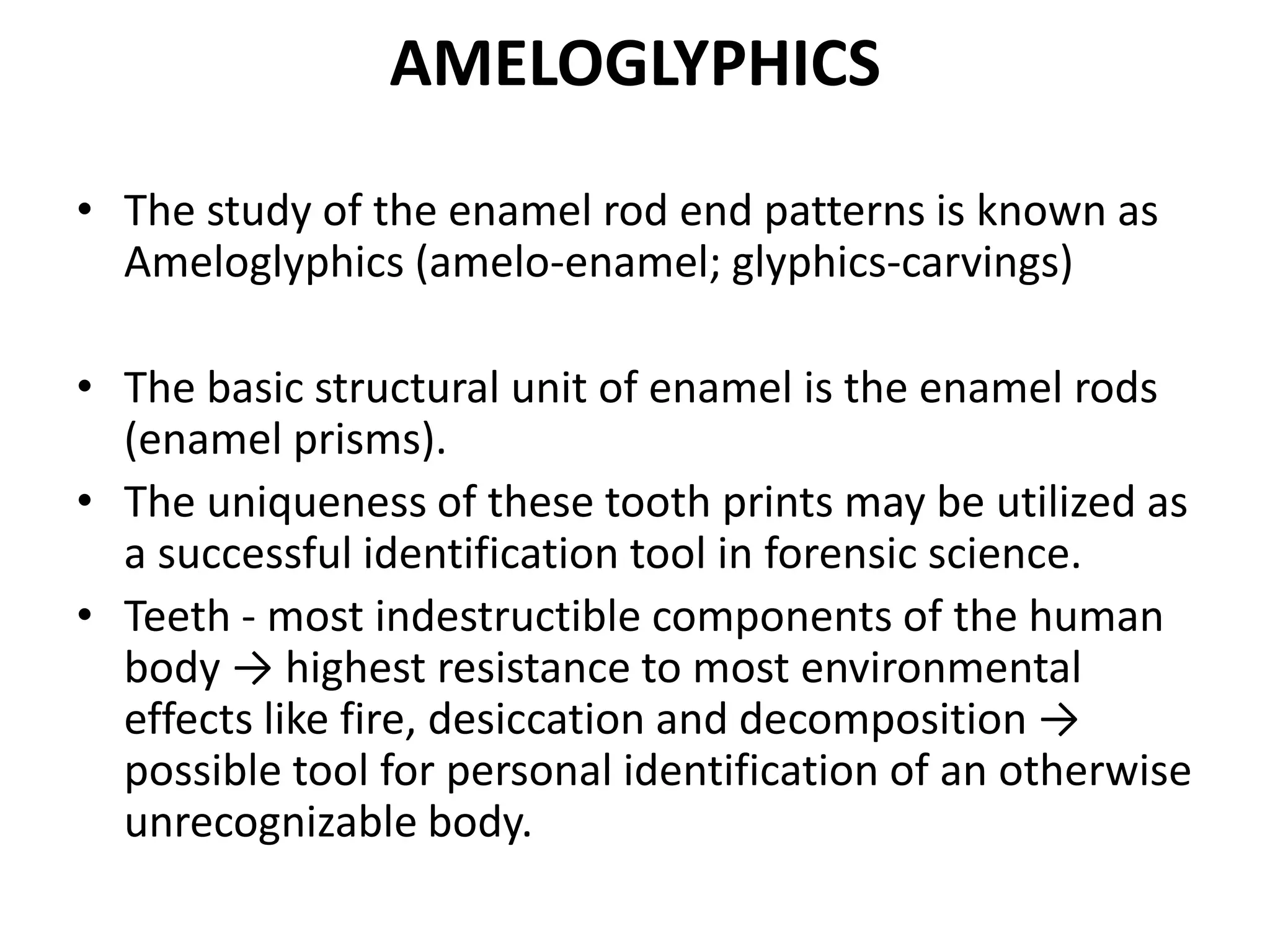
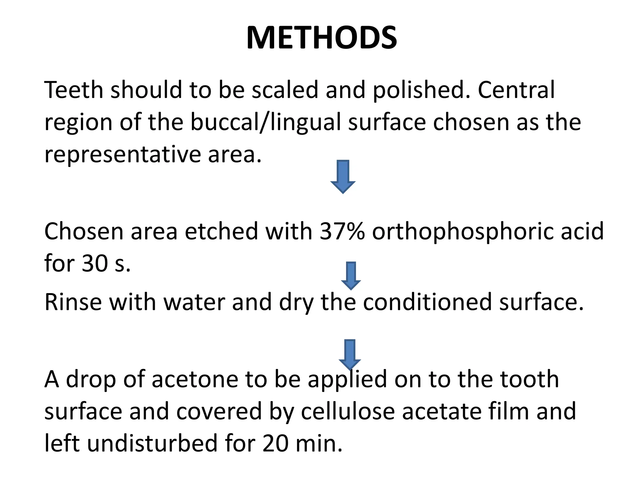
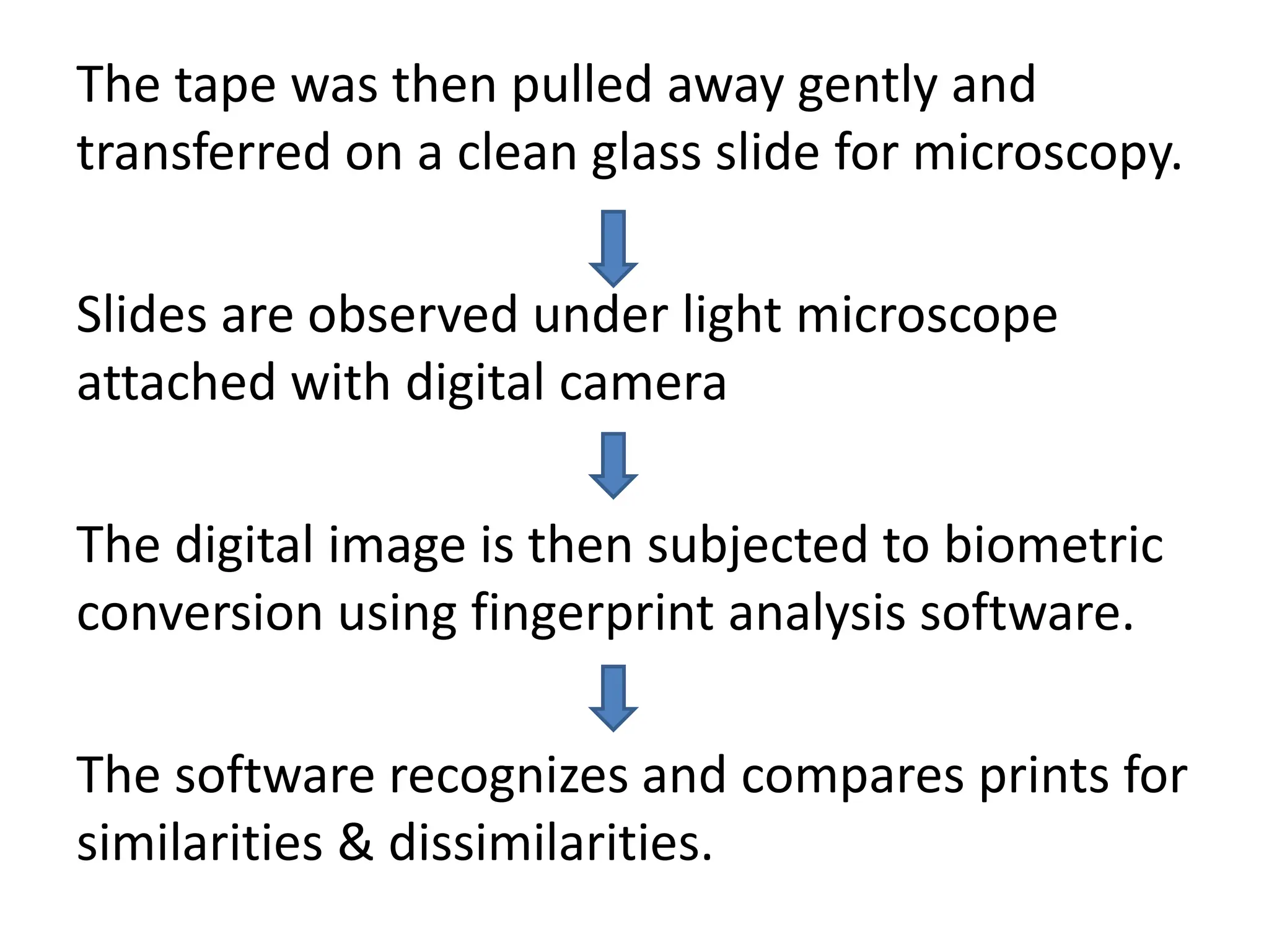
FORENSIC ODONTOLOGY
DR NONITHA S
CONTENTS
• Introduction
• Scope and purpose
• Dental identification procedures
• Identification in mass disasters
• Identification from dental DNA
• Sex determination
• Palatal rugae
• Bite marks
• Lip prints
• Dental age estimation
• Child abuse
• Ameloglyphics
• Dentist as an expert witness
• Conclusion
• References
INTRODUCTION
• It’s the area of dentistry concerned with the
correct management , examination, evaluation
and presentation of dental evidence in criminal or
civil legal proceedings in the interest of justice.
• It provides an important community services in
both the civil and criminal jurisdictions.
• Forensic dental services are of value both in
investigations and in clinical forensic medicine for
evaluation of living victims of sexual assault ,child
abuse and other domestic violence cases.
• Dr Oscar Amoedo “ Father of forensic
odontology”
SCOPE AND PURPOSE
• Identifying unknown human remains through
comparison of postmortem dental evidence with
dental records of the presumed deceased.
• Age estimation of both the living and deceased.
• Recognition and analysis of bitemarks found on
victims .
• Determining the gender of an unidentified person
/ individual.
• Identification of victims in mass disasters.
• Presenting evidence in court as expert witness.
PARAMETERS TO BE COMPARED
• Teeth
• Prosthetic appliance – bridges , partial
denture , crown , complete denture .
• Restorations
• Shape , form (morphological) pecularities.
• Genetic anomalies.
VARIOUS METHODS/TECHNIQUES IN
IDENTIFICATION
• Visual recognition by acquaintances
• Personal belongings
• Finger printing technique
• Dental identification
• DNA technique
DENTAL IDENTIFICATION PROCEDURES
1.Comparitive identification
• Oral autopsy
• Obtaining dental records
• Comparing post and
antemortem dental data
• Writing a report and
drawing conclusion
2.Reconstructive identification
• It is also known as Dental
profiling.
• To elicit population , race ,
sex , age of dead individual
• Its done when virtually no
clue exists
ORAL AUTOPSY
• It involves examination of deceased, usually with
dissection to expose the organs, to determine the
cause of death.
• It has a systemic protocol starting with critical
examination of external features of the body such as
gender, ethnicity, build, wounds, scars, tattoos.
• A thorough examination of soft tissue injuries,
fractures and presence of foreign bodies is undertaken
and samples of hard and soft tissues may be obtained
for further investigations.
• All this information is entered on to the standard
“Interpol postmortem form” which is color coded in
pink
OBTAINING DENTAL RECORDS
• Dental records contain information of
treatment and dental status of a person
during his/her lifetime.
• Such records may be in the form of dental
charts, radio graphs, casts and/or
photographs.
• The contents of all available dental records
should be transcribed onto the standard
“Interpol ante-mortem form” which is color
coded in yellow.
COMPARING POST AND ANTE-
MORTEM DENTAL DATA
• Once the post-mortem evidence and dental
records are available, the data can be
compared.
• Features compared include tooth morphology
and associated bony structures, pathology and
dental restorations.
Writing A Report And Drawing
Conclusions
• One needs to remember that any attempt at
establishing identity is addressed to the legal
authorities.
• Therefore, a detailed report and factual
conclusion, based on comparison, must be
clearly stated.
• Acharya and Taylor have suggested a range of
conclusions, which include:
• 1. Positive Identification
• 2. Probable Identification
• 3. Possible Identification
• 4. Insufficient Information
• 5. Excludes Identification
IDENTIFICATION IN MASS DISASTERS
• Disasters refer to natural calamities such as
earthquakes floods and tsunami and
accidental or man-made events as airplane
crashes/terrorist attacks that result in multiple
human fatalities.
• According to Clark, almost 50% of
identification in disasters are from dental
evidence.
• The interpol’s disaster victim identification
guide divided odontology into three sub-
sections.(Vale & Noguchi)
• 1. Postmortem unit
• 2. Antemortem unit
• 3. Comparison unit
IDENTIFICATION FROM DENTAL DNA
• Since teeth can resist extreme conditions, they are the
excellent source of DNA.
• Routinely applied technique - polymerized chain
reactions allows amplification of even highly degraded
DNA.
• This facilitates comparison with the known biological
ante-mortem sample of the person such as hair,
epithelial cells from a tooth brush or a biopsy
specimen.
• If the person’s ante-mortem sample is unavailable, the
DNA pattern may be compared to a parent or a sibling.
SEX DIFFERENTIATION
• Determining the sex of unknown human
remains is the second step in building a dental
profile.
• Gender can be determined based on:
1. morphology of skull and mandible.
2. tooth measurements.
3. DNA analysis from teeth
• Amelogenin (AMEL) is one of the major matrix
proteins secreted by the ameloblasts of the
enamel.
• The AMEL gene, coding for a highly conserved
protein, is located on X- and Y chromosomes in
humans.
• Thus the females (XX) have two identical AMEL
genes but the males (XY) have two non identical
genes.
• Preparing DNA from teeth by ultra sonification,
and subsequent PCR amplification, 100% success
in determining the sex of the individual has been
obtained
MORPHOLOGY OF SKULL AND
MANDIBLE
Cranial measurement (in mm) is useful in sex
determination
Mandible
PALATAL RUGAE IN IDENTIFICATION
Palatoscopy/ Rugoscopy:
• Study of palatal rugae, in order to establish a
person’s identity.
• The rugae pattern is as unique to a human as are
his or her fingerprints and it retains its shape
throughout life.
• The anatomical position of the rugae inside the
mouth—surrounded by cheeks, lips, tongue,
buccal pad of fat, teeth and bone—keeps them
well-protected from trauma and high
temperatures. Thus, they can be used reliably as
a reference landmark during forensic
identification.
• Useful in edentulous persons .
• Rugae patterns on the decedent’s maxilla or
maxillary dentures may be compared to old
dentures that may be recovered from the
decedent’s residence or plaster model from
dental office.
Classification of palatal rugae
1. PRIMARY RUGAE (>5mm)
2. SECONDARY RUGAE(3- 5mm)
3. FRAGMANTARY RUGAE (2-3mm)
Thomas & Khotze have further detailed primary
rugae patterns into: Branched, Unified, Cross-
linked, Annular & Papillary.
Palatal rugae
According to shape
(Kapali et al)
• 1. Straight
• 2. Curved
• 3. Wavy
• 4. Circular
• 5. Diverge
• 6. Converge
DISADVANTAGES
BITEMARKS
• A mark caused by teeth either alone or in
combination with other mouth parts ( Mac
Donald ).
• May be inflicted by humans or animals , may be
on tissue, food items or other objects.
• Human bite – Broad , U-shaped some what
circular or oval.
• Animal bite - Narrow in the anterior aspect, V
shaped & elongated; also morphology of the
teeth is different.
Concept
• Each human dentition is unique ,differing even
in identical twins.
• Its imprint in skin can show this
individualization making identification possible.
• For this reason , bite marks have been referred
to as “dental fingerprints”.
CLASSIFICATION OF BITEMARKS
Cameron & Sim’s
classification
1. Causing agents
• Human
• Animal
2. Materials
• Skin, body tissue
• Food stuff
• Other materials
Mac Donald’s classification
• Tooth pressure mark
• Tongue pressure mark
• Tooth scrape mark
CHARACTERISTICS OF HUMAN BITEMARK FOR
IDENTIFICATION
• Includes an elliptical or ovoid pattern containing
tooth & arch marks.
• An arch mark may indicate the presence of 4 to
5 teeth marks reflecting the shape of their incisal
or occlusal surfaces.
Other significant features to identify a bite mark
• i. Presence or absence of each tooth.
• ii. Peculiar shape of each tooth
• iii. Mesio-distal dimensions
• iv. Arch form and size
• v. Relationship b/w the upper and lower jaws
• vi. Any unusual features ,such as rotations,
fracture teeth ,supernumerary teeth,
microdontia, diastema etc..
BITEMARK ANALYSIS
1. Description of bitemark
• Demographic data
• Location of bitemark
• Shape
• Colour
• Type of injury
2 .Collection of evidence from victim.
• Photographs
• Salivary swabbing
• Impressions
• Tissue sample
3.physical dental profiling of the questioned evidence.
4. Physical dental profiling of the know evidence(suspect).
• Physical comparison of both of the above.
Investigations
• Preliminary questions
• Bite mark evidence collection from the victim.
Steps:
1.visual examination
2.photography
3.saliva swab
4.impression
• Evidence collection from suspect.
Physical characteristics to be studied are
Distance from cuspid to cuspid.
Tooth alignment
Teeth width, thickness, spacing
Missing teeth
Wear patterns including chips and grinding
Crowns , filling.
Conclusions in bite marks analysis
1. Definite biter/ bite mark
• There is reasonable medical certainity to indicate that the
• bite mark has been produced by the suspect’s dentition.
2. Probable biter
• Bite mark shows some degree of specificity to the suspect’s
• teeth by virtue of a sufficient number of matching points.
• Absence of any unexplainable discrepancies.
3. Possible biter
• The bite mark & the suspects dentition are consistent.
4. Not the biter
• The bite mark & the suspects dentition are not consistent.
LIP PRINTS
• Cheiloscopy is a forensic investigation
technique that deals with identification of
humans based on lips traces.
• The aim of this study is to establish the
uniqueness of lip prints which aids in personal
identification.
• Suchihashi named the wrinkles and grooves
on the lips as “sulci labiorum rubeorum”.
• The imprint produced by these grooves is
termed ‘lip print’, the examination of which is
referred to as cheiloscopy .
Santos classification
Simple wrinkles
• Straight line
• Curved line
• Angled line
Compound wrinkles
• Bifurcated
• Trifurcated
• Anomalous
• Lip prints may be left at crime scenes and can
provide a direct link to the suspect.
Disadvantages
• Doubts about the permanence of the lip
groove pattern, while they are believed to
remain unchanged through out one’s life .
• Prints produced may differ in appearance
depending on the pressure applied & direction
of pressure , hence lip prints caused by one
individual may be mistakenly identified as
those from another.
DENTAL AGE ESTIMATION
• Final step in dental profiling.
Estimation steps.
1. Age estimation in prenatal, neonatal & early
post natal child.
• By use of histologial technique
• By radiograph-non invasive
• Neonatal line indicates a live births
• By dry weight of mineralized tooth.
2. Age estimation in children & Adolscents
Eruption and tooth calcification
METHODS
1. Schown & Massler method – its based on
histological section which permits direct comparison
with radiograph
2. Demirjan’s method – based on radigraphic
illustrations of tooth developmental stages,
3. Third molar in age estimation – valuable indicator
in the age group btw 16-23 years , but questionable
now a days due to great variation in genesis, position,
morphology & time of formation.
Age estimation in adults
• challenging when compared to young age groups as numerous
endogenous & exogenous factors such as nutrition, physical strain
influences.
METHODS
A. GUSTAFSON’s method : based on morphological & histological
changes of the teeth.
AGE = 11.02 + ( 5.14A ) + (2.3S) + (4.14P) + (3.71C) +
(5.57R) + (8.98T)
where,
A – Amount of occlusion attrition
S – Coronal secondary dentin deposition
P – Loss of periodontal attachment
C – Cementum apposition at the root apex
R – Root resorption at apex
T – Dentin translucency
Each regressive changes have seven grades ( 0 , 0.5,
1, 1.5, 2, 2.5, 3 )
Attrition (A)
• • A0—no attrition
• • A1—attrition limited to enamel level
• • A2—attrition limited to dentine level
• • A3—attrition up to pulp cavity
Secondary Dentin (S)
• • S0—no secondary dentin formation
• • S1—secondary dentin up to upper part of pulp cavity
• • S2—secondary dentin up to 2/3rd of the pulp cavity
• • S3—diffuse calcification of entire pulp cavity
Periodontal attachment loss (P)
• • P0—no obvious periodontal disease
• • P1—beginning of periodontal disease but no bone loss
• • P2—periodontal disease more than 1/3rd of the root
• • P3—periodontal disease more than 2/3rd of the root
Root Translucency (T)
• •T0—no translucency
• •T1—beginning of translucency
• • T2—translucency more than 1/3rd of the apical root
• • T3—translucency more than 2/3rd of the apical root
Cementum Apposition (C)
• • C0—normal cementum
• • C1—thickness of cementum more than normal
• • C2—abnormal thickness of cementum near the apex of the root
• • C3—generalized abnormal thickness of cementum throughout the
• apex of the root
Root Resorption (R)
• • R0- no resorption
• • R1- apical 1/3rd root resorption
• •R2- apical 2/3rd root resorption
• • R3- complete root resorption
B .Dentin Translucency - Root dentine starts to
become translucent during the third decade (30s)
of life beginning at the apex and advancing
coronally .
• Root dentin starts to become translucent due to
the increased intratubular calcification. Therefore
dental root translucency increases with advancing
age.
• AGE = B0+B1X
where,
• • B0 –regression constant
• • B1-regression coefficient
• • X-length of translucency
C. Age estimation from incremental lines of
cementum
• From acellular cementum incremental lines
• Made of mineralized unstained cross section of teeth.
D. Radiographic method
• uses the pulp size measurement of six teeth.
• Measures the area of the pulp chamber / root canal
and the tooth area of canines on radiographs and
calculate their ratio.
• Based on age related secondary dentin deposition.
CHILD ABUSE
• The non-accidental, physical, mental,
emotional, or sexual trauma, exploitation or
neglect endured by a child younger than 18yrs of
age while under the care of a responsible person
such as parent , sibling , baby-sitter, teacher, or a
care taker.
Types of child abuse
• • Physical abuse
• • Educational abuse
• • Emotional abuse
• • Sexual abuse
• • Failure to thrive
• • International drugging/poisoning
• • Munchausen syndrome by proxy
Identification of possible child abuse
• Indicators of child abuse and neglect are those
signs or symptoms that should raise one’s suspicion
of the possibility of child maltreatment.
Physical indicators
• Unexplained bruises or welts in places not usually
subject to the child’s rough & tumble lifestyle.
• Unexplained injuries on face, mouth or lips,
bruises clustering to form the shape of an article
used to inflict the injury.
• Bruises at various stages of healing
• Fractures of skull , multiple fractures of skull at
various stages of healing and fractures in
children younger than 2years should raise
concerns.
• Burns – intentional cigarette burns ,
immersion burns are readily distinguishable
from accidental splash burns.
Evaluation
• Trauma to orofacial structures is frequent
manifestations of child abuse.
• Studies indicated that the incidence is as high as
50%
1. Thorough dental and general physical
examination
2. Details regarding any trauma should be complete
& obtained separately from more than one source.
3. Open ended questions should be used.
4. Communication with the parent.
Physical examination
• Face, neck should be examined for periorbital
ecchymosis , sclera hemorrhage , ptosis, deviated
nasal septum, cigarette burns, slap marks.
• Presence of adult bite mark – suggestive of
physical/sexual abuse.
• Any bruise in the shape of an object, such as belt
looped cord , hand prick or hanger.
• Color of the bruises
• Rope of burns/bruises : attempted for
strangulation, Belt marks, electric cord marks,
bruises / fractures of ribs or clavicles
Oral examination
• Torn maxillary frenum on a immobile child can
indicate trauma to the mouth from a slap,
blow or forced feeding.
• Torn lingual frenum, bruising of hard and soft
palate – sexual abuse or forced feeding.
• If evidence for ulceration & infection noted –
specimens should be cultured for evidence of
sexually transmitted diseases.
• Child presenting with extensive untreated
dental caries, infection, dental pain suspected
as a victim for physical neglect.
Management
• Clinical and medicolegal management of suspected
child abuse and neglect involve several basic steps.
1. medical and dental management
2. Documentation ( photographs )
3. Reporting
• Parental concerns
• Treatment – providing medical and dental treatment
• Referral for pediatric history taking & physical
examination.
AMELOGLYPHICS
• The study of the enamel rod end patterns is known as
Ameloglyphics (amelo-enamel; glyphics-carvings)
• The basic structural unit of enamel is the enamel rods
(enamel prisms).
• The uniqueness of these tooth prints may be utilized as
a successful identification tool in forensic science.
• Teeth - most indestructible components of the human
body → highest resistance to most environmental
effects like fire, desiccation and decomposition →
possible tool for personal identification of an otherwise
unrecognizable body.
METHODS
Teeth should to be scaled and polished. Central
region of the buccal/lingual surface chosen as the
representative area.
Chosen area etched with 37% orthophosphoric acid
for 30 s.
Rinse with water and dry the conditioned surface.
A drop of acetone to be applied on to the tooth
surface and covered by cellulose acetate film and
left undisturbed for 20 min.
The tape was then pulled away gently and
transferred on a clean glass slide for microscopy.
Slides are observed under light microscope
attached with digital camera
The digital image is then subjected to biometric
conversion using fingerprint analysis software.
The software recognizes and compares prints for
similarities & dissimilarities.
Uses
• As an adjunct with other identification
procedures
• Simple, inexpensive, rapid method.
• Comes to important in reporting antemortem
dental records of fire fighters, soldiers, jet pilots,
divers.
• Tooth prints may be used as an effective aid in
person identification even in adverse conditions
such as burn and acid attack injuries.
Radiology in forensic dentistry
• Radiographs being non destructive method play a
vital role in forensic dentistry .
Uses:
1. Identification of Victims:
• Determine age of an individual by assessing the
stage of eruption of teeth.
• Skull radiographs can be used in identification by
superimposing on antemortem radiographs or
photographs
2. Evidence in the identification of suspect:
• Identifying fractured tooth parts of victims in
the body of the suspects.
3. To determine the cause of death:
• provide evidence of bullets or foreign bodies.
4. To find faulty charting of teeth
DENTIST AS AN EXPERT WITNESS
• Expert witness is an advisor to the court and may
give opinions, draw inferences or interpret facts
about which the judge has special knowledge.
• A dentist may be required to testify in
malpractice cases, other criminal and civil cases.
In principle he should act only as an advisor to
the court and not an advocate of either side.
• Always simple and clear language should be used.
1
2
3
4
5
6
7
CONCLUSIONS
• Forensic Odontology is the forensic science that is
concerned with dental evidence.
• The roles of any forensic scientist are to collect,
preserve and interpret trace evidence, then to relay
the results to the judicial authority in a form of a
report.
• Dental records that are used to provide patients with
optimal dental service could also be very beneficial
to legal authorities during an identification process.
• Therefore, all forms of dental treatments should be
recorded and kept properly.
REFERENCES
• Dentistry for child and adolscent – dean- Mc
Donald & Avery’s
• Cameron widmer textbook of Pediatric Dentistry.
• Pinkham Text book of Pediatric Dentistry
• Text book of Pediatric Dentistry – S.G Damle
• Oral and maxillofacial pathology – Neville
DammAllen bougual.
• Shafer’s textbook of oral pathology.
• Woelfel’s – dental anatomy – Ricknec Shield,
Gabriela weiss.
FORENSIC ODONTOLOGY PPT.pptx forensic science in dentistry

FORENSIC ODONTOLOGY PPT.pptx forensic science in dentistry

  • 1.
  • 2.
    CONTENTS • Introduction • Scopeand purpose • Dental identification procedures • Identification in mass disasters • Identification from dental DNA • Sex determination • Palatal rugae • Bite marks • Lip prints • Dental age estimation • Child abuse • Ameloglyphics • Dentist as an expert witness • Conclusion • References
  • 3.
    INTRODUCTION • It’s thearea of dentistry concerned with the correct management , examination, evaluation and presentation of dental evidence in criminal or civil legal proceedings in the interest of justice. • It provides an important community services in both the civil and criminal jurisdictions. • Forensic dental services are of value both in investigations and in clinical forensic medicine for evaluation of living victims of sexual assault ,child abuse and other domestic violence cases. • Dr Oscar Amoedo “ Father of forensic odontology”
  • 4.
    SCOPE AND PURPOSE •Identifying unknown human remains through comparison of postmortem dental evidence with dental records of the presumed deceased. • Age estimation of both the living and deceased. • Recognition and analysis of bitemarks found on victims . • Determining the gender of an unidentified person / individual. • Identification of victims in mass disasters. • Presenting evidence in court as expert witness.
  • 5.
    PARAMETERS TO BECOMPARED • Teeth • Prosthetic appliance – bridges , partial denture , crown , complete denture . • Restorations • Shape , form (morphological) pecularities. • Genetic anomalies.
  • 6.
    VARIOUS METHODS/TECHNIQUES IN IDENTIFICATION •Visual recognition by acquaintances • Personal belongings • Finger printing technique • Dental identification • DNA technique
  • 7.
    DENTAL IDENTIFICATION PROCEDURES 1.Comparitiveidentification • Oral autopsy • Obtaining dental records • Comparing post and antemortem dental data • Writing a report and drawing conclusion 2.Reconstructive identification • It is also known as Dental profiling. • To elicit population , race , sex , age of dead individual • Its done when virtually no clue exists
  • 8.
    ORAL AUTOPSY • Itinvolves examination of deceased, usually with dissection to expose the organs, to determine the cause of death. • It has a systemic protocol starting with critical examination of external features of the body such as gender, ethnicity, build, wounds, scars, tattoos. • A thorough examination of soft tissue injuries, fractures and presence of foreign bodies is undertaken and samples of hard and soft tissues may be obtained for further investigations. • All this information is entered on to the standard “Interpol postmortem form” which is color coded in pink
  • 9.
    OBTAINING DENTAL RECORDS •Dental records contain information of treatment and dental status of a person during his/her lifetime. • Such records may be in the form of dental charts, radio graphs, casts and/or photographs. • The contents of all available dental records should be transcribed onto the standard “Interpol ante-mortem form” which is color coded in yellow.
  • 11.
    COMPARING POST ANDANTE- MORTEM DENTAL DATA • Once the post-mortem evidence and dental records are available, the data can be compared. • Features compared include tooth morphology and associated bony structures, pathology and dental restorations.
  • 12.
    Writing A ReportAnd Drawing Conclusions • One needs to remember that any attempt at establishing identity is addressed to the legal authorities. • Therefore, a detailed report and factual conclusion, based on comparison, must be clearly stated.
  • 13.
    • Acharya andTaylor have suggested a range of conclusions, which include: • 1. Positive Identification • 2. Probable Identification • 3. Possible Identification • 4. Insufficient Information • 5. Excludes Identification
  • 14.
    IDENTIFICATION IN MASSDISASTERS • Disasters refer to natural calamities such as earthquakes floods and tsunami and accidental or man-made events as airplane crashes/terrorist attacks that result in multiple human fatalities. • According to Clark, almost 50% of identification in disasters are from dental evidence.
  • 15.
    • The interpol’sdisaster victim identification guide divided odontology into three sub- sections.(Vale & Noguchi) • 1. Postmortem unit • 2. Antemortem unit • 3. Comparison unit
  • 17.
    IDENTIFICATION FROM DENTALDNA • Since teeth can resist extreme conditions, they are the excellent source of DNA. • Routinely applied technique - polymerized chain reactions allows amplification of even highly degraded DNA. • This facilitates comparison with the known biological ante-mortem sample of the person such as hair, epithelial cells from a tooth brush or a biopsy specimen. • If the person’s ante-mortem sample is unavailable, the DNA pattern may be compared to a parent or a sibling.
  • 18.
    SEX DIFFERENTIATION • Determiningthe sex of unknown human remains is the second step in building a dental profile. • Gender can be determined based on: 1. morphology of skull and mandible. 2. tooth measurements. 3. DNA analysis from teeth
  • 19.
    • Amelogenin (AMEL)is one of the major matrix proteins secreted by the ameloblasts of the enamel. • The AMEL gene, coding for a highly conserved protein, is located on X- and Y chromosomes in humans. • Thus the females (XX) have two identical AMEL genes but the males (XY) have two non identical genes. • Preparing DNA from teeth by ultra sonification, and subsequent PCR amplification, 100% success in determining the sex of the individual has been obtained
  • 20.
    MORPHOLOGY OF SKULLAND MANDIBLE Cranial measurement (in mm) is useful in sex determination
  • 22.
  • 23.
    PALATAL RUGAE INIDENTIFICATION Palatoscopy/ Rugoscopy: • Study of palatal rugae, in order to establish a person’s identity. • The rugae pattern is as unique to a human as are his or her fingerprints and it retains its shape throughout life. • The anatomical position of the rugae inside the mouth—surrounded by cheeks, lips, tongue, buccal pad of fat, teeth and bone—keeps them well-protected from trauma and high temperatures. Thus, they can be used reliably as a reference landmark during forensic identification.
  • 24.
    • Useful inedentulous persons . • Rugae patterns on the decedent’s maxilla or maxillary dentures may be compared to old dentures that may be recovered from the decedent’s residence or plaster model from dental office.
  • 25.
    Classification of palatalrugae 1. PRIMARY RUGAE (>5mm) 2. SECONDARY RUGAE(3- 5mm) 3. FRAGMANTARY RUGAE (2-3mm) Thomas & Khotze have further detailed primary rugae patterns into: Branched, Unified, Cross- linked, Annular & Papillary. Palatal rugae
  • 26.
    According to shape (Kapaliet al) • 1. Straight • 2. Curved • 3. Wavy • 4. Circular • 5. Diverge • 6. Converge
  • 28.
  • 29.
    BITEMARKS • A markcaused by teeth either alone or in combination with other mouth parts ( Mac Donald ). • May be inflicted by humans or animals , may be on tissue, food items or other objects. • Human bite – Broad , U-shaped some what circular or oval. • Animal bite - Narrow in the anterior aspect, V shaped & elongated; also morphology of the teeth is different.
  • 30.
    Concept • Each humandentition is unique ,differing even in identical twins. • Its imprint in skin can show this individualization making identification possible. • For this reason , bite marks have been referred to as “dental fingerprints”.
  • 31.
    CLASSIFICATION OF BITEMARKS Cameron& Sim’s classification 1. Causing agents • Human • Animal 2. Materials • Skin, body tissue • Food stuff • Other materials Mac Donald’s classification • Tooth pressure mark • Tongue pressure mark • Tooth scrape mark
  • 32.
    CHARACTERISTICS OF HUMANBITEMARK FOR IDENTIFICATION • Includes an elliptical or ovoid pattern containing tooth & arch marks. • An arch mark may indicate the presence of 4 to 5 teeth marks reflecting the shape of their incisal or occlusal surfaces.
  • 33.
    Other significant featuresto identify a bite mark • i. Presence or absence of each tooth. • ii. Peculiar shape of each tooth • iii. Mesio-distal dimensions • iv. Arch form and size • v. Relationship b/w the upper and lower jaws • vi. Any unusual features ,such as rotations, fracture teeth ,supernumerary teeth, microdontia, diastema etc..
  • 34.
    BITEMARK ANALYSIS 1. Descriptionof bitemark • Demographic data • Location of bitemark • Shape • Colour • Type of injury
  • 35.
    2 .Collection ofevidence from victim. • Photographs • Salivary swabbing • Impressions • Tissue sample 3.physical dental profiling of the questioned evidence. 4. Physical dental profiling of the know evidence(suspect). • Physical comparison of both of the above.
  • 36.
    Investigations • Preliminary questions •Bite mark evidence collection from the victim. Steps: 1.visual examination 2.photography 3.saliva swab 4.impression • Evidence collection from suspect.
  • 37.
    Physical characteristics tobe studied are Distance from cuspid to cuspid. Tooth alignment Teeth width, thickness, spacing Missing teeth Wear patterns including chips and grinding Crowns , filling.
  • 38.
    Conclusions in bitemarks analysis 1. Definite biter/ bite mark • There is reasonable medical certainity to indicate that the • bite mark has been produced by the suspect’s dentition. 2. Probable biter • Bite mark shows some degree of specificity to the suspect’s • teeth by virtue of a sufficient number of matching points. • Absence of any unexplainable discrepancies. 3. Possible biter • The bite mark & the suspects dentition are consistent. 4. Not the biter • The bite mark & the suspects dentition are not consistent.
  • 39.
    LIP PRINTS • Cheiloscopyis a forensic investigation technique that deals with identification of humans based on lips traces. • The aim of this study is to establish the uniqueness of lip prints which aids in personal identification.
  • 40.
    • Suchihashi namedthe wrinkles and grooves on the lips as “sulci labiorum rubeorum”. • The imprint produced by these grooves is termed ‘lip print’, the examination of which is referred to as cheiloscopy .
  • 41.
    Santos classification Simple wrinkles •Straight line • Curved line • Angled line Compound wrinkles • Bifurcated • Trifurcated • Anomalous
  • 44.
    • Lip printsmay be left at crime scenes and can provide a direct link to the suspect. Disadvantages • Doubts about the permanence of the lip groove pattern, while they are believed to remain unchanged through out one’s life . • Prints produced may differ in appearance depending on the pressure applied & direction of pressure , hence lip prints caused by one individual may be mistakenly identified as those from another.
  • 45.
    DENTAL AGE ESTIMATION •Final step in dental profiling. Estimation steps. 1. Age estimation in prenatal, neonatal & early post natal child. • By use of histologial technique • By radiograph-non invasive • Neonatal line indicates a live births • By dry weight of mineralized tooth.
  • 46.
    2. Age estimationin children & Adolscents Eruption and tooth calcification METHODS 1. Schown & Massler method – its based on histological section which permits direct comparison with radiograph 2. Demirjan’s method – based on radigraphic illustrations of tooth developmental stages, 3. Third molar in age estimation – valuable indicator in the age group btw 16-23 years , but questionable now a days due to great variation in genesis, position, morphology & time of formation.
  • 49.
    Age estimation inadults • challenging when compared to young age groups as numerous endogenous & exogenous factors such as nutrition, physical strain influences. METHODS A. GUSTAFSON’s method : based on morphological & histological changes of the teeth. AGE = 11.02 + ( 5.14A ) + (2.3S) + (4.14P) + (3.71C) + (5.57R) + (8.98T) where, A – Amount of occlusion attrition S – Coronal secondary dentin deposition P – Loss of periodontal attachment C – Cementum apposition at the root apex R – Root resorption at apex T – Dentin translucency Each regressive changes have seven grades ( 0 , 0.5, 1, 1.5, 2, 2.5, 3 )
  • 50.
    Attrition (A) • •A0—no attrition • • A1—attrition limited to enamel level • • A2—attrition limited to dentine level • • A3—attrition up to pulp cavity Secondary Dentin (S) • • S0—no secondary dentin formation • • S1—secondary dentin up to upper part of pulp cavity • • S2—secondary dentin up to 2/3rd of the pulp cavity • • S3—diffuse calcification of entire pulp cavity Periodontal attachment loss (P) • • P0—no obvious periodontal disease • • P1—beginning of periodontal disease but no bone loss • • P2—periodontal disease more than 1/3rd of the root • • P3—periodontal disease more than 2/3rd of the root
  • 51.
    Root Translucency (T) ••T0—no translucency • •T1—beginning of translucency • • T2—translucency more than 1/3rd of the apical root • • T3—translucency more than 2/3rd of the apical root Cementum Apposition (C) • • C0—normal cementum • • C1—thickness of cementum more than normal • • C2—abnormal thickness of cementum near the apex of the root • • C3—generalized abnormal thickness of cementum throughout the • apex of the root Root Resorption (R) • • R0- no resorption • • R1- apical 1/3rd root resorption • •R2- apical 2/3rd root resorption • • R3- complete root resorption
  • 52.
    B .Dentin Translucency- Root dentine starts to become translucent during the third decade (30s) of life beginning at the apex and advancing coronally . • Root dentin starts to become translucent due to the increased intratubular calcification. Therefore dental root translucency increases with advancing age. • AGE = B0+B1X where, • • B0 –regression constant • • B1-regression coefficient • • X-length of translucency
  • 53.
    C. Age estimationfrom incremental lines of cementum • From acellular cementum incremental lines • Made of mineralized unstained cross section of teeth. D. Radiographic method • uses the pulp size measurement of six teeth. • Measures the area of the pulp chamber / root canal and the tooth area of canines on radiographs and calculate their ratio. • Based on age related secondary dentin deposition.
  • 54.
    CHILD ABUSE • Thenon-accidental, physical, mental, emotional, or sexual trauma, exploitation or neglect endured by a child younger than 18yrs of age while under the care of a responsible person such as parent , sibling , baby-sitter, teacher, or a care taker.
  • 55.
    Types of childabuse • • Physical abuse • • Educational abuse • • Emotional abuse • • Sexual abuse • • Failure to thrive • • International drugging/poisoning • • Munchausen syndrome by proxy
  • 56.
    Identification of possiblechild abuse • Indicators of child abuse and neglect are those signs or symptoms that should raise one’s suspicion of the possibility of child maltreatment. Physical indicators • Unexplained bruises or welts in places not usually subject to the child’s rough & tumble lifestyle. • Unexplained injuries on face, mouth or lips, bruises clustering to form the shape of an article used to inflict the injury. • Bruises at various stages of healing
  • 57.
    • Fractures ofskull , multiple fractures of skull at various stages of healing and fractures in children younger than 2years should raise concerns. • Burns – intentional cigarette burns , immersion burns are readily distinguishable from accidental splash burns.
  • 58.
    Evaluation • Trauma toorofacial structures is frequent manifestations of child abuse. • Studies indicated that the incidence is as high as 50% 1. Thorough dental and general physical examination 2. Details regarding any trauma should be complete & obtained separately from more than one source. 3. Open ended questions should be used. 4. Communication with the parent.
  • 59.
    Physical examination • Face,neck should be examined for periorbital ecchymosis , sclera hemorrhage , ptosis, deviated nasal septum, cigarette burns, slap marks. • Presence of adult bite mark – suggestive of physical/sexual abuse. • Any bruise in the shape of an object, such as belt looped cord , hand prick or hanger. • Color of the bruises • Rope of burns/bruises : attempted for strangulation, Belt marks, electric cord marks, bruises / fractures of ribs or clavicles
  • 60.
    Oral examination • Tornmaxillary frenum on a immobile child can indicate trauma to the mouth from a slap, blow or forced feeding. • Torn lingual frenum, bruising of hard and soft palate – sexual abuse or forced feeding. • If evidence for ulceration & infection noted – specimens should be cultured for evidence of sexually transmitted diseases. • Child presenting with extensive untreated dental caries, infection, dental pain suspected as a victim for physical neglect.
  • 61.
    Management • Clinical andmedicolegal management of suspected child abuse and neglect involve several basic steps. 1. medical and dental management 2. Documentation ( photographs ) 3. Reporting • Parental concerns • Treatment – providing medical and dental treatment • Referral for pediatric history taking & physical examination.
  • 63.
    AMELOGLYPHICS • The studyof the enamel rod end patterns is known as Ameloglyphics (amelo-enamel; glyphics-carvings) • The basic structural unit of enamel is the enamel rods (enamel prisms). • The uniqueness of these tooth prints may be utilized as a successful identification tool in forensic science. • Teeth - most indestructible components of the human body → highest resistance to most environmental effects like fire, desiccation and decomposition → possible tool for personal identification of an otherwise unrecognizable body.
  • 64.
    METHODS Teeth should tobe scaled and polished. Central region of the buccal/lingual surface chosen as the representative area. Chosen area etched with 37% orthophosphoric acid for 30 s. Rinse with water and dry the conditioned surface. A drop of acetone to be applied on to the tooth surface and covered by cellulose acetate film and left undisturbed for 20 min.
  • 65.
    The tape wasthen pulled away gently and transferred on a clean glass slide for microscopy. Slides are observed under light microscope attached with digital camera The digital image is then subjected to biometric conversion using fingerprint analysis software. The software recognizes and compares prints for similarities & dissimilarities.
  • 66.
    Uses • As anadjunct with other identification procedures • Simple, inexpensive, rapid method. • Comes to important in reporting antemortem dental records of fire fighters, soldiers, jet pilots, divers. • Tooth prints may be used as an effective aid in person identification even in adverse conditions such as burn and acid attack injuries.
  • 67.
    Radiology in forensicdentistry • Radiographs being non destructive method play a vital role in forensic dentistry . Uses: 1. Identification of Victims: • Determine age of an individual by assessing the stage of eruption of teeth. • Skull radiographs can be used in identification by superimposing on antemortem radiographs or photographs
  • 68.
    2. Evidence inthe identification of suspect: • Identifying fractured tooth parts of victims in the body of the suspects. 3. To determine the cause of death: • provide evidence of bullets or foreign bodies. 4. To find faulty charting of teeth
  • 69.
    DENTIST AS ANEXPERT WITNESS • Expert witness is an advisor to the court and may give opinions, draw inferences or interpret facts about which the judge has special knowledge. • A dentist may be required to testify in malpractice cases, other criminal and civil cases. In principle he should act only as an advisor to the court and not an advocate of either side. • Always simple and clear language should be used.
  • 70.
  • 71.
    CONCLUSIONS • Forensic Odontologyis the forensic science that is concerned with dental evidence. • The roles of any forensic scientist are to collect, preserve and interpret trace evidence, then to relay the results to the judicial authority in a form of a report. • Dental records that are used to provide patients with optimal dental service could also be very beneficial to legal authorities during an identification process. • Therefore, all forms of dental treatments should be recorded and kept properly.
  • 72.
    REFERENCES • Dentistry forchild and adolscent – dean- Mc Donald & Avery’s • Cameron widmer textbook of Pediatric Dentistry. • Pinkham Text book of Pediatric Dentistry • Text book of Pediatric Dentistry – S.G Damle • Oral and maxillofacial pathology – Neville DammAllen bougual. • Shafer’s textbook of oral pathology. • Woelfel’s – dental anatomy – Ricknec Shield, Gabriela weiss.