
More Related Content
What's hot
What's hot (20)
Similar to Biological Membrane
Similar to Biological Membrane (20)
More from SurendraMarasini1
Recently uploaded
Recently uploaded (20)
Biological Membrane
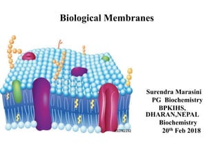
- 1. Biological Membranes Surendra Marasini PG Biochemistry BPKIHS, DHARAN,NEPAL Biochemistry 20th Feb 2018
- 2. Objectives • Introduction • Structure and chemical composition • In reference to Lipids, Protein and Carbohydrate moieties in the membrane • Example of Typical Biomembrane (RBC membrane)
- 3. Introduction: Biological membranes • Barrier that separates the cellular contents from external environments • Eukaryotic cells contain internal membrane systems also
- 5. Why do we need the Biological membranes??? • Boundaries around cells • Boundaries around sub cellular compartments • Compartmentalize and segregate intracellular events and separate cells • Mediates regulation of cellular functions by Acting as selective barriers Allowing inside environment of cells or organelles to differ from outside
- 6. Why do we need Biological membranes Contd… • Signaling processes- specific receptors, chemical and electrical signal generation • Specific enzyme systems are located • Plasma membrane contains- specific systems ,pumps, channels, transporters • Normal cellular function starts with cell membrane
- 7. Structure of Biological membrane
- 8. Structure of membrane contd…
- 9. Components of Membranes • Basic components-lipids, proteins & carbohydrates
- 10. Lipids of the Biomembranes Provide basic structure of all membranes Most abundant: Phospholipids others : Sphingolipids and Sterols FA chain length of 14-24C and with at least one cis double bond and other saturated FA component provides hydrophobic barrier and others provide hydrophilic making amphiphilic Cis double bond provides kink which affects in fluidity
- 11. Phospholipids on the Biomembranes
- 14. Glycosphingolipids • Simplest glycolipid –cerebrosides • More complex glycolipids-gangliosides
- 16. Sterols • Common sterol-cholesterol • Mainly in plasma membranes but lesser in organelle membranes • Maintains fluidity in high as well as low temperatures
- 17. Role of cholesterol in membrane fluidity • Below Tm –interferes with the hydrocarbon tails of fatty acids thus increasing the fluidity • Above Tm _ limits the disorder because it is more rigid than hydrocarbon tails of fatty acids.
- 18. Membrane fluidity controlled by type of lipid content • Fluidity depends on lipid composition at certain temperature • Transition from rigid to fluid occurs above transition temperature (Tm) • Saturated FA interact strongly to increase higher Tm • Unsaturated cis configuration increase fluidity of bilayer by decreasing compactness of side chain
- 20. 3 types of lipid aggregates • Micelles, Bilayer sheet & vesicle
- 21. Lipid rafts
- 23. Membrane proteins • Specific membrane functions • Approximately 50% by weight, larger molecule, 1 protein per 50-100 lipid molecules • Two class of membrane proteins Peripheral proteins: dissociate from membrane by polar reagents such as extreme pH or high salt concentration Integral proteins: only released by disruption of phospholipid bilayer by reagents S/A detergents ( Amphipathic molecules). : Most of them are transmembrane proteins.
- 26. Transmembrane proteins Fig: transbilayer disposition of Glycophorin across the RBC membrane
- 28. Beta sheet barrels • 8-22 beta sheets fold into barrel like structure with hydrophilic pore • Seen in mitochondrial membrane • Examples: Porins, 16 strands antiparallel beta sheet
- 29. RBC membrane • Spectrin, ankyrin, actin provides support and structure to the membrane • Most integral proteins are glycoproteins
- 30. Major membrane proteins of Human RBCs
- 31. Principle proteins of the red cell membrane
- 32. Interactions of the proteins of Red cell membrane
- 33. Membrane carbohydrates • Play a key role in cell-cell recognition • Ability of a cell to distinguish from one another • Basis of rejection of foreign cells by immune system
- 34. To be continued with Different transport mechanisms…….
- 35. References • Lehninger´s Biochemistry • Harper´s Illustrated Biochemistry 30th Edition • Biochemistry (Stryers) 6th Edition
Editor's Notes
- Membrane trafficking/barrier or boundary
- Beside the plasma membrane there are subcellular membranes too.....
- Boundaries around cells-Plasma membrane, boundaries around cellular compartments- Mitochondria, Nucleus, Golgi complex ,ER etc Plasma membrane is selectively permeable outer boundary of the cell
- Damage to membrane can affect the water balance and ion influx and therefore grossly alter most processes in the cell. Membrane contains specific receptors for external stimuli.
- Discovered by Singer and Nicolson in 1972 after studying the electron microscopic view, physical and chemical studies…… Protein Iceberg in the sea of lipid…..proteins are floating on the ocean of the lipids….. 50-80 A thick trillaminar structure...when viewed from electron microscope…Protein icebergs in the sea of lipid bilayers that is proteins float in the ocean of the 2D structure of lipid bilayers…….combined study of electron microscopy, physical and chemical evidences --integral proteins float in the sea of lipid by hydrophobic interactions with their non polar amino acid side chains… Carbohydrate moieties are exposed to the extracellular face of the membrane…
- Inner leaflet= cytoplasmic leaflet…..
- Composition of Lipid, protein and carbohydrate varies from one membrane to another
- Cis double bonds provides kinks which affects in fluidity….more the double bonds more will be the fluidity
- Simplest phospholipid is the phosphatidic acid….
- Ethanolamine/cephalin--- white matter of brain, Serine- apoptosis,….inositol--- precursor of second messenger
- Inner leaflet contains phosphatidylserine, phosphatidylinositol & phosphatidylethanolamine…….outer leaflet contains sphingomyelin,g lycolipid & choline…
- Sugar containing lipids built on backbone of ceramide…..sphingosine+ fatty acid= ceramide… Common in the animal muscle cell membrane and nerve cell membrane Galactosyl ceramides & glucosyl ceramides…
- Even in the single leaflet the lipid distribution is not uniform. Cholesterol is prominent in the plasma membrane………inositol,ethanolamine and choline etc are very importanet for signal transduction triggered by hormones……glycolipid is virtually absent in plasma membrane of animal cell.
- Cholesterol is the third major type of membrane lipid..structure is quite different from phospholipids…. In membranes, hydroxyl group interacts w ith PL and the hydrocarbon chain interacts with the fatty acid chain .. Hydrocarbon chain is attached to one end of the steroid nucleus and hydroxyl group attached to other end. Cholesterol is the lipid based on steroid nucleus… ----Ergosterol, Sitosterol & Stigmasterols are other sterols of plants//
- Above the Tm ,limits the disorder because it is more rigid than the hydrocarbon tails so cannot move in the membrane in the same extent as that of the fatty acid , thus limiting the Fluidity….????/?????????????
- -At least one unsaturated FA with at least 1cis double bond is seen in cell membrane to maintain fluidity. Cholesterol at temp. below Tm, interfers with acyl tail interaction to increase fluidity as well as at above Tm, rigidity of ring limits fluidity.
- Responsible for maintaining lipid asymmetry……catalyzed and uncatalyzed.lateral diffusion within the leaflet is very rapid whereas one leaflet to other is very slow…..Flippase- catalyzes the translocation of phosphatidyl serine,ethanolamine & inositol from non cytosolic leaflet to the cytosolic leaflet Floppase—ABC transporter family.Transports PLs from cytosolic to outer leaflet. Scramblases- moves the lipid in either direction,toward equilibrium
- Amphipathic lipid aggregates that form in water……In micelles the hydrophobic chains of the fatty acids are sequestered inside the core/spherical The bilayer sheet is relatively unstable and spontaneously form a third type of aggregate k/a vesicle or liposome…
- Even in the single leaflet the distribution is not uniform…….stable association of sphingolipids and cholesterol produces thicker region than other region called lipid rafts…
- Caveolin is an integral membrane protein with two globular domains connected by a hairpin-shaped hydrophobic domain, which binds the protein to the cytoplasmic leaflet of the plasma membrane. Three palmitoyl groups attached to the carboxyl-terminal globular domain further anchor it to the membrane. Caveolin (actually, a family of related caveolins) forms dimers and associates with cholesterol-rich regions in the membrane, and the presence of caveolin dimers forces the associated lipid bilayer to curve inward, forming caveolae (“little caves”) in the surface of the cell Enriched with specific type of membrane protein called caveolin
- X-ray Crystallography enable the 3D structure of membrane proteins Proteins determine membranes specific function…..cell membrane & organelle membranes each have unique collection of proteins…. ….Carbohydrates present as a prt of glycoproteins and glycolipids…
- The amount of proteins equals or exceeds the quantity of lipids in the membrane….the outstanding exception is myelin an electrical insulator found on nerve fibres…
- Integral/Intrinsic and peripheral/extrinsic…….GPI ( Glycosylphosphatidylinositol) can be released by using phospholipase C…….Integral proteins can be extracted with the detergents which disrupts the hydrophobic interactions with lipid bilayers and form Micelle like clusters…
- Transbilayer deposition of glycophorin across the RBC membrane….Hydrophilic domain containing all the sugar residues is on the outer leaflet….Hexagon ( sialic acid & tetrasaccharide)…. Segment of hydrophobic region ( 75-93) forms an alpha helix that traverse the bilayer…. 20-30 non polar amino acid sequence can pass through lipid bilayer as alpha helix & can be identified by hydropathy plots
- For known proteins of the plasma membranes the spatial relationship of protein domains to lipids falls under six categories…. Single pass and multipass transmembrane proteins Type Iand II-have single transmembrane helix…type I has amino terminus outside the helix and carboxy terminus in typeII Type III-Multiple transmembrane helices in a single a polypeptide Type Iv- transmembrane domain of several polypeptide assemble to form channels… Type V- proteins covalently linked to the lipids Type vi- Proteins have both transmembrane helices and GPI anchors….
- a class of proteins whose molecules can form channels…….specially large pores are formed….
- Good example for peripheral and integral proteins….deficiency of spectrin leads to hereditary spherocytosis…. Glycophorin and band 3 are integral proteins
- In SDS PAGE electrophoresis the proteins are separated on the basis of molecular wt…Spectrin being the high molecular weight proteins appears near the point of application and known as Band 1. Spectrin- most abundant in the erythrocyte membrane cytoskeleton..alpha & beta….
- This interaction helps to determine the shape and flexibility of cells…the alpha and beta chains of spectrin rolled each other an antiparallel orientation to form highly extended structural unit=100nm….Ankyrin:is a pyramid shaped protein that binds spectrin and tightly with band 3…..Actin: short double helical structure that binds with band 4.1….Cytoskeletal assembly
- Carbohydrates covalently linked to proteins ( Glycoproteins) and lipids ( Glycolipids) are also part of membranes… Helps to protect the cell from outside world…also helps in cell –cell recognition…