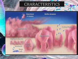
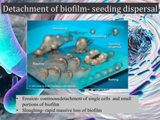
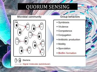
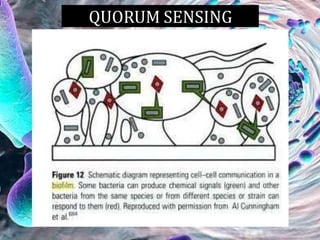
Dr. K. PALANI SELVI MDS
• Introduction
• Definition
• Basic criteria
• Characteristics
• Ultrastructure of a biofilm
• Formation and factors affecting its formation
• Endodontic biofilms
• Role of E.feacalis
• Microbial diversity in endodontic biofilm
• Current therapeutic options in endodontic
biofilm
• Eradication of biofilm
• Biofilm assessment methods
• Conclusion
• From an ecological perspective,
the root canal can be considered a
highly controlled environment
with a limited number of niches.
• The main limiting factors in root
canal niches that influence
bacterial colonization are, for
instance, oxygen and nutrient
availability .
INTRODUCTION
JOE - Redefining the Persistent Infection
in Root Canals: Possible Role of Biofilm
Communities - Luis Chávez 2014
 Biofilm is defined as a sessile multi cellular microbial
community characterized by cells that are firmly attached
to a surface and embedded in a self produced matrix of
extracellular polymeric substances
 Biofilms are formed whenever there is free flow of fluid ,
microorganisms and a solid surface. It is one of the basic
survival strategies employed by bacteria
DEFINITION
•
Costerton JW, Lewandowski Z, DeBeer D, Caldwell D, Korber D, James G. Biofilms, the customized microniche.
J Bacteriol. 1994;176:2137-42.
Caldwell DE, Atuku E, Wilkie DC, Wivcharuk KP, Karthikeyan S, Korber DR, et al. Germ theory
vs. community theory in understanding and controlling the proliferation of biofilms. Adv Dent Res.
1997;11:4-13.
Biofilms should possess
1. AUTOPOIESIS- ability to self organize
2. HOMEOSTASIS-resist
environmental pertubations
3.SYNERGY- effective in association
than in isolation
4.COMMUNITY- respond to environmental
changes as a unit rather than single individual
ULTRASTRUCTURE OF A BIOFILM
 Basic structure - heterogenous arrangement of microbial
cells on a solid surface
 Glycocalyx matrix - Extrapolymeric substance surrounds
the microcolonies and anchors the bacterial cell to the
substrate
 85% - matrix and 15% - cells
 Mushroom shape/ tower shape
 Water channels are primitive circulatory system in
biofilms
ULTRASTRUCTURE OF A BIOFILM
85% MATRIX
15% CELLS
 First stage - a conditioning film forms-
transport of microbe to the substrate surface
 Second stage – initial non-specific
microbial-substrate adherence phase
 Third stage - Bacterial growth and biofilm
expansion
 Fourth stage - Detachment of biofilm
STAGES OF BIOFILM FORMATION
 PH, temperature, surface energy of substrate, flow rate
of fluid, nutrient availability, bacterial growth stage,
surface hydrophobicity
• Erosion- continousdetachment of single cells and small
portions of biofilm
• Sloughing- rapid massive loss of biofilm
 Forms on root canal
dentin of an infected tooth
 Identified by Nair 1987
 Cocci, rods , filaments and
spirochetes are seen
 Morphologically distinct type
of bacteria are seen
 Eg- E.faecalis
 Root surface biofilms - adjacent to root
apex of endodontically treated teeth
 Seen in teeth with asymptomatic
periapical periodontitis and chronic
apical abscess with sinus tract
 Multispecies in nature- F. nucleatum,
Po. gingivalis, and Tannerella forsythensis
 Dominated by cocci and short rods
with cocci attached to tooth substrate
 Isolated biofilms in the
periapical area of endodontically
involved teeth
• Eg- Actinomyces , P.propionicum
 Bacteria adheres to artificial biomaterial
surface and forms biofilms
 Opportunistic infection- nosocomial
organisms
 Eg- coagulase negative staphylococcus, s.
aureus, enterococci, streptococci,
p.aeruginosa
 Serum plays a significant role in biofilm
formation
 GP coated with biofilm
E. FEACALIS & BIOFILM
• E. faecalis is a gram-positive,
facultative anaerobic cocci that
is strongly associated with
endodontic infections.
• It causes nosocomial infections
• Frequently isolated from the
failed root canals undergoing
retreatment.
Duggan JM, Sedgley CM. Biofilm formation of oral and endodontic Enterococcus faecalis. J Endod.
2007;33:815-8. Al-Ahmad A, Muller N, Wiedmann-Al-Ahmad M, Sava I, Hubner J, Follo M, et al.
Endodontic and salivary isolates of Enterococcus faecalis integrate into biofilm from human salivary
bacteria cultivated in vitro. J Endod. 2009;35:986-91.
• They can grow in
- extremely Alkaline pH,
- salt concentrated environment
- survive a temperature of 60°C for 30 min.
• E. faecalis is able to suppress the action of
lymphocytes, potentially contributing to
endodontic failure.
• E.faecalis has the ability to form biofilm that
can resist calcium hydroxide dressing by
maintaining pH homeostasis.
The development of E. faecalis biofilm on the
root canal dentin involves three stages as follows:
Stage 1:
Microcolonies are formed
as E. faecalis cells adhere
on the root canal dentin
surface
Stage 2:
Bacterial-mediated dissolution of
the mineral fraction from the dentin
substrate leads to localized increase
in the calcium and phosphate ions
causing mineralization (or
calcification) of the E. faecalis
biofilm
Stage 3:
Due to this interaction of
bacteria and their metabolic
products on dentin, E. faecalis
biofilm is mineralized.
CURRENT THREUPATIC OPTIONS FOR ENDODONTIC BIOFILM
OZONE
ULTRASONIC
IRRIGATION
• Micromanipulator-assisted analysis
• Green fluorescent protein (GFP) tagging
• Confocal laser scanning microscopy (CLSM)
• Flow cytometry
• Fluorescence in situ hybridization (FISH)
- have made biofilm characterization very comprehensive.
• It is foremost to understand that how the
biofilm formed by root canal bacteria
resists endodontic treatment measures.
• Elimination or significant reduction of
endodontic bacterial biofilms and
prevention of recontamination of the root
canal after treatment are the essential
elements for successful outcomes of
endodontic treatment.
CONCLUSION
Biofilms
Biofilms

Biofilms

  • 1.
    Dr. K. PALANISELVI MDS
  • 2.
    • Introduction • Definition •Basic criteria • Characteristics • Ultrastructure of a biofilm • Formation and factors affecting its formation • Endodontic biofilms • Role of E.feacalis • Microbial diversity in endodontic biofilm • Current therapeutic options in endodontic biofilm • Eradication of biofilm • Biofilm assessment methods • Conclusion
  • 3.
    • From anecological perspective, the root canal can be considered a highly controlled environment with a limited number of niches. • The main limiting factors in root canal niches that influence bacterial colonization are, for instance, oxygen and nutrient availability . INTRODUCTION JOE - Redefining the Persistent Infection in Root Canals: Possible Role of Biofilm Communities - Luis Chávez 2014
  • 4.
     Biofilm isdefined as a sessile multi cellular microbial community characterized by cells that are firmly attached to a surface and embedded in a self produced matrix of extracellular polymeric substances  Biofilms are formed whenever there is free flow of fluid , microorganisms and a solid surface. It is one of the basic survival strategies employed by bacteria DEFINITION • Costerton JW, Lewandowski Z, DeBeer D, Caldwell D, Korber D, James G. Biofilms, the customized microniche. J Bacteriol. 1994;176:2137-42.
  • 5.
    Caldwell DE, AtukuE, Wilkie DC, Wivcharuk KP, Karthikeyan S, Korber DR, et al. Germ theory vs. community theory in understanding and controlling the proliferation of biofilms. Adv Dent Res. 1997;11:4-13. Biofilms should possess 1. AUTOPOIESIS- ability to self organize 2. HOMEOSTASIS-resist environmental pertubations 3.SYNERGY- effective in association than in isolation 4.COMMUNITY- respond to environmental changes as a unit rather than single individual
  • 6.
  • 7.
     Basic structure- heterogenous arrangement of microbial cells on a solid surface  Glycocalyx matrix - Extrapolymeric substance surrounds the microcolonies and anchors the bacterial cell to the substrate  85% - matrix and 15% - cells  Mushroom shape/ tower shape  Water channels are primitive circulatory system in biofilms ULTRASTRUCTURE OF A BIOFILM 85% MATRIX 15% CELLS
  • 8.
     First stage- a conditioning film forms- transport of microbe to the substrate surface  Second stage – initial non-specific microbial-substrate adherence phase  Third stage - Bacterial growth and biofilm expansion  Fourth stage - Detachment of biofilm
  • 9.
  • 10.
     PH, temperature,surface energy of substrate, flow rate of fluid, nutrient availability, bacterial growth stage, surface hydrophobicity
  • 14.
    • Erosion- continousdetachmentof single cells and small portions of biofilm • Sloughing- rapid massive loss of biofilm
  • 18.
     Forms onroot canal dentin of an infected tooth  Identified by Nair 1987  Cocci, rods , filaments and spirochetes are seen  Morphologically distinct type of bacteria are seen  Eg- E.faecalis
  • 19.
     Root surfacebiofilms - adjacent to root apex of endodontically treated teeth  Seen in teeth with asymptomatic periapical periodontitis and chronic apical abscess with sinus tract  Multispecies in nature- F. nucleatum, Po. gingivalis, and Tannerella forsythensis  Dominated by cocci and short rods with cocci attached to tooth substrate
  • 20.
     Isolated biofilmsin the periapical area of endodontically involved teeth • Eg- Actinomyces , P.propionicum
  • 21.
     Bacteria adheresto artificial biomaterial surface and forms biofilms  Opportunistic infection- nosocomial organisms  Eg- coagulase negative staphylococcus, s. aureus, enterococci, streptococci, p.aeruginosa  Serum plays a significant role in biofilm formation  GP coated with biofilm
  • 22.
    E. FEACALIS &BIOFILM • E. faecalis is a gram-positive, facultative anaerobic cocci that is strongly associated with endodontic infections. • It causes nosocomial infections • Frequently isolated from the failed root canals undergoing retreatment. Duggan JM, Sedgley CM. Biofilm formation of oral and endodontic Enterococcus faecalis. J Endod. 2007;33:815-8. Al-Ahmad A, Muller N, Wiedmann-Al-Ahmad M, Sava I, Hubner J, Follo M, et al. Endodontic and salivary isolates of Enterococcus faecalis integrate into biofilm from human salivary bacteria cultivated in vitro. J Endod. 2009;35:986-91. • They can grow in - extremely Alkaline pH, - salt concentrated environment - survive a temperature of 60°C for 30 min. • E. faecalis is able to suppress the action of lymphocytes, potentially contributing to endodontic failure. • E.faecalis has the ability to form biofilm that can resist calcium hydroxide dressing by maintaining pH homeostasis.
  • 23.
    The development ofE. faecalis biofilm on the root canal dentin involves three stages as follows: Stage 1: Microcolonies are formed as E. faecalis cells adhere on the root canal dentin surface Stage 2: Bacterial-mediated dissolution of the mineral fraction from the dentin substrate leads to localized increase in the calcium and phosphate ions causing mineralization (or calcification) of the E. faecalis biofilm Stage 3: Due to this interaction of bacteria and their metabolic products on dentin, E. faecalis biofilm is mineralized.
  • 25.
    CURRENT THREUPATIC OPTIONSFOR ENDODONTIC BIOFILM
  • 26.
  • 29.
    • Micromanipulator-assisted analysis •Green fluorescent protein (GFP) tagging • Confocal laser scanning microscopy (CLSM) • Flow cytometry • Fluorescence in situ hybridization (FISH) - have made biofilm characterization very comprehensive.
  • 30.
    • It isforemost to understand that how the biofilm formed by root canal bacteria resists endodontic treatment measures. • Elimination or significant reduction of endodontic bacterial biofilms and prevention of recontamination of the root canal after treatment are the essential elements for successful outcomes of endodontic treatment. CONCLUSION