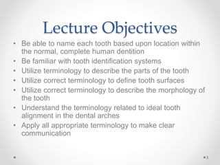
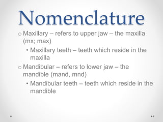
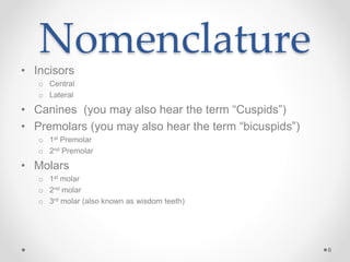
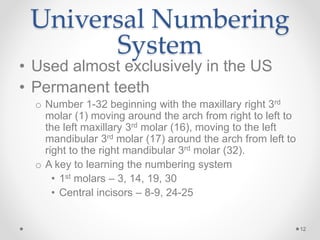
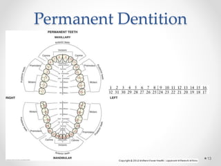
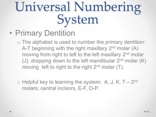
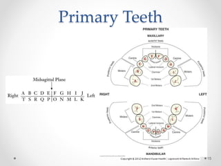
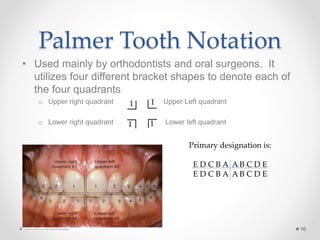
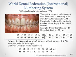
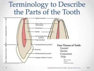
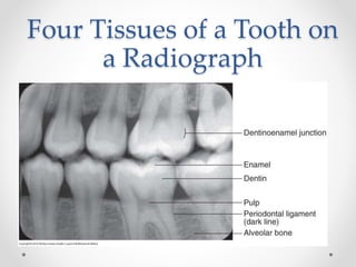
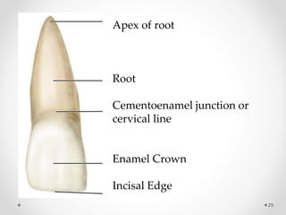
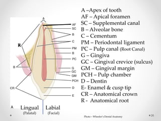
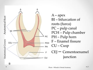
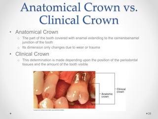
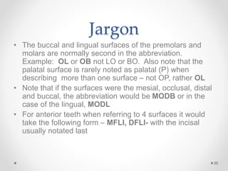
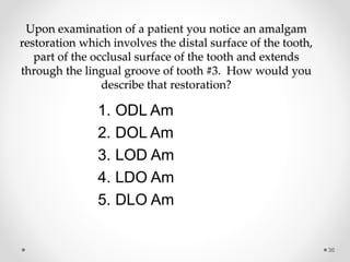
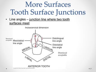
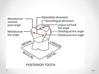
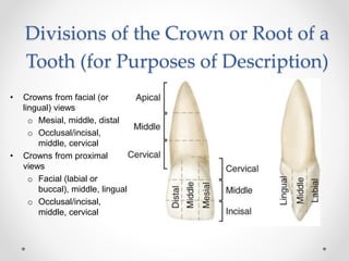
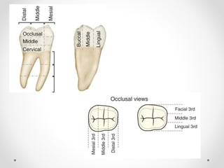
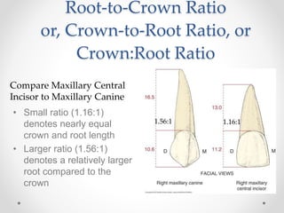
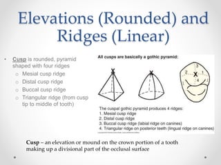
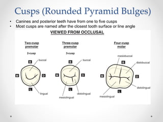
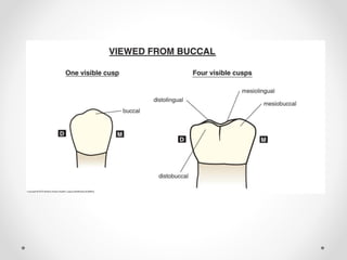
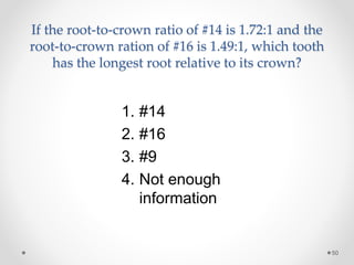
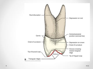
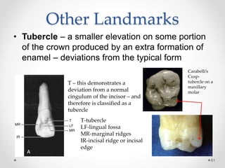
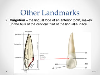
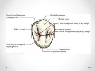
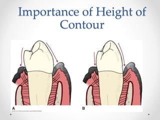
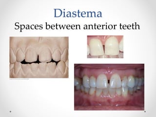
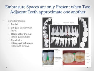
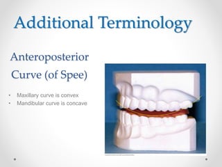
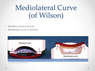
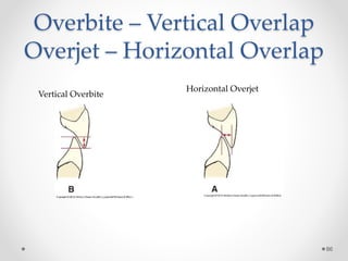
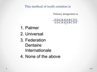
This document provides an introduction to dental terminology used to describe tooth anatomy and identification. It begins with an overview of terminology for describing the primary and permanent dentition, including dental formulas. Tooth identification systems including the Universal Numbering System and Palmer Notation are explained. Terminology is defined for identifying tooth surfaces, landmarks, and structures. Descriptions are provided for anatomical crowns and roots as well as root-to-crown ratios. Landmarks such as cusps, ridges, and line angles are defined. The document aims to familiarize students with standard dental terminology.
Coronal and radicular pulp
Apical foramen
Accessory canal
Functions of dental pulp
Components of dental pulp
Functions of pulpal extracellular matrix
Organization of cells in the pulp
The principle cells of the pulp
The pathways of collagen synthesis
Matrix and ground substances
Vasculature and lymphatic supply
Innervation of Dentin- pulp complex
Disorders of the dental pulp
Advances in pulp vitality testing
The Indian Dental Academy is the Leader in continuing dental education , training dentists in all aspects of dentistry and
offering a wide range of dental certified courses in different formats.
A Complete presentation explaining the complete morphology of Maxillary first molar, for the benefit of people like me who tried and failed to find everything in one package
Upload By : Ahmed Ali Abbas
Babylon University College of Dentistry
download this file from Website on google theoptimalsmile.wix.com/dentistry
Oral histology
Knee anatomy and clinical tests 2024.pdfvimalpl1234
This includes all relevant anatomy and clinical tests compiled from standard textbooks, Campbell,netter etc..It is comprehensive and best suited for orthopaedicians and orthopaedic residents.
Ethanol (CH3CH2OH), or beverage alcohol, is a two-carbon alcohol
that is rapidly distributed in the body and brain. Ethanol alters many
neurochemical systems and has rewarding and addictive properties. It
is the oldest recreational drug and likely contributes to more morbidity,
mortality, and public health costs than all illicit drugs combined. The
5th edition of the Diagnostic and Statistical Manual of Mental Disorders
(DSM-5) integrates alcohol abuse and alcohol dependence into a single
disorder called alcohol use disorder (AUD), with mild, moderate,
and severe subclassifications (American Psychiatric Association, 2013).
In the DSM-5, all types of substance abuse and dependence have been
combined into a single substance use disorder (SUD) on a continuum
from mild to severe. A diagnosis of AUD requires that at least two of
the 11 DSM-5 behaviors be present within a 12-month period (mild
AUD: 2–3 criteria; moderate AUD: 4–5 criteria; severe AUD: 6–11 criteria).
The four main behavioral effects of AUD are impaired control over
drinking, negative social consequences, risky use, and altered physiological
effects (tolerance, withdrawal). This chapter presents an overview
of the prevalence and harmful consequences of AUD in the U.S.,
the systemic nature of the disease, neurocircuitry and stages of AUD,
comorbidities, fetal alcohol spectrum disorders, genetic risk factors, and
pharmacotherapies for AUD.
Title: Sense of Smell
Presenter: Dr. Faiza, Assistant Professor of Physiology
Qualifications:
MBBS (Best Graduate, AIMC Lahore)
FCPS Physiology
ICMT, CHPE, DHPE (STMU)
MPH (GC University, Faisalabad)
MBA (Virtual University of Pakistan)
Learning Objectives:
Describe the primary categories of smells and the concept of odor blindness.
Explain the structure and location of the olfactory membrane and mucosa, including the types and roles of cells involved in olfaction.
Describe the pathway and mechanisms of olfactory signal transmission from the olfactory receptors to the brain.
Illustrate the biochemical cascade triggered by odorant binding to olfactory receptors, including the role of G-proteins and second messengers in generating an action potential.
Identify different types of olfactory disorders such as anosmia, hyposmia, hyperosmia, and dysosmia, including their potential causes.
Key Topics:
Olfactory Genes:
3% of the human genome accounts for olfactory genes.
400 genes for odorant receptors.
Olfactory Membrane:
Located in the superior part of the nasal cavity.
Medially: Folds downward along the superior septum.
Laterally: Folds over the superior turbinate and upper surface of the middle turbinate.
Total surface area: 5-10 square centimeters.
Olfactory Mucosa:
Olfactory Cells: Bipolar nerve cells derived from the CNS (100 million), with 4-25 olfactory cilia per cell.
Sustentacular Cells: Produce mucus and maintain ionic and molecular environment.
Basal Cells: Replace worn-out olfactory cells with an average lifespan of 1-2 months.
Bowman’s Gland: Secretes mucus.
Stimulation of Olfactory Cells:
Odorant dissolves in mucus and attaches to receptors on olfactory cilia.
Involves a cascade effect through G-proteins and second messengers, leading to depolarization and action potential generation in the olfactory nerve.
Quality of a Good Odorant:
Small (3-20 Carbon atoms), volatile, water-soluble, and lipid-soluble.
Facilitated by odorant-binding proteins in mucus.
Membrane Potential and Action Potential:
Resting membrane potential: -55mV.
Action potential frequency in the olfactory nerve increases with odorant strength.
Adaptation Towards the Sense of Smell:
Rapid adaptation within the first second, with further slow adaptation.
Psychological adaptation greater than receptor adaptation, involving feedback inhibition from the central nervous system.
Primary Sensations of Smell:
Camphoraceous, Musky, Floral, Pepperminty, Ethereal, Pungent, Putrid.
Odor Detection Threshold:
Examples: Hydrogen sulfide (0.0005 ppm), Methyl-mercaptan (0.002 ppm).
Some toxic substances are odorless at lethal concentrations.
Characteristics of Smell:
Odor blindness for single substances due to lack of appropriate receptor protein.
Behavioral and emotional influences of smell.
Transmission of Olfactory Signals:
From olfactory cells to glomeruli in the olfactory bulb, involving lateral inhibition.
Primitive, less old, and new olfactory systems with different path
Flu Vaccine Alert in Bangalore Karnatakaaddon Scans
As flu season approaches, health officials in Bangalore, Karnataka, are urging residents to get their flu vaccinations. The seasonal flu, while common, can lead to severe health complications, particularly for vulnerable populations such as young children, the elderly, and those with underlying health conditions.
Dr. Vidisha Kumari, a leading epidemiologist in Bangalore, emphasizes the importance of getting vaccinated. "The flu vaccine is our best defense against the influenza virus. It not only protects individuals but also helps prevent the spread of the virus in our communities," he says.
This year, the flu season is expected to coincide with a potential increase in other respiratory illnesses. The Karnataka Health Department has launched an awareness campaign highlighting the significance of flu vaccinations. They have set up multiple vaccination centers across Bangalore, making it convenient for residents to receive their shots.
To encourage widespread vaccination, the government is also collaborating with local schools, workplaces, and community centers to facilitate vaccination drives. Special attention is being given to ensuring that the vaccine is accessible to all, including marginalized communities who may have limited access to healthcare.
Residents are reminded that the flu vaccine is safe and effective. Common side effects are mild and may include soreness at the injection site, mild fever, or muscle aches. These side effects are generally short-lived and far less severe than the flu itself.
Healthcare providers are also stressing the importance of continuing COVID-19 precautions. Wearing masks, practicing good hand hygiene, and maintaining social distancing are still crucial, especially in crowded places.
Protect yourself and your loved ones by getting vaccinated. Together, we can help keep Bangalore healthy and safe this flu season. For more information on vaccination centers and schedules, residents can visit the Karnataka Health Department’s official website or follow their social media pages.
Stay informed, stay safe, and get your flu shot today!
Recomendações da OMS sobre cuidados maternos e neonatais para uma experiência pós-natal positiva.
Em consonância com os ODS – Objetivos do Desenvolvimento Sustentável e a Estratégia Global para a Saúde das Mulheres, Crianças e Adolescentes, e aplicando uma abordagem baseada nos direitos humanos, os esforços de cuidados pós-natais devem expandir-se para além da cobertura e da simples sobrevivência, de modo a incluir cuidados de qualidade.
Estas diretrizes visam melhorar a qualidade dos cuidados pós-natais essenciais e de rotina prestados às mulheres e aos recém-nascidos, com o objetivo final de melhorar a saúde e o bem-estar materno e neonatal.
Uma “experiência pós-natal positiva” é um resultado importante para todas as mulheres que dão à luz e para os seus recém-nascidos, estabelecendo as bases para a melhoria da saúde e do bem-estar a curto e longo prazo. Uma experiência pós-natal positiva é definida como aquela em que as mulheres, pessoas que gestam, os recém-nascidos, os casais, os pais, os cuidadores e as famílias recebem informação consistente, garantia e apoio de profissionais de saúde motivados; e onde um sistema de saúde flexível e com recursos reconheça as necessidades das mulheres e dos bebês e respeite o seu contexto cultural.
Estas diretrizes consolidadas apresentam algumas recomendações novas e já bem fundamentadas sobre cuidados pós-natais de rotina para mulheres e neonatos que recebem cuidados no pós-parto em unidades de saúde ou na comunidade, independentemente dos recursos disponíveis.
É fornecido um conjunto abrangente de recomendações para cuidados durante o período puerperal, com ênfase nos cuidados essenciais que todas as mulheres e recém-nascidos devem receber, e com a devida atenção à qualidade dos cuidados; isto é, a entrega e a experiência do cuidado recebido. Estas diretrizes atualizam e ampliam as recomendações da OMS de 2014 sobre cuidados pós-natais da mãe e do recém-nascido e complementam as atuais diretrizes da OMS sobre a gestão de complicações pós-natais.
O estabelecimento da amamentação e o manejo das principais intercorrências é contemplada.
Recomendamos muito.
Vamos discutir essas recomendações no nosso curso de pós-graduação em Aleitamento no Instituto Ciclos.
Esta publicação só está disponível em inglês até o momento.
Prof. Marcus Renato de Carvalho
www.agostodourado.com
Tom Selleck Health: A Comprehensive Look at the Iconic Actor’s Wellness Journeygreendigital
Tom Selleck, an enduring figure in Hollywood. has captivated audiences for decades with his rugged charm, iconic moustache. and memorable roles in television and film. From his breakout role as Thomas Magnum in Magnum P.I. to his current portrayal of Frank Reagan in Blue Bloods. Selleck's career has spanned over 50 years. But beyond his professional achievements. fans have often been curious about Tom Selleck Health. especially as he has aged in the public eye.
Follow us on: Pinterest
Introduction
Many have been interested in Tom Selleck health. not only because of his enduring presence on screen but also because of the challenges. and lifestyle choices he has faced and made over the years. This article delves into the various aspects of Tom Selleck health. exploring his fitness regimen, diet, mental health. and the challenges he has encountered as he ages. We'll look at how he maintains his well-being. the health issues he has faced, and his approach to ageing .
Early Life and Career
Childhood and Athletic Beginnings
Tom Selleck was born on January 29, 1945, in Detroit, Michigan, and grew up in Sherman Oaks, California. From an early age, he was involved in sports, particularly basketball. which played a significant role in his physical development. His athletic pursuits continued into college. where he attended the University of Southern California (USC) on a basketball scholarship. This early involvement in sports laid a strong foundation for his physical health and disciplined lifestyle.
Transition to Acting
Selleck's transition from an athlete to an actor came with its physical demands. His first significant role in "Magnum P.I." required him to perform various stunts and maintain a fit appearance. This role, which he played from 1980 to 1988. necessitated a rigorous fitness routine to meet the show's demands. setting the stage for his long-term commitment to health and wellness.
Fitness Regimen
Workout Routine
Tom Selleck health and fitness regimen has evolved. adapting to his changing roles and age. During his "Magnum, P.I." days. Selleck's workouts were intense and focused on building and maintaining muscle mass. His routine included weightlifting, cardiovascular exercises. and specific training for the stunts he performed on the show.
Selleck adjusted his fitness routine as he aged to suit his body's needs. Today, his workouts focus on maintaining flexibility, strength, and cardiovascular health. He incorporates low-impact exercises such as swimming, walking, and light weightlifting. This balanced approach helps him stay fit without putting undue strain on his joints and muscles.
Importance of Flexibility and Mobility
In recent years, Selleck has emphasized the importance of flexibility and mobility in his fitness regimen. Understanding the natural decline in muscle mass and joint flexibility with age. he includes stretching and yoga in his routine. These practices help prevent injuries, improve posture, and maintain mobilit
Title: Sense of Taste
Presenter: Dr. Faiza, Assistant Professor of Physiology
Qualifications:
MBBS (Best Graduate, AIMC Lahore)
FCPS Physiology
ICMT, CHPE, DHPE (STMU)
MPH (GC University, Faisalabad)
MBA (Virtual University of Pakistan)
Learning Objectives:
Describe the structure and function of taste buds.
Describe the relationship between the taste threshold and taste index of common substances.
Explain the chemical basis and signal transduction of taste perception for each type of primary taste sensation.
Recognize different abnormalities of taste perception and their causes.
Key Topics:
Significance of Taste Sensation:
Differentiation between pleasant and harmful food
Influence on behavior
Selection of food based on metabolic needs
Receptors of Taste:
Taste buds on the tongue
Influence of sense of smell, texture of food, and pain stimulation (e.g., by pepper)
Primary and Secondary Taste Sensations:
Primary taste sensations: Sweet, Sour, Salty, Bitter, Umami
Chemical basis and signal transduction mechanisms for each taste
Taste Threshold and Index:
Taste threshold values for Sweet (sucrose), Salty (NaCl), Sour (HCl), and Bitter (Quinine)
Taste index relationship: Inversely proportional to taste threshold
Taste Blindness:
Inability to taste certain substances, particularly thiourea compounds
Example: Phenylthiocarbamide
Structure and Function of Taste Buds:
Composition: Epithelial cells, Sustentacular/Supporting cells, Taste cells, Basal cells
Features: Taste pores, Taste hairs/microvilli, and Taste nerve fibers
Location of Taste Buds:
Found in papillae of the tongue (Fungiform, Circumvallate, Foliate)
Also present on the palate, tonsillar pillars, epiglottis, and proximal esophagus
Mechanism of Taste Stimulation:
Interaction of taste substances with receptors on microvilli
Signal transduction pathways for Umami, Sweet, Bitter, Sour, and Salty tastes
Taste Sensitivity and Adaptation:
Decrease in sensitivity with age
Rapid adaptation of taste sensation
Role of Saliva in Taste:
Dissolution of tastants to reach receptors
Washing away the stimulus
Taste Preferences and Aversions:
Mechanisms behind taste preference and aversion
Influence of receptors and neural pathways
Impact of Sensory Nerve Damage:
Degeneration of taste buds if the sensory nerve fiber is cut
Abnormalities of Taste Detection:
Conditions: Ageusia, Hypogeusia, Dysgeusia (parageusia)
Causes: Nerve damage, neurological disorders, infections, poor oral hygiene, adverse drug effects, deficiencies, aging, tobacco use, altered neurotransmitter levels
Neurotransmitters and Taste Threshold:
Effects of serotonin (5-HT) and norepinephrine (NE) on taste sensitivity
Supertasters:
25% of the population with heightened sensitivity to taste, especially bitterness
Increased number of fungiform papillae
- Video recording of this lecture in English language: https://youtu.be/lK81BzxMqdo
- Video recording of this lecture in Arabic language: https://youtu.be/Ve4P0COk9OI
- Link to download the book free: https://nephrotube.blogspot.com/p/nephrotube-nephrology-books.html
- Link to NephroTube website: www.NephroTube.com
- Link to NephroTube social media accounts: https://nephrotube.blogspot.com/p/join-nephrotube-on-social-media.html
Explore natural remedies for syphilis treatment in Singapore. Discover alternative therapies, herbal remedies, and lifestyle changes that may complement conventional treatments. Learn about holistic approaches to managing syphilis symptoms and supporting overall health.
263778731218 Abortion Clinic /Pills In Harare ,sisternakatoto
263778731218 Abortion Clinic /Pills In Harare ,ABORTION WOMEN’S CLINIC +27730423979 IN women clinic we believe that every woman should be able to make choices in her pregnancy. Our job is to provide compassionate care, safety,affordable and confidential services. That’s why we have won the trust from all generations of women all over the world. we use non surgical method(Abortion pills) to terminate…Dr.LISA +27730423979women Clinic is committed to providing the highest quality of obstetrical and gynecological care to women of all ages. Our dedicated staff aim to treat each patient and her health concerns with compassion and respect.Our dedicated group ABORTION WOMEN’S CLINIC +27730423979 IN women clinic we believe that every woman should be able to make choices in her pregnancy. Our job is to provide compassionate care, safety,affordable and confidential services. That’s why we have won the trust from all generations of women all over the world. we use non surgical method(Abortion pills) to terminate…Dr.LISA +27730423979women Clinic is committed to providing the highest quality of obstetrical and gynecological care to women of all ages. Our dedicated staff aim to treat each patient and her health concerns with compassion and respect.Our dedicated group of receptionists, nurses, and physicians have worked together as a teamof receptionists, nurses, and physicians have worked together as a team wwww.lisywomensclinic.co.za/
MANAGEMENT OF ATRIOVENTRICULAR CONDUCTION BLOCK.pdfJim Jacob Roy
Cardiac conduction defects can occur due to various causes.
Atrioventricular conduction blocks ( AV blocks ) are classified into 3 types.
This document describes the acute management of AV block.
MANAGEMENT OF ATRIOVENTRICULAR CONDUCTION BLOCK.pdf
Dana/Roseman/Terminology
1. Introduction
Form and Function of
the Human Dentition
-Terminology -
Dr. George F. Richards
1
2. 2
References:
Wheeler’s Dental Anatomy, Physiology,
and Occlusion
Chapter 1
Woefel’s Dental Anatomy
Chapter 1
Assessment may cover any information in these two chapters as well
as information in ppt. and lecture.
3. Lecture Objectives
• Be able to name each tooth based upon location within
the normal, complete human dentition
• Be familiar with tooth identification systems
• Utilize terminology to describe the parts of the tooth
• Utilize correct terminology to define tooth surfaces
• Utilize correct terminology to describe the morphology of
the tooth
• Understand the terminology related to ideal tooth
alignment in the dental arches
• Apply all appropriate terminology to make clear
communication
3
4. Nomenclature
o Maxillary – refers to upper jaw – the maxilla
(mx; max)
• Maxillary teeth – teeth which reside in the
maxilla
o Mandibular – refers to lower jaw – the
mandible (mand, mnd)
• Mandibular teeth – teeth which reside in the
mandible
4
5. Nomenclature
• Primary Teeth – Deciduous Teeth
o “primary can mean “constituting or belonging to the first stage in
any process.” The term deciduous can mean “not permanent,
transitory.” (Nelson, Stanley J.. Wheeler's Dental Anatomy, Physiology and Occlusion, 9th Edition. W.B. Saunders
Company, 042009. p. 2).
• Permanent Teeth – Succedaneous Teeth
o “ succedaneous can be used to describe a successor dentition
and does not suggest permanence, whereas the term permanent
suggests a permanent dentition, which may not be the case due
to dental caries, periodontal diseases, and trauma.”(Nelson, Stanley J..
Wheeler's Dental Anatomy, Physiology and Occlusion, 9th Edition. W.B. Saunders Company, 042009. p. 2).
5
6. • Incisors
o Central
o Lateral
• Canines (you may also hear the term “Cuspids”)
• Premolars (you may also hear the term “bicuspids”)
o 1st Premolar
o 2nd Premolar
• Molars
o 1st molar
o 2nd molar
o 3rd molar (also known as wisdom teeth)
6
Nomenclature
7. Nomenclature
• Dental formula for primary/deciduous teeth per quadrant
7
o I=Incisors - 2 maxillary, 2 mandibular
• Central
• Lateral
o C=Canines – 1 maxillary, 1 mandibular
o M=Molars – 2 maxillary, 2 mandibular
• 1st Molar
• 2nd Molar
o There are no primary/deciduous premolars
X 2 = 20 teeth in all
9. 9
Nomenclature
• Dental formula for permanent dentition per quadrant
• I=Incisors – 2 maxillary, 2 mandibular
o Central
o Lateral
• C=Canines – 2 maxillary, 2 mandibular
• P=Premolars – 2 maxillary, 2 mandibular
o 1st Premolar
o 2nd Premolar
• M=Molars – 3 maxillary, 3 mandibular
o 1st Molar
o 2nd Molar
o 3rd Molar (wisdom teeth)
32 teeth in all
11. Tooth Identification
(Numbering) Systems
• Universal Numbering System
• Zsigmondy/Palmer Tooth Notation
System (Palmer)
• Fédération Dentaire Internationale
(World Dental Federation
International) Numbering System
(FDI)
11
12. Universal Numbering
System
• Used almost exclusively in the US
• Permanent teeth
o Number 1-32 beginning with the maxillary right 3rd
molar (1) moving around the arch from right to left to
the left maxillary 3rd molar (16), moving to the left
mandibular 3rd molar (17) around the arch from left to
right to the right mandibular 3rd molar (32).
o A key to learning the numbering system
• 1st molars – 3, 14, 19, 30
• Central incisors – 8-9, 24-25
12
14. • Primary Dentition
o The alphabet is used to number the primary dentition:
A-T beginning with the right maxillary 2nd molar (A)
moving from right to left to the left maxillary 2nd molar
(J), dropping down to the left mandibular 2nd molar (K)
moving left to right to the right 2nd molar (T).
o Helpful key to learning the system: A, J, K, T – 2nd
molars; central incisors, E-F, O-P.
14
Universal Numbering
System
16. Palmer Tooth Notation
• Used mainly by orthodontists and oral surgeons. It
utilizes four different bracket shapes to denote each of
the four quadrants
o Upper right quadrant Upper Left quadrant
1 1
o Lower right quadrant Lower left quadrant
16
∟
∟
∟
∟
Primary designation is:
E D C B A A B C D E
E D C B A A B C D E
1
1
17. World Dental Federation (International)
Numbering System
17
Fédération Dentaire Internationale (FDI)
This system numbers each quadrant
with a number – #1 Maxillary R, #2
Maxillary L, #3 Mandibular L, #4
Mandibular R followed by the tooth
number 1-8 starting with the central
incisors.
Example: Upper Right Canine = 13,
Upper Left Canine = 23, etc.
Primary teeth are prefixed with 5 for upper right, 6 for upper left, 7 for
lower left, and 8 for lower right.
Example: Lower left canine would be 73
Photo
18. The arrow points to which tooth?
18
1. 11
2. C
3. H
4. I
5. M
6. None of the above
20. The permanent dental formula of man is
20
A
B
C
D
1. A
2. B
3. C
4. D
This question is right from Dental
Decks 2012-2013 – Do you notice some
change in nomenclature?
21. Assuming the x-ray is mounted correctly, which tooth is
this?
21
You are looking from the buccal to the
lingual
1. 14
2. 36
3. 19
4. 18
5. 2 & 3
22. This symbol and notation equates to which tooth?
22
5 ∟
1. Maxillary second
premolar
2. Maxillary left first
premolar
3. Mandibular right
second premolar
4. Mandibular left
second premolar
23. Terminology to Describe
the Parts of the Tooth
23
Four Tissues of Tooth:
Enamel
Dentin
Cementum
Pulp
Blood vessels
Nerves
Connective tissues
(Pulp Canal)
25. 25
Apex of root
Root
Cementoenamel junction or
cervical line
Enamel Crown
Incisal Edge
26. A –Apex of tooth
AF – Apical foramen
SC – Supplemental canal
B – Alveolar bone
C – Cementum
PM – Periodontal ligament
PC – Pulp canal (Root Canal)
G – Gingiva
GC – Gingival crevice (sulcus)
GM – Gingival margin
PCH – Pulp chamber
D – Dentin
E- Enamel & cusp tip
CR – Anatomical crown
R - Anatomical root
26
R
Lingual Labial
(Palatal) (Facial)
Photo – Wheeler’s Dental Anatomy
27. A – apex
BI – bifurcation of
roots (furca)
PC – pulp canal
PCH – Pulp chamber
PH – Pulp horn
F – Enamel fissure
CU – Cusp
CEJ – Cementoenamel
junction
Photo – Wheeler’s Dental Anatomy 27
Anatomical Crown Anatomical Root
28. Anatomical Crown vs.
Clinical Crown
• Anatomical Crown
o The part of the tooth covered with enamel extending to the cementoenamel
junction of the tooth
o Its dimension only changes due to wear or trauma
• Clinical Crown
o This determination is made depending upon the position of the periodontal
tissues and the amount of the tooth visible
28
29. Anatomic Terms
• Occlusal surface – the chewing
surface of premolars and molars
• Mesial surface – that surface closest
to the midline of the mouth
• Distal surface – that surface farthest
from the midline of the mouth
29
All of these terms can also be used to indicate navigation through the dental arches and
mouth. Ex: It appears that the facial lesion on tooth #30 started in the buccal groove and
continued mesially at the cervical line into the interproximal space.
30. Anatomic Terms
• Lingual surface – that surface closest to the
tongue
• Palatal surface – that surface closest to the
palate
• Labial surface – the “facial” or cheeks and lip
surface of the anterior teeth - incisors and
canines
• Buccal surface – the “facial” or cheek surface
of premolars and molars
30
31. The chewing surface of
31
1. The mesial surface
2. The occlusal
surface
3. The incisal edges
4. The buccal surface
32. Surfaces
32
Mesial Surface
Distal Surface
Interproximal space:
Space formed where 2 teeth
approximate one another
Question:
Is there ever
a situation
when two
mesial
surfaces
approximate
one another?
When two
distal
surfaces
approximate
one another?
Figure courtesy Wheeler’s Dental Anatomy
Occlusal
Occlusal
Occlusal
Incisal
Incisal
Incisal
Occlusal
33. Learn the Jargon
• When referring to more than one surface of the tooth at a
time, particularly when referring to surfaces that are restored
or are to be restored, there is an accepted protocol. Often
times computers confuse this issue, but the sequence is this:
• When referring to the occlusal and mesial surfaces, the
abbreviation is MO, not OM
• When referring to the occlusal and distal surfaces, the
abbreviation is DO, not OD
• When referring to the occlusal, mesial and distal surfaces, the
abbreviation is MOD, not DOM or ODM or DMO or OMD
33
34. Jargon
• When referring to the distal and lingual surfaces of the
anterior teeth, the abbreviation is DL, not LD
• When referring to the mesial and lingual surfaces of
anterior teeth, the abbreviation is ML, not LM
• The rule, if there were a rule, would be the interproximal
surface is stated first then the facial (labial) or lingual
surface. Generally the labial surface of the anterior teeth
when referring to more than one surface of the tooth is
referred to as the “facial” surface. For example MF, or
DF. Referring to that surface as the “labial” surface in
this instance would confuse “labial” with “lingual”.
34
35. Jargon
• The buccal and lingual surfaces of the premolars and
molars are normally second in the abbreviation.
Example: OL or OB not LO or BO. Also note that the
palatal surface is rarely noted as palatal (P) when
describing more than one surface – not OP, rather OL
• Note that if the surfaces were the mesial, occlusal, distal
and buccal, the abbreviation would be MODB or in the
case of the lingual, MODL
• For anterior teeth when referring to 4 surfaces it would
take the following form – MFLI, DFLI- with the incisal
usually notated last
35
36. Upon examination of a patient you notice an amalgam
restoration which involves the distal surface of the tooth,
part of the occlusal surface of the tooth and extends
through the lingual groove of tooth #3. How would you
describe that restoration?
36
1. ODL Am
2. DOL Am
3. LOD Am
4. LDO Am
5. DLO Am
37. More Surfaces
Tooth Surface Junctions
• Line angles – junction line where two tooth
surfaces meet
37
Mesiofacial
acceptable
Distofacial
acceptable
39. • Surface junctions = two surfaces that join, such
39
as:
o Distolabial (Distofacial acceptable)
o Mesiobuccal (Mesiofacial acceptable)
• Dimension = the distance between two opposite
walls, such as:
o Mesiodistal dimension
40. Point Angles
• A point angle is formed by the junction of three surfaces.
40 All teeth have 4 point angles
41. Point Angles
41
From the schematic it appears that the point angle may be the cusp tip. That is
not the case. The point angle derives it name from the combination of names of
the surfaces forming it. For example, the junction of the mesial, buccal and
occlusal surfaces of a molar is called the mesiobucco-occlusal point angle.
42. Each tooth has :
42
1. Two point angles
2. Six point angles
3. Four point angles
4. Eight point angles
43. Divisions of the Crown or Root of a
Tooth (for Purposes of Description)
• Crowns from facial (or
lingual) views
o Mesial, middle, distal
o Occlusal/incisal,
middle, cervical
• Crowns from proximal
views
o Facial (labial or
buccal), middle, lingual
o Occlusal/incisal,
middle, cervical
44.
45. Root-to-Crown Ratio
or, Crown-to-Root Ratio, or
Crown:Root Ratio
Compare Maxillary Central
Incisor to Maxillary Canine
• Small ratio (1.16:1)
denotes nearly equal
crown and root length
• Larger ratio (1.56:1)
denotes a relatively larger
root compared to the
crown
1.56:1 1.16:1
47. Elevations (Rounded) and
Ridges (Linear)
• Cusp is rounded, pyramid
shaped with four ridges
o Mesial cusp ridge
o Distal cusp ridge
o Buccal cusp ridge
o Triangular ridge (from cusp
tip to middle of tooth)
Cusp – an elevation or mound on the crown portion of a tooth
making up a divisional part of the occlusal surface
48. Cusps (Rounded Pyramid Bulges)
• Canines and posterior teeth have from one to five cusps
• Most cusps are named after the closest tooth surface or line angle
49.
50. If the root-to-crown ratio of #14 is 1.72:1 and the
root-to-crown ration of #16 is 1.49:1, which tooth
has the longest root relative to its crown?
50
1. #14
2. #16
3. #9
4. Not enough
information
51. Other Landmarks
• Ridge – any linear elevation on the surface of a
tooth and is named according to its location
(e.g., buccal ridge, incisal ridge, marginal ridge)
• Marginal ridges – those rounded borders of
enamel that form the mesial and distal margins
of the occlusal surfaces of premolars and molars
and the mesial and distal margins of the lingual
surfaces of the incisors and canines
51
52. Other Landmarks
• Triangular ridges – descend from the tips of
cusps of molars and premolars toward the
central part of the occlusal surfaces and are so
named because the slopes of each side of the
ridge are inclined to resemble two sides of a
triangle. They are named after the cusps to
which they belong. Ex: triangular ridge of the
buccal cusp of the maxillary first premolar.
52
53. Ridges (Linear Bulges): Mesial and
Distal Cusp Ridges (also Called Cusp Slopes or Cusp Arms)
Note the
term
“slope”
substituted
with
“ridge”
54. 54
Note the term “slope” substituted with “ridge”
56. Any linear elevation on the surface of a
tooth is called
56
1. An incline
2. A Prominence
3. A ridge
4. A tuberosity
57. Other Landmarks
• Transverse ridge – when buccal and lingual
triangular ridges join transversely across the
occlusal surface
• Oblique ridge – a ridge crossing obliquely the
occlusal surface of the maxillary molars –
formed by the union of the triangular ridge of the
distobuccal cusp and the distal cusp ridge of the
mesiolingual cusp
57
58. Ridges: Two Ridges May Join to
Form One Transverse (or Oblique)
Ridge
The oblique ridge is
a ridge crossing
obliquely the
occlusal surfaces of
maxillary molars
and formed by the
union of the
triangular ridge of
the distobuccal cusp
and the distal cusp
ridge of the
mesiolingual cusp
60. Any union of two triangular ridges
produces a single ridge which is called
60
1. A cusp ridge
2. A marginal ridge
3. A transverse ridge
4. A proximal ridge
5. A oblique ridge
61. Other Landmarks
• Tubercle – a smaller elevation on some portion
of the crown produced by an extra formation of
enamel – deviations from the typical form
61
T – this demonstrates a
deviation from a normal
cingulum of the incisor – and
therefore is classified as a
tubercle
T-tubercle
LF-lingual fossa
MR-marginal ridges
IR-incisal ridge or incisal
edge
Carabelli’s
Cusp-tubercle
on a
maxillary
molar
62. Other Landmarks
• Cingulum – the lingual lobe of an anterior tooth, makes
up the bulk of the cervical third of the lingual surface
62
63. Fossa & Pits
• Fossa – an irregular depression or concavity
o Lingual fossae – lingual surface of incisors
o Central fossae – on the occlusal surface of molars
o Triangular fossae – on molars and premolars on the occlusal surfaces
mesial or distal to marginal ridges
63
65. 65
The unique occlusal feature of this molar is
the
1. Transverse ridge of a
lower left first molar
2. The oblique ridge of a
maxillary right molar
3. The oblique ridge of a
maxillary left molar
4. The transverse ridge of a
maxillary right molar
66. Fossae & Pits
66
• Three-cusped
premolars and most
molars have three
fossae
o Mesial
o Central
o Distal
67. Sulcus
• Sulcus – a long depression
or valley in the surface of a
tooth between ridges and
cusps, the inclines of which
meet at an angle. A sulcus
has a developmental
groove at the junction of its
inclines (The term sulcus
should not be confused
with the term groove.)
67
The sulcus is the broad trough running mesio-distally with the
developmental groove at its base.
69. Other Landmarks
• Developmental groove – a shallow groove or
line between the primary parts of the crown or
root
o Supplemental groove – less distinct, a shallow linear depression on
the surface of a tooth - supplemental to a developmental groove
o Buccal and lingual grooves – developmental grooves found on the
buccal and lingual surfaces of posterior teeth
• Pits – small pinpoint depressions located at the
junction of developmental grooves or at
terminals of those grooves
o Central pit - a term used to describe a landmark in the central fossa of
molars where developmental grooves join
69
70. Grooves (Linear
Depressions)
• Major (developmental)
o Central groove
o Fossa grooves
• Minor (supplemental)
o Marginal ridge grooves
o Extra grooves
A developmental groove is a shallow groove or
line between the primary parts of the crown or root.
A supplemental groove, less distinct, is also a
shallow linear depression on the surface of a tooth,
but it is supplemental to a developmental groove
and does not mark the junction of primary parts.
Buccal and lingual grooves are developmental
grooves found on the buccal and lingual surfaces
of posterior teeth.
71. Grooves (Linear Depressions)
• Central grooves run
mesiodistally
• Major grooves are
named for the
surface or line angle
they “aim” toward
72. Pits are Often Found within
Fossae (Depressions)
• Two-cusped premolars have two
fossae
o Mesial
o Distal
• Incisors (especially maxillary) have
one broad, shallow fossa
(sometimes with a lingual pit)
Pits are located at the junction of developmental grooves or at the
terminus of these grooves
74. Tooth Development from
Lobes
• Lobe – one of the primary
sections of formation in the
development of the crown.
Cusps and mamelons are
representatives of lobes
• Anterior teeth form from four
lobes (three facial and one
lingual)
• Premolars form from three facial
lobes and one lobe per lingual
cusp
• Molars form from one lobe per
major cusp
75. Mamelon
• Mamelon – any one of the three rounded protuberances
found on the incisal ridges a newly erupted incisor teeth-maxillary
and mandibular
75
76. Upon examination the dentist notes an anomalous
enamel outcropping on a molar. This enamel
projection would be classified as a
76
1. Mamelon
2. Ridge
3. Developmental
depression
4. Tubercle
82. Embrasure Spaces are only Present when Two
Adjacent Teeth approximate one another
• Four embrasures
o Facial
o Lingual (larger than
facial)
o Occlusal or incisal
(often quite small)
o Cervical =
interproximal space
(filled with gingiva)
83. The sulcus of a premolar or molar and the
central groove are the same anatomical
landmark.
83
1. True
2. False
87. This method of tooth notation is
87
Primary designation is:
E D C B A A B C D E
E D C B A A B C D E
1. Palmer
2. Universal
3. Federation
Dentaire
Internationale
4. None of the above
88. Mesial, distal, buccal, lingual, palatal, facial
indicate
88
1. Surfaces of the
teeth
2. Types of
restoration
3. Types of cusps
4. Direction
5. 1 & 4
89. Using all tooth numbering systems, the arrow
points to which tooth?
89
1. 7
2. 22
3. 10
4. 25
5. 2&3
6. 1
90. The line marks the mesial surface
90
1. 24
2. 25
3. 24 & 25
4. 23
5. 26
91. This formula is for
91
1. Deciduous teeth
2. Permanent teeth
3. Succedaneous
teeth
4. Mixed dentition
92. Which numbering system is used
almost exclusively in the United States?
92
1. FDI
2. Universal
3. Zsigmondy/
Palmer
4. Palmer
5. Federation
Dentaire
Internationale
93. The universal system for tooth numbering
93
1. Counts from the
midline 1-16
2. Counts from the
max left 3rd molar
moving to the right
1-16
3. Counts 1-32
4. Counts from left to
right
94. What is the universal notation
94
1. 23
2. 25
3. 42
4. 81
5. 1
95. The Palmer numbering system is
frequently used by
95
1. Pedodontists
2. Orthodontists
3. Oral surgeons
4. None of the above
5. 2 & 3
96. The FDI numbering system
96
1. Is used by most of
the world
2. Uses numbers and
letters
3. Is used primarily in
the US
4. None of the above
97. 97
Horizontal overlap of the anterior teeth is
also called:
1. Overlap
2. Overbite
3. Overjut
4. Overjet
5. None of the
above