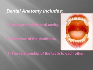
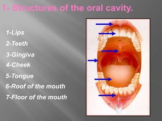
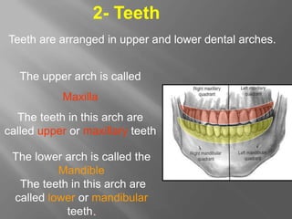
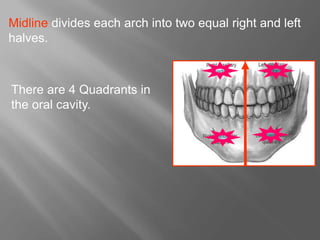
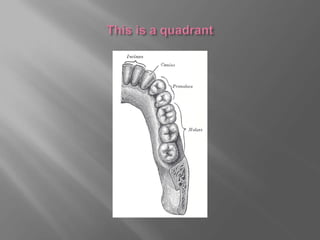
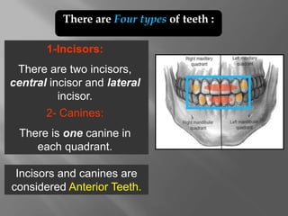
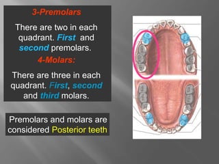
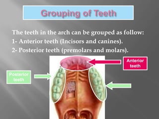
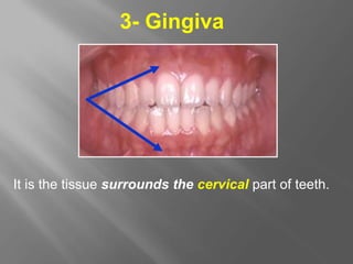
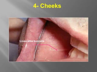
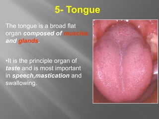
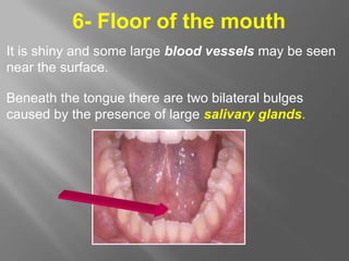
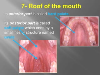
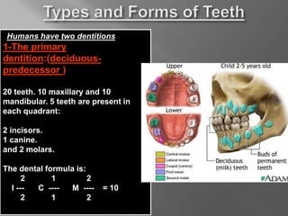
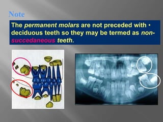
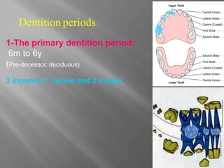
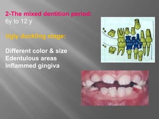
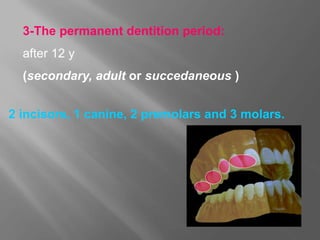
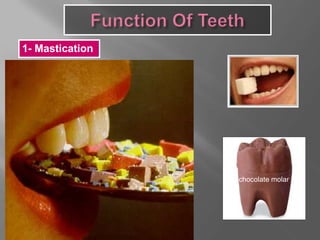
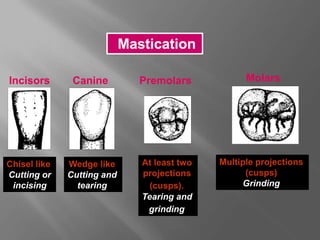
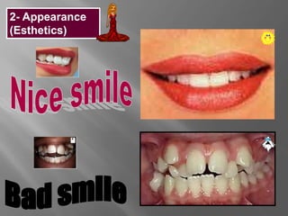
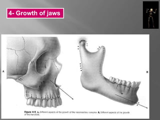
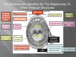
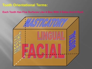
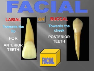
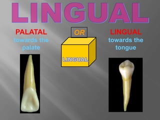
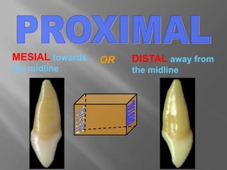
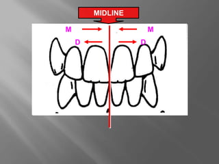
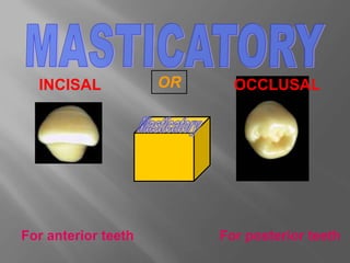
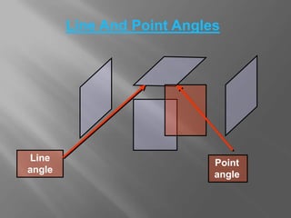
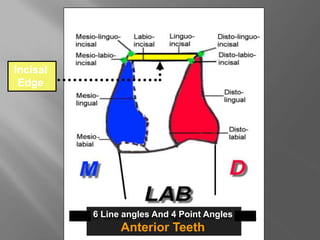
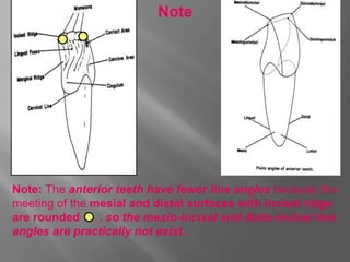
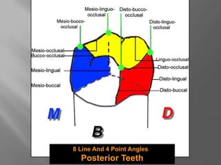
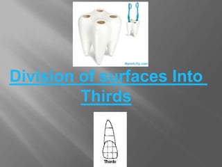
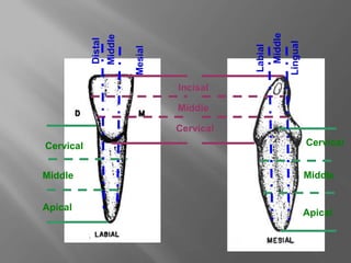
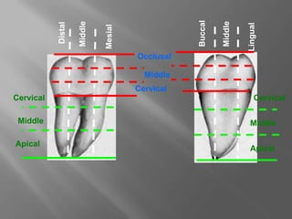
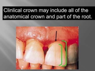
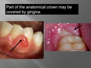
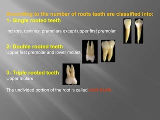
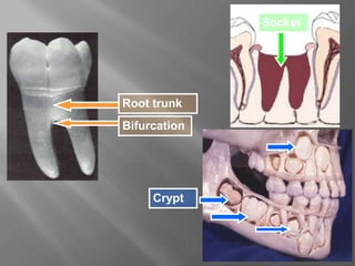
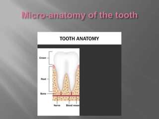
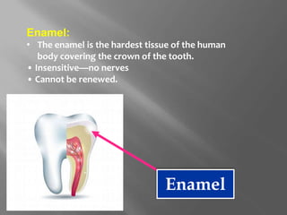
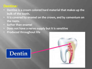
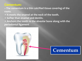
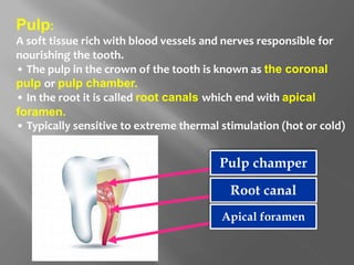
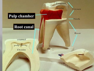
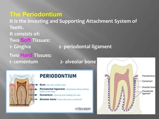
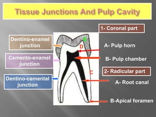
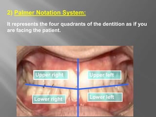
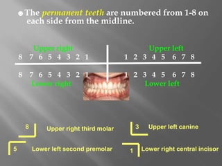
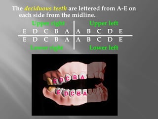
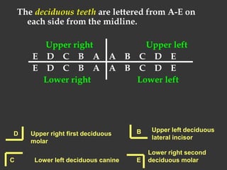
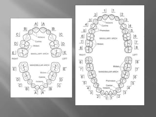
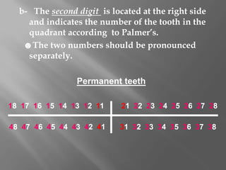
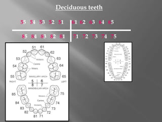
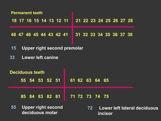
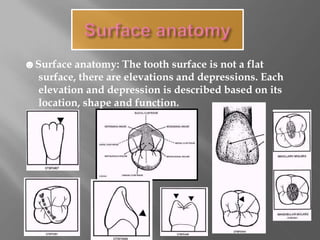
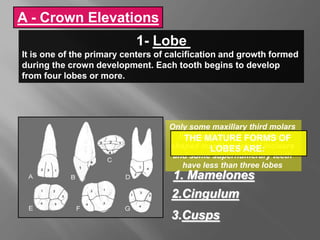
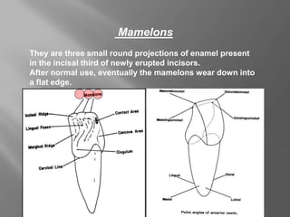
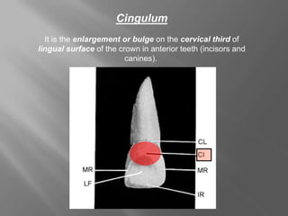
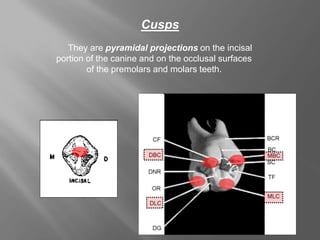
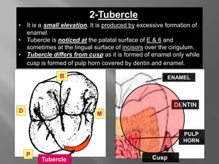
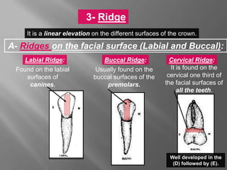
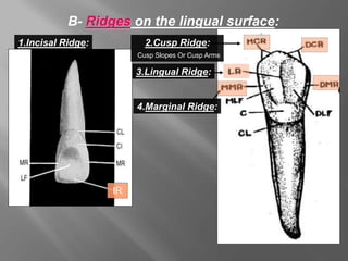
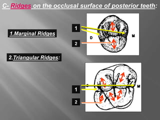
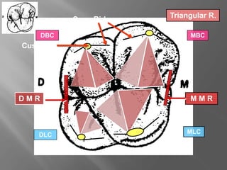
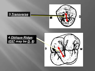
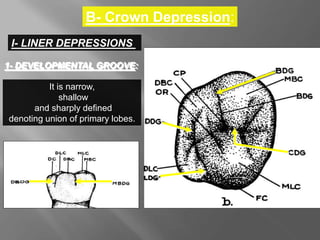
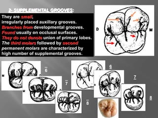
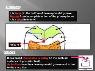
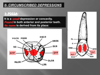
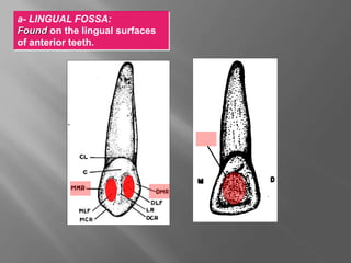
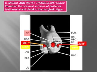
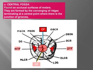
molar children
Dental Anatomy Includes:
2- Overview of the dentitions
3- The relationship of the teeth to each other.
1- Structures of the oral cavity.
STRUCTURES OF THE
ORAL CAVITY
1- Structures of the oral cavity.
2-Teeth
3-Gingiva
4-Cheek
5-Tongue
6-Roof of the mouth
7-Floor of the mouth
1-Lips
Two Lips Upper Lip and Lower
Lip.
The Corner of the mouth where
the two lips meet is called
Commissure
Two Grooves are distinguished
around lips.
*Nasolabial groove
*Labiomental groove.
1- Lips
Teeth are arranged in upper and lower dental arches.
The upper arch is called
Maxilla
The teeth in this arch are
called upper or maxillary teeth
The lower arch is called the
Mandible
The teeth in this arch are
called lower or mandibular
teeth.
2- Teeth
Midline divides each arch into two equal right and left
halves.
There are 4 Quadrants in
the oral cavity.
1-Incisors:
There are two incisors,
central incisor and lateral
incisor.
2- Canines:
There is one canine in
each quadrant.
Incisors and canines are
considered Anterior Teeth.
There are Four types of teeth :
3-Premolars
There are two in each
quadrant. First and
second premolars.
4-Molars:
There are three in each
quadrant. First, second
and third molars.
Premolars and molars are
considered Posterior teeth
The teeth in the arch can be grouped as follow:
1- Anterior teeth (Incisors and canines).
2- Posterior teeth (premolars and molars).
Anterior
teeth
Posterior
teeth
3- Gingiva
It is the tissue surrounds the cervical part of teeth.
4- Cheeks
Linea alba buccalis
The tongue is a broad flat
organ composed of muscles
and glands.
•It is the principle organ of
taste and is most important
in speech,mastication and
swallowing.
5- Tongue
6- Floor of the mouth
It is shiny and some large blood vessels may be seen
near the surface.
Beneath the tongue there are two bilateral bulges
caused by the presence of large salivary glands.
7- Roof of the mouth
Its anterior part is called hard palate.
Its posterior part is called
soft palate which ends by a
small fleshy structure named
uvula
TYPES OF DENTITION
Humans have two dentitions
1-The primary
dentition:(deciduous-
predecessor )
20 teeth. 10 maxillary and 10
mandibular. 5 teeth are present in
each quadrant:
2 incisors.
1 canine.
and 2 molars.
The dental formula is:
2 1 2
I --- C ---- M ---- = 10
2 1 2
2-The permanent dentition:
(secondary, adult or
succedaneous )
32 teeth, 16 maxillary and
16 mandibular.8 teeth in each
quadrant are Present:
2 incisors.
1 canine.
2 premolars.
and 3 molars.
The dental formula is :
2 1 2 3
I ---- C ---- PM --- M ---- = 16
2 1 2 3
•
The permanent molars are not preceded with
deciduous teeth so they may be termed as non-
succedaneous teeth.
Note
1-The primary dentition period:
6m to 6y
(Pre-decessor, deciduous)
2 incisors, 1 canine and 2 molars.
Dentition periods
2-The mixed dentition period:
6y to 12 y
Ugly duckling stage:
Different color & size
Edentulous areas
Inflammed gingiva
3-The permanent dentition period:
after 12 y
(secondary, adult or succedaneous )
2 incisors, 1 canine, 2 premolars and 3 molars.
FUNCTIONS OF
TEETH
1- Mastication
chocolate molar
Incisors Canine Premolars Molars
Chisel like
Cutting or
incising
Wedge like
Cutting and
tearing
At least two
projections
(cusps).
Tearing and
grinding
Multiple projections
(cusps)
Grinding
Mastication
2- Appearance
(Esthetics)
3- Speech
4- Growth of jaws
TOOTH SURFACES
AND ASPECTS
The Surfaces Are Identified By The Relationship To
Other Orofacial Structures
Distal surface
M
M
D
D
Distal surface
LIP
CHEEK
(buccal)
TONGUE
PALATE
Anterior
teeth
Posterior
teeth
Maxillary
teeth
Mandibular
teeth
Posterior teeth
Anterior teeth
MIDLINE
Proximal
surfaces
for all the
teeth
Tooth Orientational Terms:
Each Tooth Has Five Surfaces Like A Box With 4 Sides And A Roof.
LABIAL
Towards the
lip
FOR
ANTERIOR
TEETH
BUCCAL
Towards the
cheek
POSTERIOR
TEETH
OR
PALATAL
towards the
palate
LINGUAL
towards the
tongue
OR
MESIAL towards
the midline
DISTAL away from
the midline
OR
MIDLINE
M
D
M
D
For anterior teeth For posterior teeth
INCISAL OCCLUSAL
OR
Line And Point Angles
Line
angle
Point
angle
6 Line angles And 4 Point Angles
Anterior Teeth
Incisal
Edge
Note: The anterior teeth have fewer line angles because the
meeting of the mesial and distal surfaces with incisal ridge
are rounded , so the mesio-incisal and disto-incisal line
angles are practically not exist. .
Note
8 Line And 4 Point Angles
Posterior Teeth
Division of surfaces Into
Thirds
Incisal
Cervical
Cervical
Middle
Middle
Apical
Middle
Mesial
Distal
Cervical
Middle
Apical
Middle
Lingual
Labial
Occlusal
Middle
Cervical
Cervical
Cervical
Middle
Middle
Apical
Apical
Mesial
Middle
Distal
Buccal
Middle
Lingual
MACRO AND MICRO
ANATOMY OF TEETH
Each tooth has three
anatomical parts:
I.Crown
II.The Neck
II.Root (s)
1- Macro-anatomy of the tooth
Enamel
Cementum
Macro-anatomy of the Tooth
Each tooth has three anatomical parts:
I.Crown part of the tooth which is seen in the
oral cavity and is called Clincal crown
Part of the tooth covered by enamel is called
Anatomical crown
II.The Neck: cervical line
Meating between crown and root (cemento-
enamel junction)
II.Root (s) part of the tooth embedded in the
jaw bones and is called Clinical root
Part of the tooth covered by cementum is called
Anatomical root
•Anatomical and clinical crown.
Clinical
Crown
Anatomical
Crown
Anatomical
crown
Clinical
crown
Clinlical crown may include all of the
anatomical crown and part of the root.
Part of the anatomical crown may be
covered by gingiva.
According to the number of roots teeth are classified into:
1- Single rooted teeth
Incisors, canines, premolars except upper first premolar
2- Double rooted teeth
Upper first premolar and lower molars
3- Triple rooted teeth
Upper molars
The undivided portion of the root is called root trunk
Root trunk
Bifurcation
Socket
Crypt
Enamel:
• The enamel is the hardest tissue of the human
body covering the crown of the tooth.
• Insensitive—no nerves
• Cannot be renewed.
Enamel
Dentine:
• Dentine is a cream colored hard material that makes up the
bulk of the tooth.
• It is covered by enamel on the crown, and by cementum on
the roots.
• Softer than enamel
• Does not have a nerve supply but it is sensitive
• Produced throughout life
Dentin
Cementum:
• The cementum is a thin calcified tissue covering of the
roots.
• It meets the enamel at the neck of the tooth.
• Softer than enamel and dentin.
• Anchors the tooth to the alveolar bone along with the
periodontal ligament
Cementum
Pulp:
A soft tissue rich with blood vessels and nerves responsible for
nourishing the tooth.
• The pulp in the crown of the tooth is known as the coronal
pulp or pulp chamber.
• In the root it is called root canals which end with apical
foramen.
• Typically sensitive to extreme thermal stimulation (hot or cold)
Pulp champer
Root canal
Apical foramen
•Microanatomy
Three calcified tissues:
Enamel, Dentin and Cementum
And one soft tissue, Pulp.
Pulp cavity consists of :
Pulp chamber and pulp canal or canals
Pulp chamber
Root canal
The Periodontium
It is the investing and Supporting Attachment System of
Teeth.
It consists of:
Two Soft Tissues:
1- Gingiva 2- periodontal ligament
Two Hard Tissues:
1- cementum 2- alveolar bone
Dentino-enamel
junction
Cemento-enamel
junction
Dentino-cemental
junction
E
D
C
A- Pulp horn
B- Pulp chamber
1- Coronal part
2- Radicular part
A- Root canal
B-Apical foramen
1)The universal numbering system:.
The number is always preceded by the sign # to
designate that the system is used for universal system.
Permanent teeth (1 – 32)
1 2 3 4 5 6 7 8 9 10 11 12 13 14 15 16
32 31 30 29 28 27 26 25 24 23 22 21 20 19 18 17
Numbering and coding systems of the teeth
#28 Lower right first
premolar
#16 Upper left third molar
Deciduous teeth (1 - 20)
1 2 3 4 5 6 7 8 9 10
20 19 18 17 16 15 14 13 12 11
The letter is preceded by #d
molar clock
d# 14 Lower left deciduous
lateral incisor
Deciduous teeth (A - T)
A B C D E F G H I J
T S R Q P O N M L K
The letter is preceded by # only
molar clock
#B Upper right
deciduous first molar
2) Palmer Notation System:
It represents the four quadrants of the dentition as if you
are facing the patient.
Upper right Upper left
Lower right Lower left
☻The permanent teeth are numbered from 1-8 on
each side from the midline.
Upper right Upper left
8 7 6 5 4 3 2 1 1 2 3 4 5 6 7 8
8 7 6 5 4 3 2 1 1 2 3 4 5 6 7 8
Lower right Lower left
8 Upper right third molar
5 Lower left second premolar
3 Upper left canine
1 Lower right central incisor
The deciduous teeth are lettered from A-E on
each side from the midline.
Upper right Upper left
E D C B A A B C D E
E D C B A A B C D E
Lower right Lower left
A
B
C
D
E
A
B
C
D
E
The deciduous teeth are lettered from A-E on
each side from the midline.
Upper right Upper left
E D C B A A B C D E
E D C B A A B C D E
Lower right Lower left
D Upper right first deciduous
molar
Lower left deciduous canine
C
Upper left deciduous
lateral incisor
B
Lower right second
deciduous molar
E
The teeth are designed by using binomial systems:
a. The first digit of the code is located at the left
side and indicates the quadrant.
In the permanent dentition.
U.R. 1 2 U.L.
L.R. 4 3 L.L.
In the deciduous dentition.
U.R 5 6 U.L.
L.R. 8 7 L.L.
3- The Federation Dentaire international (FDI)
gold molar
b- The second digit is located at the right side
and indicates the number of the tooth in the
quadrant according to Palmer’s.
☻The two numbers should be pronounced
separately.
18 17 16 15 14 13 12 11 21 22 23 24 25 26 27 28
48 47 46 45 44 43 42 41 31 32 33 34 35 36 37 38
Permanent teeth
Deciduous teeth
55 54 53 52 51 61 62 63 64 65
85 84 83 82 81 71 72 73 74 75
Permanent teeth
21 22 23 24 25 26 27 28
18 17 16 15 14 13 12 11
48 47 46 45 44 43 42 41 31 32 33 34 35 36 37 38
Deciduous teeth
55 54 53 52 51 61 62 63 64 65
71 72 73 74 75
85 84 83 82 81
15 Upper right second premolar
33 Lower left canine
55 Upper right second
deciduous molar
72 Lower left lateral deciduous
incisor
☻Surface anatomy: The tooth surface is not a flat
surface, there are elevations and depressions. Each
elevation and depression is described based on its
location, shape and function.
Anatomical Landmarks
Of The Crown
1- Lobe
It is one of the primary centers of calcification and growth formed
during the crown development. Each tooth begins to develop
from four lobes or more.
THE MATURE FORMS OF
LOBES ARE:
1. Mamelones
2.Cingulum
3.Cusps
A - Crown Elevations
Mamelons
They are three small round projections of enamel present
in the incisal third of newly erupted incisors.
After normal use, eventually the mamelons wear down into
a flat edge.
Cingulum
It is the enlargement or bulge on the cervical third of
lingual surface of the crown in anterior teeth (incisors and
canines).
Cusps
They are pyramidal projections on the incisal
portion of the canine and on the occlusal surfaces
of the premolars and molars teeth.
2-Tubercle
• It is a small elevation. It is produced by excessive formation of
enamel.
• Tubercle is noticed at the palatal surface of E & 6 and
sometimes at the lingual surface of incisors over the cingulum.
• Tubercle differs from cusp as it is formed of enamel only while
cusp is formed of pulp horn covered by dentin and enamel.
PULP
HORN
Cusp
ENAMEL
DENTIN
Tubercle
M
B
D
P
3- Ridge
It is a linear elevation on the different surfaces of the crown.
A- Ridges on the facial surface (Labial and Buccal):
Labial Ridge: Buccal Ridge: Cervical Ridge:
Found on the labial
surfaces of
canines.
Usually found on the
buccal surfaces of the
premolars.
It is found on the
cervical one third of
the facial surfaces of
all the teeth.
Well developed in the
(D) followed by (E).
B- Ridges on the lingual surface:
1.Incisal Ridge:
3.Lingual Ridge:
4.Marginal Ridge:
2.Cusp Ridge:
Cusp Slopes Or Cusp Arms
IR
C- Ridges on the occlusal surface of posterior teeth:
1.Marginal Ridges
2.Triangular Ridges:
1
2
1
2
Triangular R.
Cusp Tip
M M R
D M R
Cusp Ridges
MBC
MLC
DLC
DBC
3.Transverse
4.Oblique Ridge:
(E67 may be D, 8)
B- Crown Depression:
I- LINER DEPRESSIONS
1- DEVELOPMENTAL GROOVE:
It is narrow,
shallow
and sharply defined
denoting union of primary lobes.
They are small,
irregularly placed auxillary grooves.
Branches from developmental grooves.
Found usually on occlusal surfaces.
They do not denote union of primary lobes.
The third molars followed by second
permanent molars are characterized by
high number of supplemental grooves.
2- SUPPLEMENTAL GROOVES:
_
6
6
_
7
8
_
8
7
It is found in the bottom of developmental groove.
Results from incomplete union of the primary lobes.
It is a fault in enamel.
3- FISSURE:
It is a broad depression or valley on the occlusal
surfaces of posterior teeth.
Its inclines meet in a developmental groove and extend
to the cusp tips.
SULCUS:
Cusp tip
Cusp tip
Dentin
Enamel
Fissure
Developmental
groove
II- CIRCUMSCRIBED DEPRESSIONS
It is a small depression or concavity.
Found in both anterior and posterior teeth.
Its name is derived from its place:
1- FOSSA:
a- LINGUAL FOSSA:
Found on the lingual surfaces
of anterior teeth.
LF
b- MESIAL AND DISTAL TRIANGULAR FOSSA:
Found on the occlusal surfaces of posterior
teeth mesial and distal to the marginal ridges.
DMR MMR
MTF
DTF
c- CENTRAL FOSSA:
Found on occlusal surfaces of molars.
They are formed by the converging of ridges
terminating at a central point where there is the
junction of grooves.
CF
CP
a- TRUE PITS:
These are small pinpoint depression.
present at the junction / or at the
ends
of the developmental grooves.
They may be found at the bottom
of the central fossa (central pit)
or at the bottom of the mesial and
distal triangular fossae
(mesial and distal pits).
2- PITS:
b- FAULTY PIT
Usually develops as a result
of incomplete formation of enamel.
It is located at the end of the buccal
developmental grooves of the lower
molars or palatal developmental
grooves
of the upper molars.
c.p
.
d.p
.
m.p
.
p.p. b.p.
Introduction.ppt
Introduction.ppt

Introduction.ppt

  • 1.
  • 2.
    Dental Anatomy Includes: 2-Overview of the dentitions 3- The relationship of the teeth to each other. 1- Structures of the oral cavity.
  • 3.
  • 4.
    1- Structures ofthe oral cavity. 2-Teeth 3-Gingiva 4-Cheek 5-Tongue 6-Roof of the mouth 7-Floor of the mouth 1-Lips
  • 5.
    Two Lips UpperLip and Lower Lip. The Corner of the mouth where the two lips meet is called Commissure Two Grooves are distinguished around lips. *Nasolabial groove *Labiomental groove. 1- Lips
  • 6.
    Teeth are arrangedin upper and lower dental arches. The upper arch is called Maxilla The teeth in this arch are called upper or maxillary teeth The lower arch is called the Mandible The teeth in this arch are called lower or mandibular teeth. 2- Teeth
  • 7.
    Midline divides eacharch into two equal right and left halves. There are 4 Quadrants in the oral cavity.
  • 9.
    1-Incisors: There are twoincisors, central incisor and lateral incisor. 2- Canines: There is one canine in each quadrant. Incisors and canines are considered Anterior Teeth. There are Four types of teeth :
  • 10.
    3-Premolars There are twoin each quadrant. First and second premolars. 4-Molars: There are three in each quadrant. First, second and third molars. Premolars and molars are considered Posterior teeth
  • 11.
    The teeth inthe arch can be grouped as follow: 1- Anterior teeth (Incisors and canines). 2- Posterior teeth (premolars and molars). Anterior teeth Posterior teeth
  • 12.
    3- Gingiva It isthe tissue surrounds the cervical part of teeth.
  • 13.
  • 14.
    The tongue isa broad flat organ composed of muscles and glands. •It is the principle organ of taste and is most important in speech,mastication and swallowing. 5- Tongue
  • 15.
    6- Floor ofthe mouth It is shiny and some large blood vessels may be seen near the surface. Beneath the tongue there are two bilateral bulges caused by the presence of large salivary glands.
  • 16.
    7- Roof ofthe mouth Its anterior part is called hard palate. Its posterior part is called soft palate which ends by a small fleshy structure named uvula
  • 17.
  • 18.
    Humans have twodentitions 1-The primary dentition:(deciduous- predecessor ) 20 teeth. 10 maxillary and 10 mandibular. 5 teeth are present in each quadrant: 2 incisors. 1 canine. and 2 molars. The dental formula is: 2 1 2 I --- C ---- M ---- = 10 2 1 2
  • 19.
    2-The permanent dentition: (secondary,adult or succedaneous ) 32 teeth, 16 maxillary and 16 mandibular.8 teeth in each quadrant are Present: 2 incisors. 1 canine. 2 premolars. and 3 molars. The dental formula is : 2 1 2 3 I ---- C ---- PM --- M ---- = 16 2 1 2 3
  • 20.
    • The permanent molarsare not preceded with deciduous teeth so they may be termed as non- succedaneous teeth. Note
  • 21.
    1-The primary dentitionperiod: 6m to 6y (Pre-decessor, deciduous) 2 incisors, 1 canine and 2 molars. Dentition periods
  • 22.
    2-The mixed dentitionperiod: 6y to 12 y Ugly duckling stage: Different color & size Edentulous areas Inflammed gingiva
  • 23.
    3-The permanent dentitionperiod: after 12 y (secondary, adult or succedaneous ) 2 incisors, 1 canine, 2 premolars and 3 molars.
  • 24.
  • 25.
  • 26.
    Incisors Canine PremolarsMolars Chisel like Cutting or incising Wedge like Cutting and tearing At least two projections (cusps). Tearing and grinding Multiple projections (cusps) Grinding Mastication
  • 27.
  • 28.
  • 29.
  • 30.
  • 31.
    The Surfaces AreIdentified By The Relationship To Other Orofacial Structures Distal surface M M D D Distal surface LIP CHEEK (buccal) TONGUE PALATE Anterior teeth Posterior teeth Maxillary teeth Mandibular teeth Posterior teeth Anterior teeth MIDLINE Proximal surfaces for all the teeth
  • 32.
    Tooth Orientational Terms: EachTooth Has Five Surfaces Like A Box With 4 Sides And A Roof.
  • 33.
  • 34.
  • 35.
    MESIAL towards the midline DISTALaway from the midline OR
  • 36.
  • 37.
    For anterior teethFor posterior teeth INCISAL OCCLUSAL OR
  • 38.
    Line And PointAngles Line angle Point angle
  • 39.
    6 Line anglesAnd 4 Point Angles Anterior Teeth Incisal Edge
  • 40.
    Note: The anteriorteeth have fewer line angles because the meeting of the mesial and distal surfaces with incisal ridge are rounded , so the mesio-incisal and disto-incisal line angles are practically not exist. . Note
  • 41.
    8 Line And4 Point Angles Posterior Teeth
  • 42.
  • 43.
  • 44.
  • 45.
  • 46.
    Each tooth hasthree anatomical parts: I.Crown II.The Neck II.Root (s) 1- Macro-anatomy of the tooth Enamel Cementum
  • 47.
    Macro-anatomy of theTooth Each tooth has three anatomical parts: I.Crown part of the tooth which is seen in the oral cavity and is called Clincal crown Part of the tooth covered by enamel is called Anatomical crown II.The Neck: cervical line Meating between crown and root (cemento- enamel junction) II.Root (s) part of the tooth embedded in the jaw bones and is called Clinical root Part of the tooth covered by cementum is called Anatomical root
  • 48.
    •Anatomical and clinicalcrown. Clinical Crown Anatomical Crown
  • 49.
  • 50.
    Clinlical crown mayinclude all of the anatomical crown and part of the root.
  • 51.
    Part of theanatomical crown may be covered by gingiva.
  • 52.
    According to thenumber of roots teeth are classified into: 1- Single rooted teeth Incisors, canines, premolars except upper first premolar 2- Double rooted teeth Upper first premolar and lower molars 3- Triple rooted teeth Upper molars The undivided portion of the root is called root trunk
  • 53.
  • 55.
    Enamel: • The enamelis the hardest tissue of the human body covering the crown of the tooth. • Insensitive—no nerves • Cannot be renewed. Enamel
  • 56.
    Dentine: • Dentine isa cream colored hard material that makes up the bulk of the tooth. • It is covered by enamel on the crown, and by cementum on the roots. • Softer than enamel • Does not have a nerve supply but it is sensitive • Produced throughout life Dentin
  • 57.
    Cementum: • The cementumis a thin calcified tissue covering of the roots. • It meets the enamel at the neck of the tooth. • Softer than enamel and dentin. • Anchors the tooth to the alveolar bone along with the periodontal ligament Cementum
  • 58.
    Pulp: A soft tissuerich with blood vessels and nerves responsible for nourishing the tooth. • The pulp in the crown of the tooth is known as the coronal pulp or pulp chamber. • In the root it is called root canals which end with apical foramen. • Typically sensitive to extreme thermal stimulation (hot or cold) Pulp champer Root canal Apical foramen
  • 59.
    •Microanatomy Three calcified tissues: Enamel,Dentin and Cementum And one soft tissue, Pulp. Pulp cavity consists of : Pulp chamber and pulp canal or canals Pulp chamber Root canal
  • 60.
    The Periodontium It isthe investing and Supporting Attachment System of Teeth. It consists of: Two Soft Tissues: 1- Gingiva 2- periodontal ligament Two Hard Tissues: 1- cementum 2- alveolar bone
  • 61.
    Dentino-enamel junction Cemento-enamel junction Dentino-cemental junction E D C A- Pulp horn B-Pulp chamber 1- Coronal part 2- Radicular part A- Root canal B-Apical foramen
  • 62.
    1)The universal numberingsystem:. The number is always preceded by the sign # to designate that the system is used for universal system. Permanent teeth (1 – 32) 1 2 3 4 5 6 7 8 9 10 11 12 13 14 15 16 32 31 30 29 28 27 26 25 24 23 22 21 20 19 18 17 Numbering and coding systems of the teeth #28 Lower right first premolar #16 Upper left third molar
  • 63.
    Deciduous teeth (1- 20) 1 2 3 4 5 6 7 8 9 10 20 19 18 17 16 15 14 13 12 11 The letter is preceded by #d molar clock d# 14 Lower left deciduous lateral incisor
  • 64.
    Deciduous teeth (A- T) A B C D E F G H I J T S R Q P O N M L K The letter is preceded by # only molar clock #B Upper right deciduous first molar
  • 66.
    2) Palmer NotationSystem: It represents the four quadrants of the dentition as if you are facing the patient. Upper right Upper left Lower right Lower left
  • 67.
    ☻The permanent teethare numbered from 1-8 on each side from the midline. Upper right Upper left 8 7 6 5 4 3 2 1 1 2 3 4 5 6 7 8 8 7 6 5 4 3 2 1 1 2 3 4 5 6 7 8 Lower right Lower left 8 Upper right third molar 5 Lower left second premolar 3 Upper left canine 1 Lower right central incisor
  • 68.
    The deciduous teethare lettered from A-E on each side from the midline. Upper right Upper left E D C B A A B C D E E D C B A A B C D E Lower right Lower left A B C D E A B C D E
  • 69.
    The deciduous teethare lettered from A-E on each side from the midline. Upper right Upper left E D C B A A B C D E E D C B A A B C D E Lower right Lower left D Upper right first deciduous molar Lower left deciduous canine C Upper left deciduous lateral incisor B Lower right second deciduous molar E
  • 71.
    The teeth aredesigned by using binomial systems: a. The first digit of the code is located at the left side and indicates the quadrant. In the permanent dentition. U.R. 1 2 U.L. L.R. 4 3 L.L. In the deciduous dentition. U.R 5 6 U.L. L.R. 8 7 L.L. 3- The Federation Dentaire international (FDI) gold molar
  • 72.
    b- The seconddigit is located at the right side and indicates the number of the tooth in the quadrant according to Palmer’s. ☻The two numbers should be pronounced separately. 18 17 16 15 14 13 12 11 21 22 23 24 25 26 27 28 48 47 46 45 44 43 42 41 31 32 33 34 35 36 37 38 Permanent teeth
  • 73.
    Deciduous teeth 55 5453 52 51 61 62 63 64 65 85 84 83 82 81 71 72 73 74 75
  • 74.
    Permanent teeth 21 2223 24 25 26 27 28 18 17 16 15 14 13 12 11 48 47 46 45 44 43 42 41 31 32 33 34 35 36 37 38 Deciduous teeth 55 54 53 52 51 61 62 63 64 65 71 72 73 74 75 85 84 83 82 81 15 Upper right second premolar 33 Lower left canine 55 Upper right second deciduous molar 72 Lower left lateral deciduous incisor
  • 75.
    ☻Surface anatomy: Thetooth surface is not a flat surface, there are elevations and depressions. Each elevation and depression is described based on its location, shape and function.
  • 76.
  • 77.
    1- Lobe It isone of the primary centers of calcification and growth formed during the crown development. Each tooth begins to develop from four lobes or more. THE MATURE FORMS OF LOBES ARE: 1. Mamelones 2.Cingulum 3.Cusps A - Crown Elevations
  • 78.
    Mamelons They are threesmall round projections of enamel present in the incisal third of newly erupted incisors. After normal use, eventually the mamelons wear down into a flat edge.
  • 79.
    Cingulum It is theenlargement or bulge on the cervical third of lingual surface of the crown in anterior teeth (incisors and canines).
  • 80.
    Cusps They are pyramidalprojections on the incisal portion of the canine and on the occlusal surfaces of the premolars and molars teeth.
  • 81.
    2-Tubercle • It isa small elevation. It is produced by excessive formation of enamel. • Tubercle is noticed at the palatal surface of E & 6 and sometimes at the lingual surface of incisors over the cingulum. • Tubercle differs from cusp as it is formed of enamel only while cusp is formed of pulp horn covered by dentin and enamel. PULP HORN Cusp ENAMEL DENTIN Tubercle M B D P
  • 82.
    3- Ridge It isa linear elevation on the different surfaces of the crown. A- Ridges on the facial surface (Labial and Buccal): Labial Ridge: Buccal Ridge: Cervical Ridge: Found on the labial surfaces of canines. Usually found on the buccal surfaces of the premolars. It is found on the cervical one third of the facial surfaces of all the teeth. Well developed in the (D) followed by (E).
  • 83.
    B- Ridges onthe lingual surface: 1.Incisal Ridge: 3.Lingual Ridge: 4.Marginal Ridge: 2.Cusp Ridge: Cusp Slopes Or Cusp Arms IR
  • 84.
    C- Ridges onthe occlusal surface of posterior teeth: 1.Marginal Ridges 2.Triangular Ridges: 1 2 1 2
  • 85.
    Triangular R. Cusp Tip MM R D M R Cusp Ridges MBC MLC DLC DBC
  • 86.
  • 87.
    B- Crown Depression: I-LINER DEPRESSIONS 1- DEVELOPMENTAL GROOVE: It is narrow, shallow and sharply defined denoting union of primary lobes.
  • 88.
    They are small, irregularlyplaced auxillary grooves. Branches from developmental grooves. Found usually on occlusal surfaces. They do not denote union of primary lobes. The third molars followed by second permanent molars are characterized by high number of supplemental grooves. 2- SUPPLEMENTAL GROOVES: _ 6 6 _ 7 8 _ 8 7
  • 89.
    It is foundin the bottom of developmental groove. Results from incomplete union of the primary lobes. It is a fault in enamel. 3- FISSURE: It is a broad depression or valley on the occlusal surfaces of posterior teeth. Its inclines meet in a developmental groove and extend to the cusp tips. SULCUS: Cusp tip Cusp tip Dentin Enamel Fissure Developmental groove
  • 90.
    II- CIRCUMSCRIBED DEPRESSIONS Itis a small depression or concavity. Found in both anterior and posterior teeth. Its name is derived from its place: 1- FOSSA:
  • 91.
    a- LINGUAL FOSSA: Foundon the lingual surfaces of anterior teeth. LF
  • 92.
    b- MESIAL ANDDISTAL TRIANGULAR FOSSA: Found on the occlusal surfaces of posterior teeth mesial and distal to the marginal ridges. DMR MMR MTF DTF
  • 93.
    c- CENTRAL FOSSA: Foundon occlusal surfaces of molars. They are formed by the converging of ridges terminating at a central point where there is the junction of grooves. CF CP
  • 94.
    a- TRUE PITS: Theseare small pinpoint depression. present at the junction / or at the ends of the developmental grooves. They may be found at the bottom of the central fossa (central pit) or at the bottom of the mesial and distal triangular fossae (mesial and distal pits). 2- PITS: b- FAULTY PIT Usually develops as a result of incomplete formation of enamel. It is located at the end of the buccal developmental grooves of the lower molars or palatal developmental grooves of the upper molars. c.p . d.p . m.p . p.p. b.p.