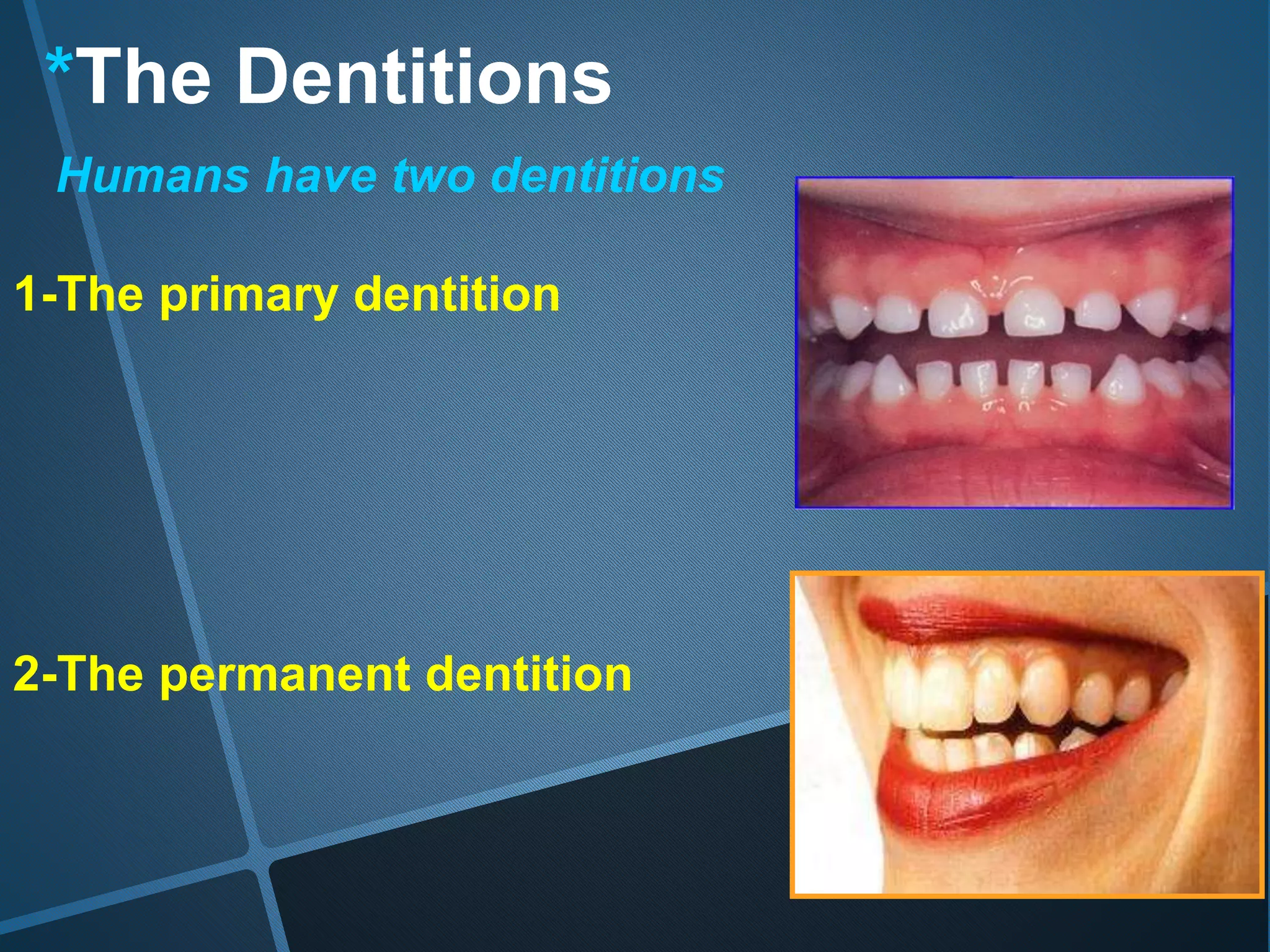
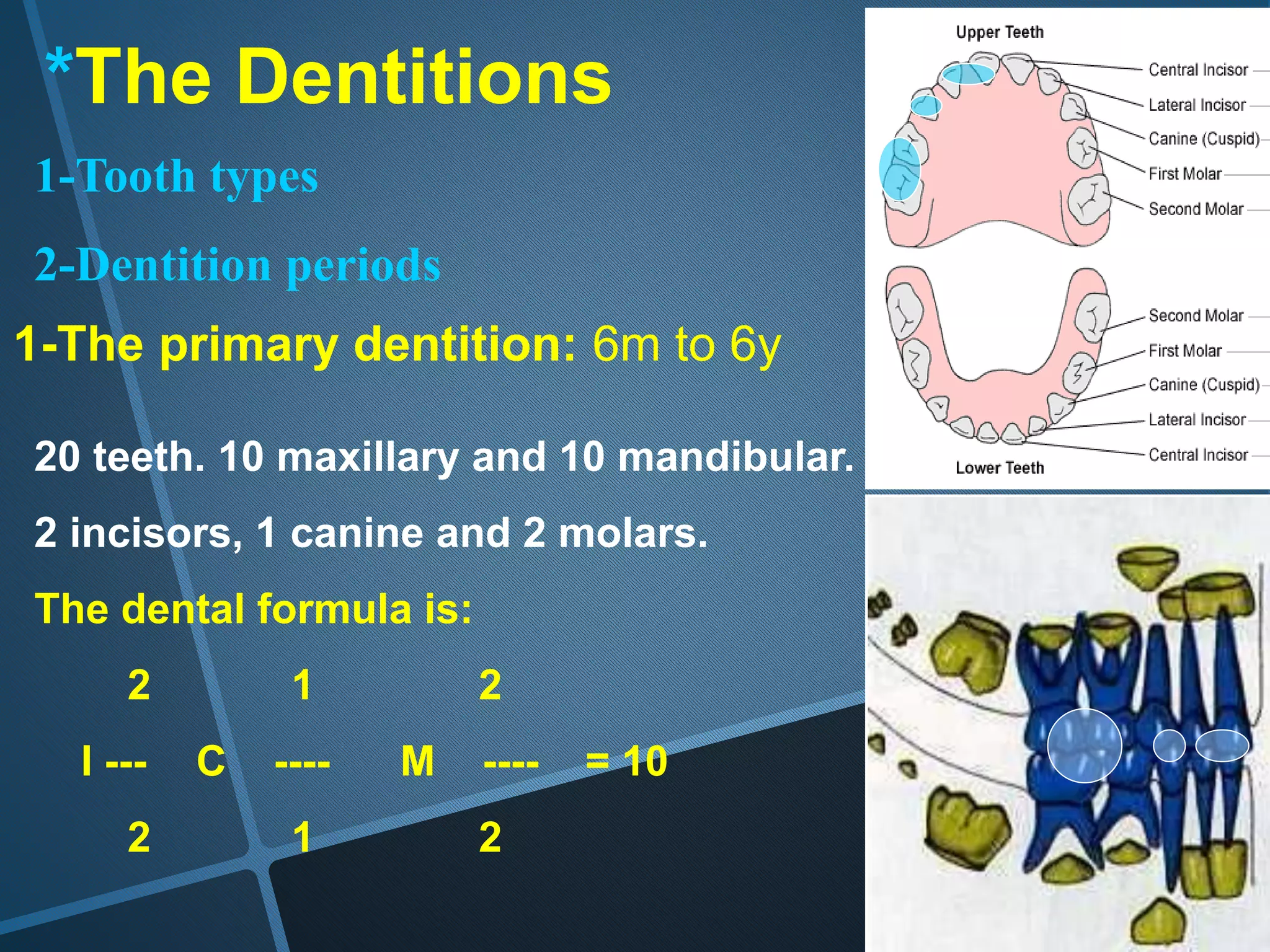
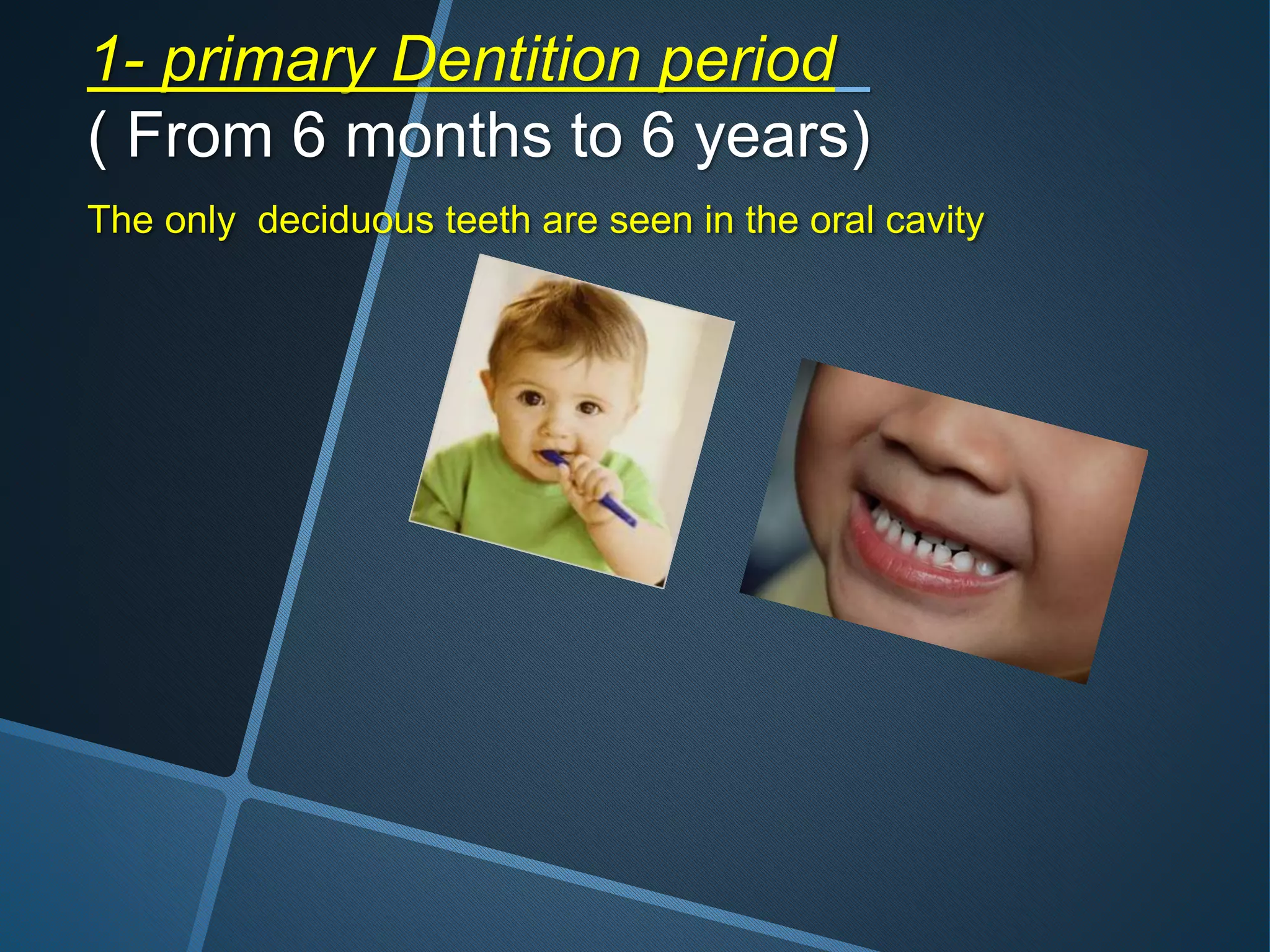
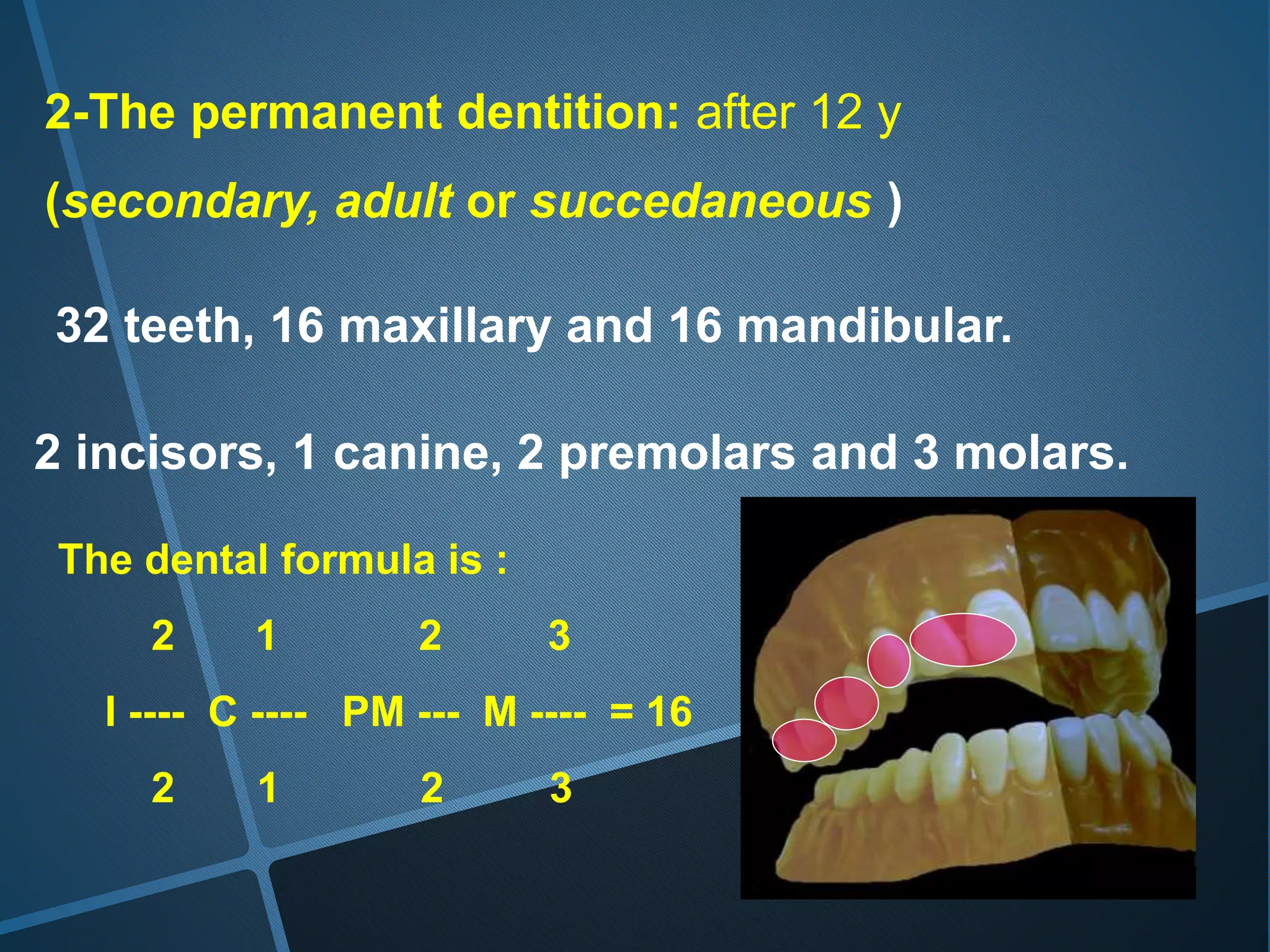
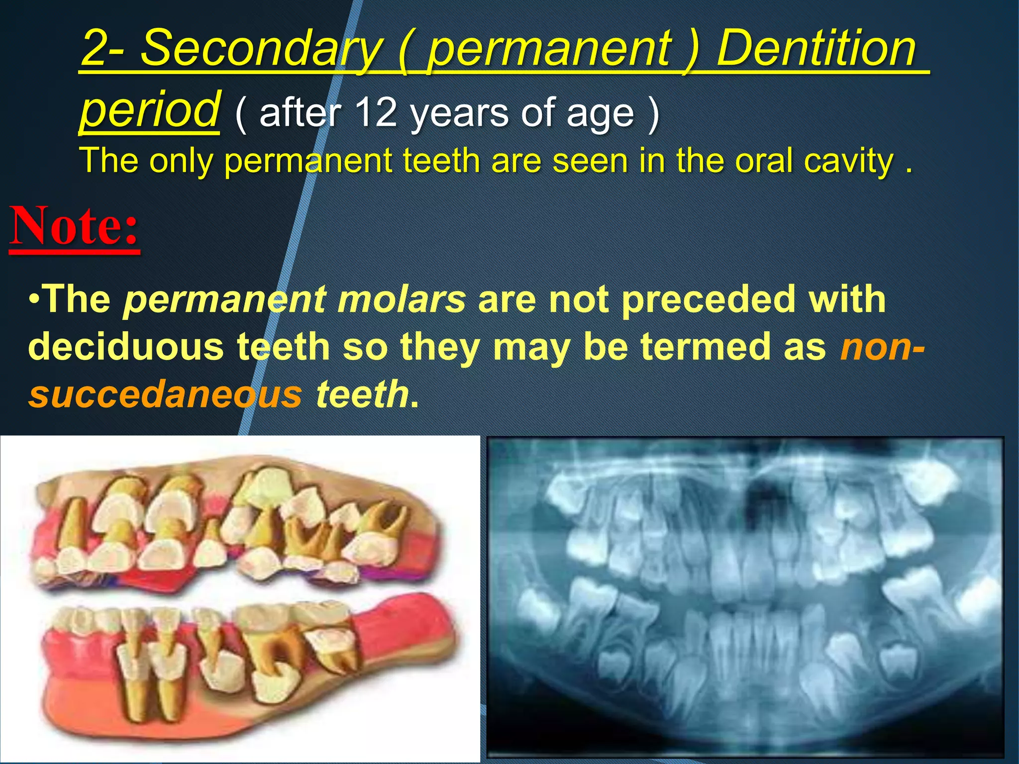
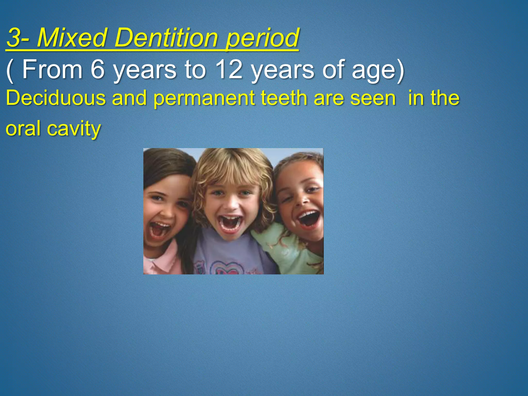
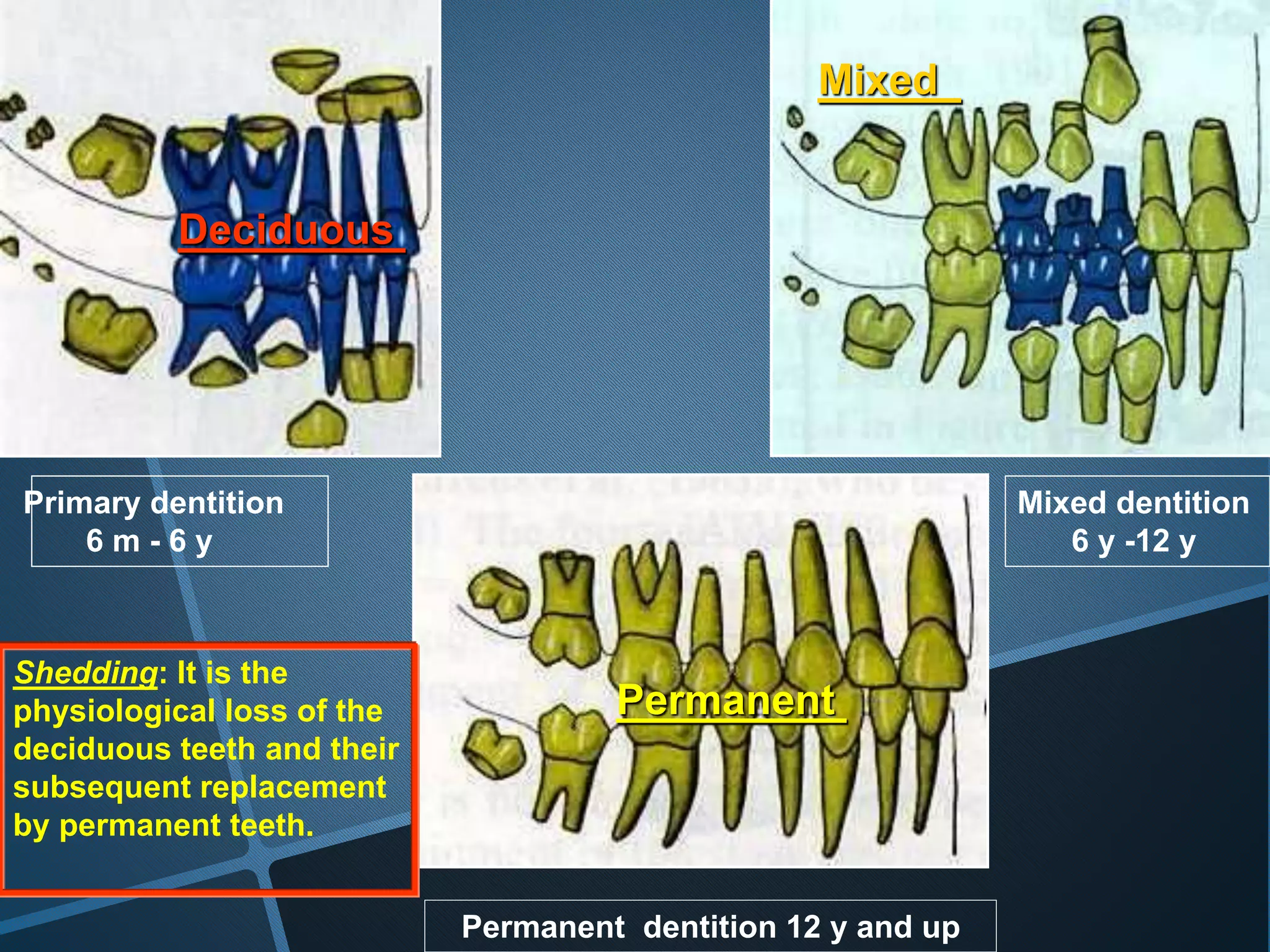
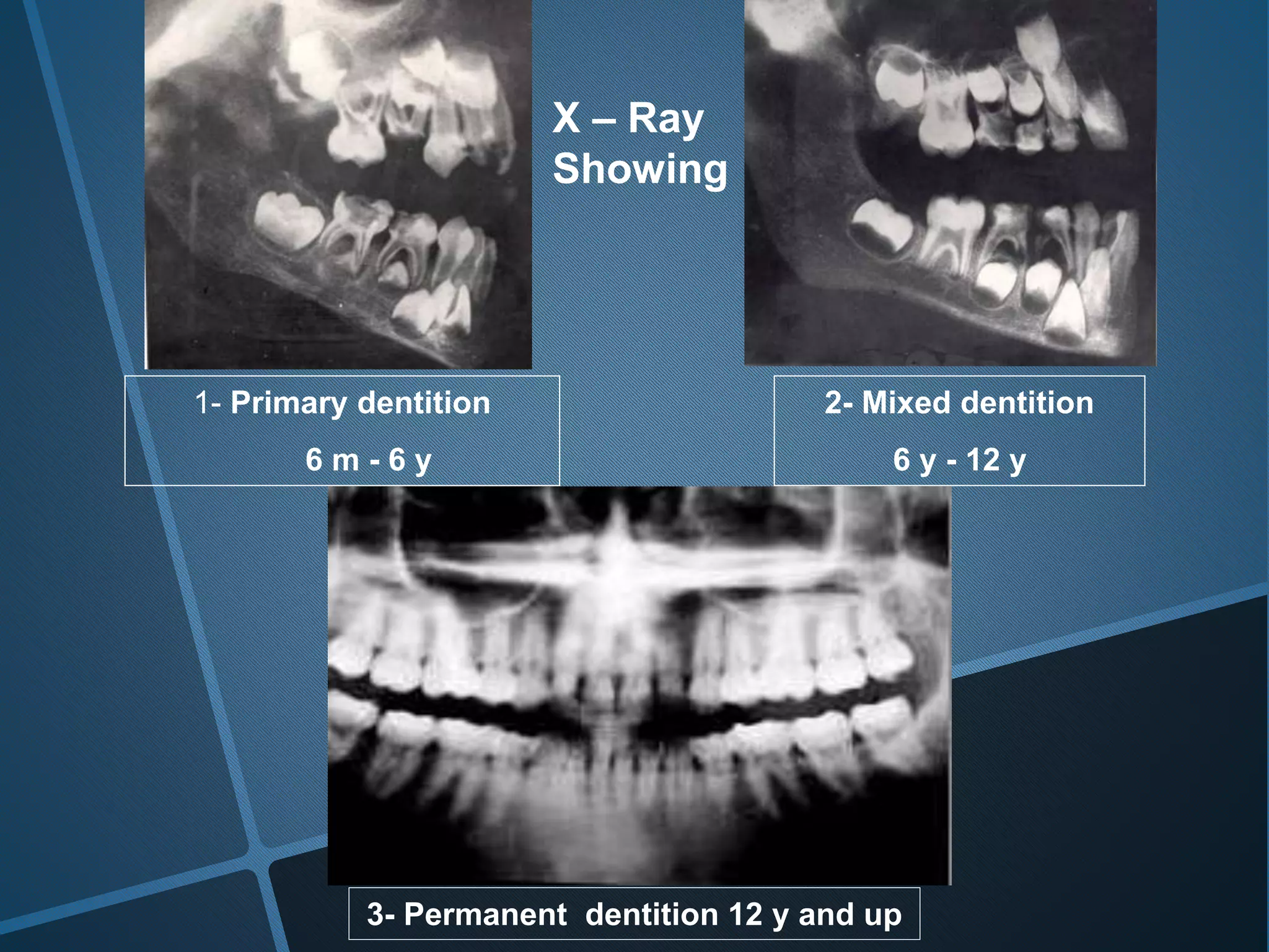
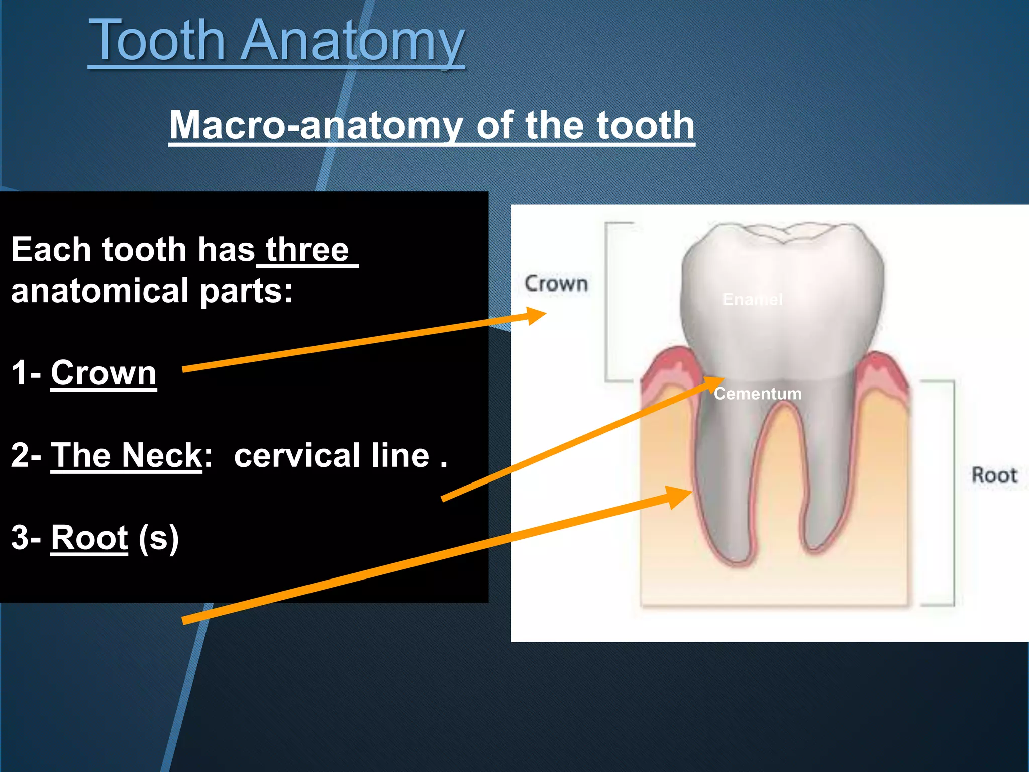
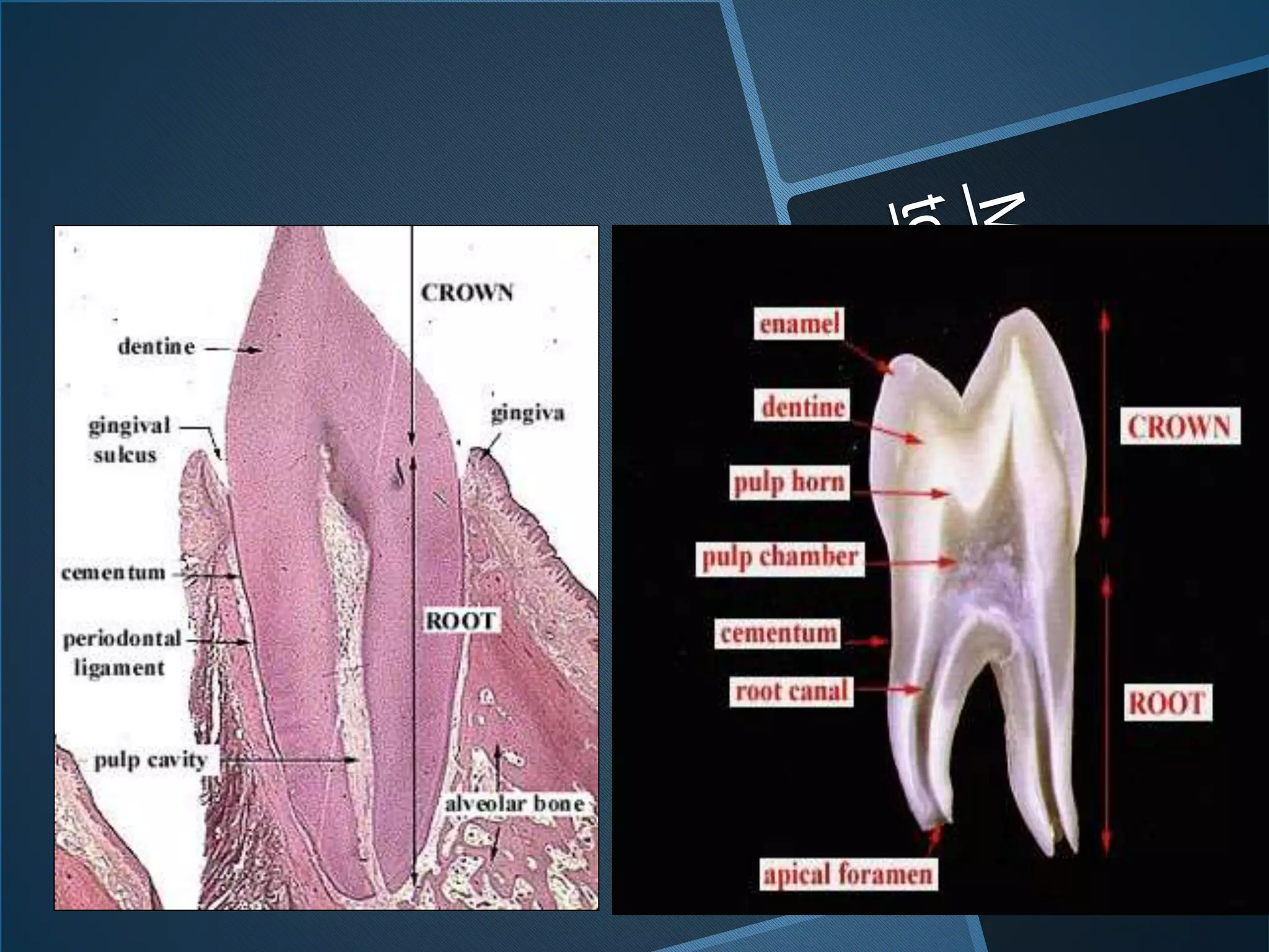
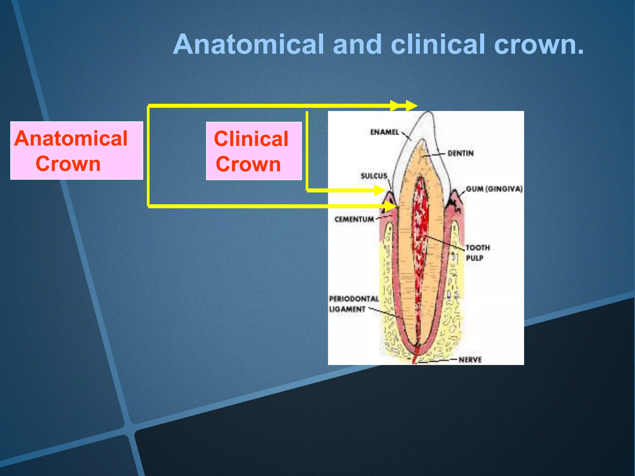
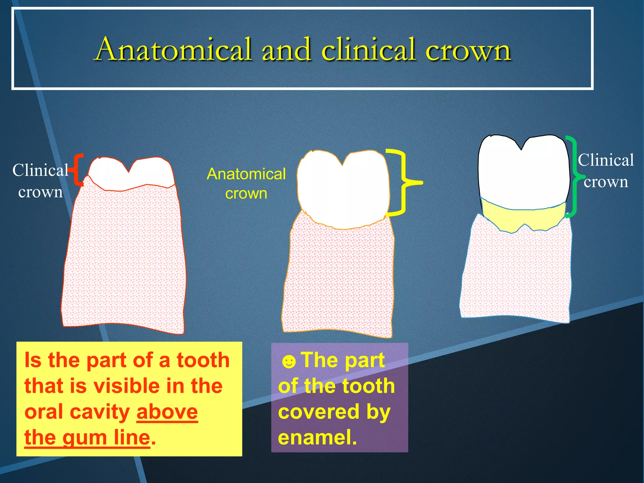
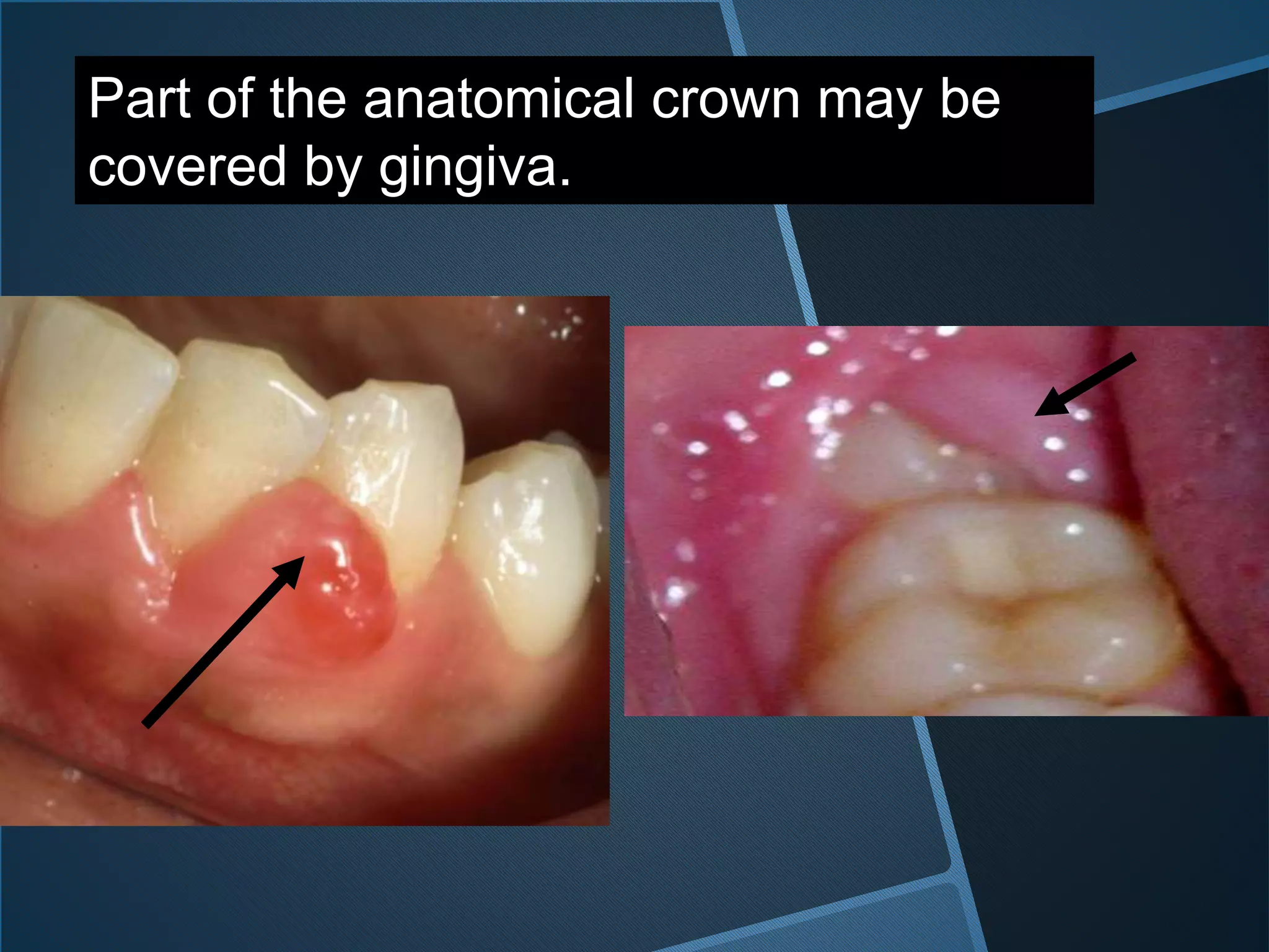
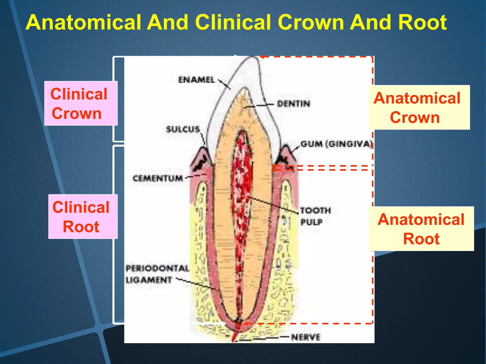
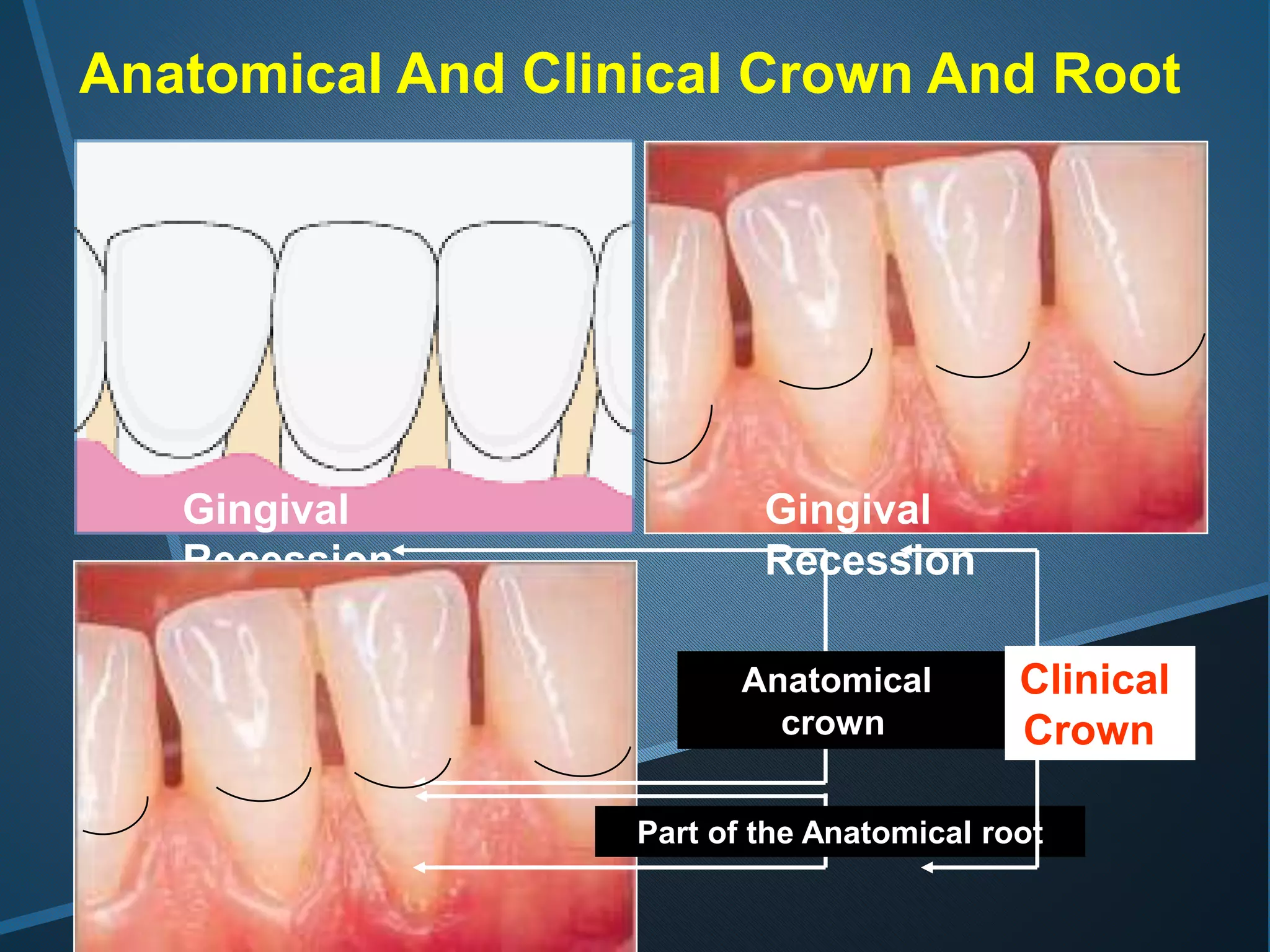
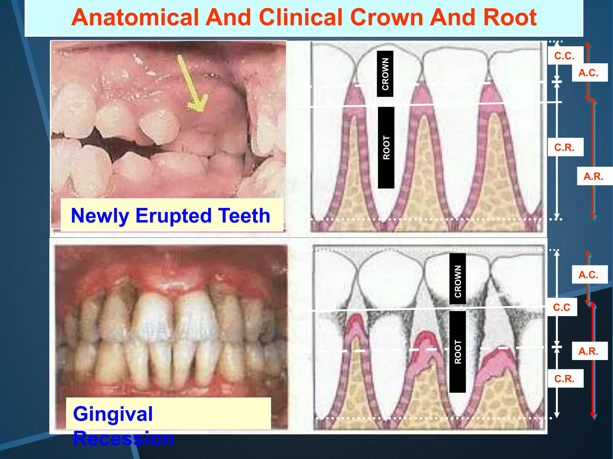
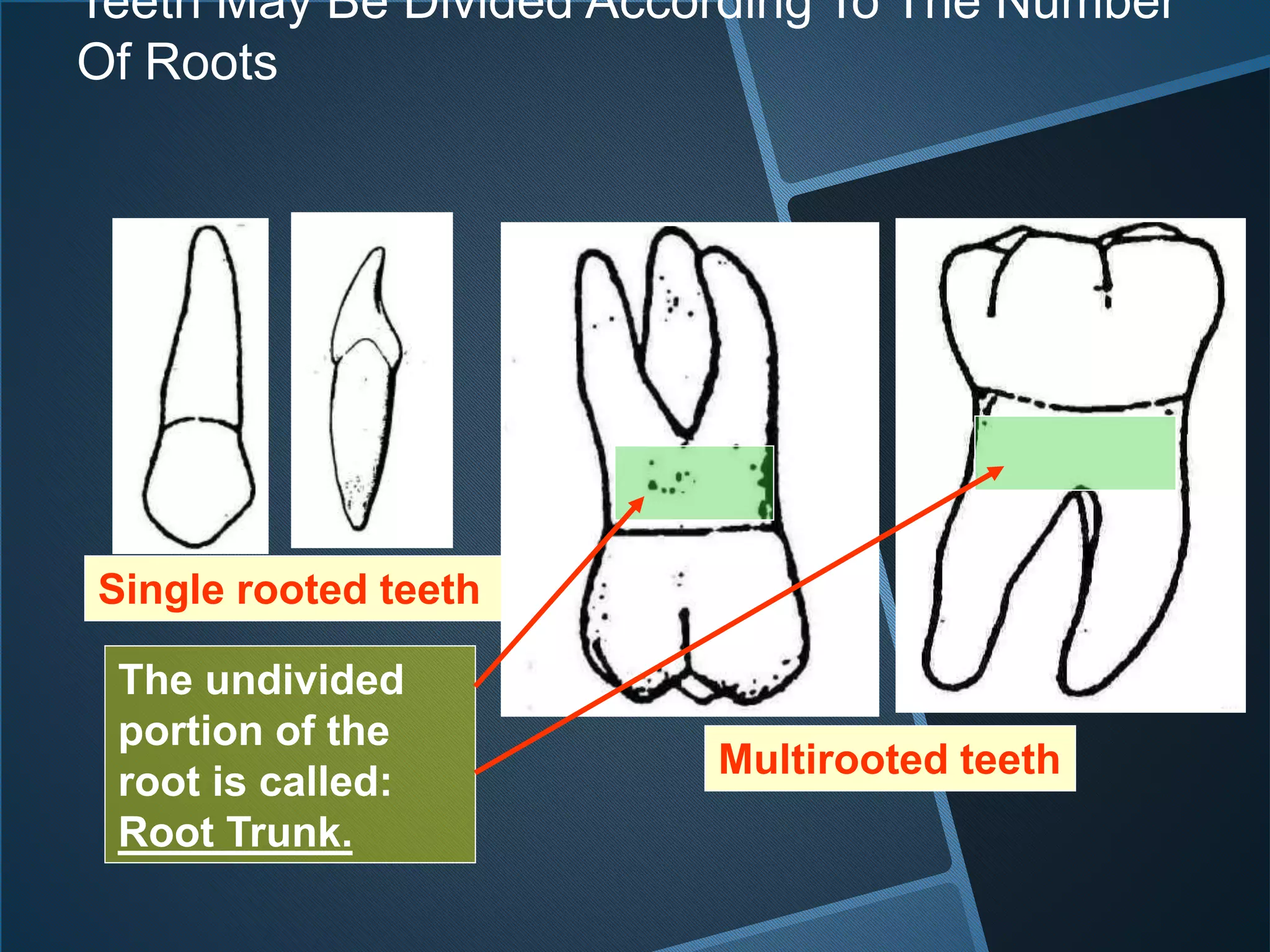
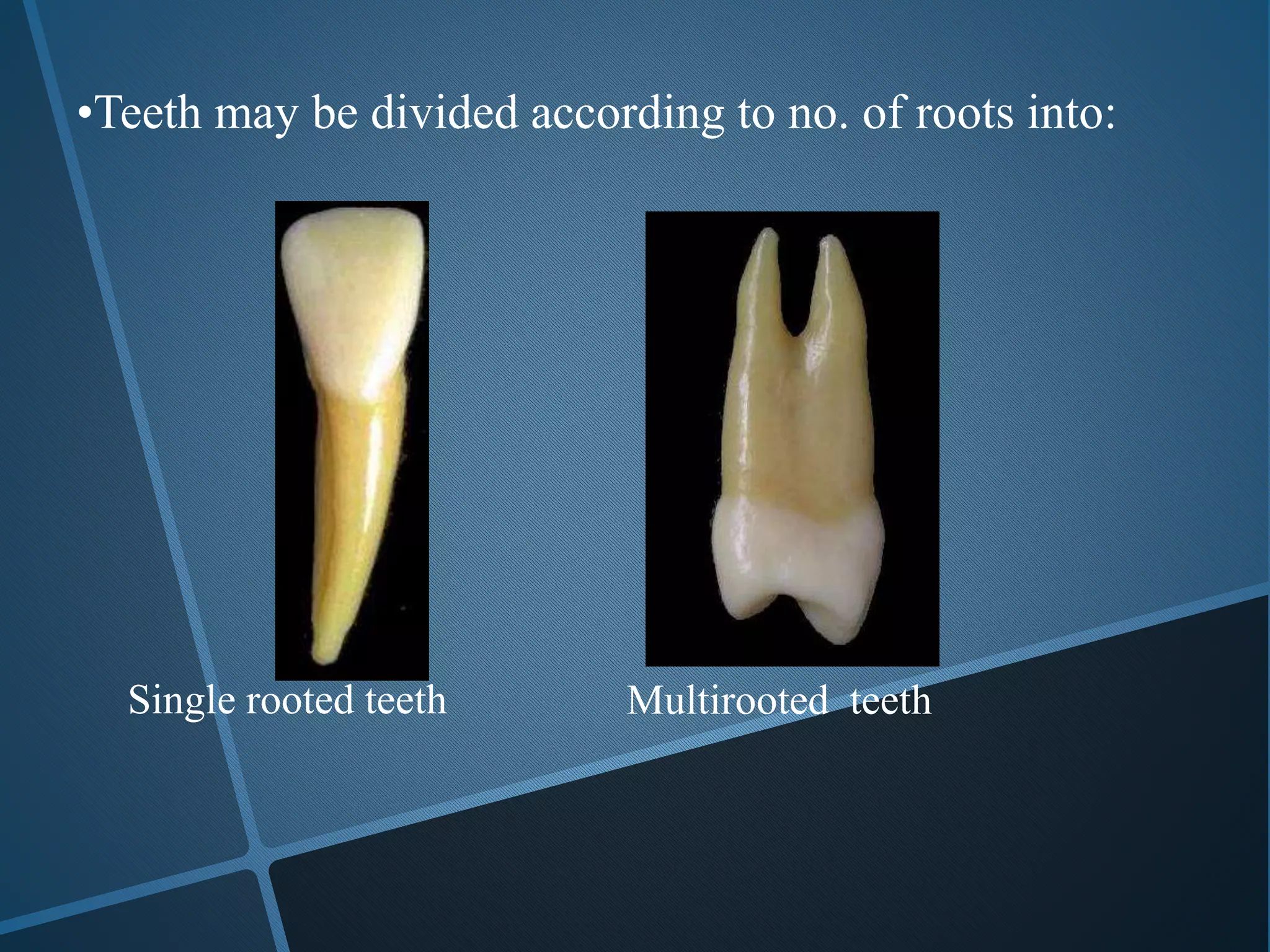
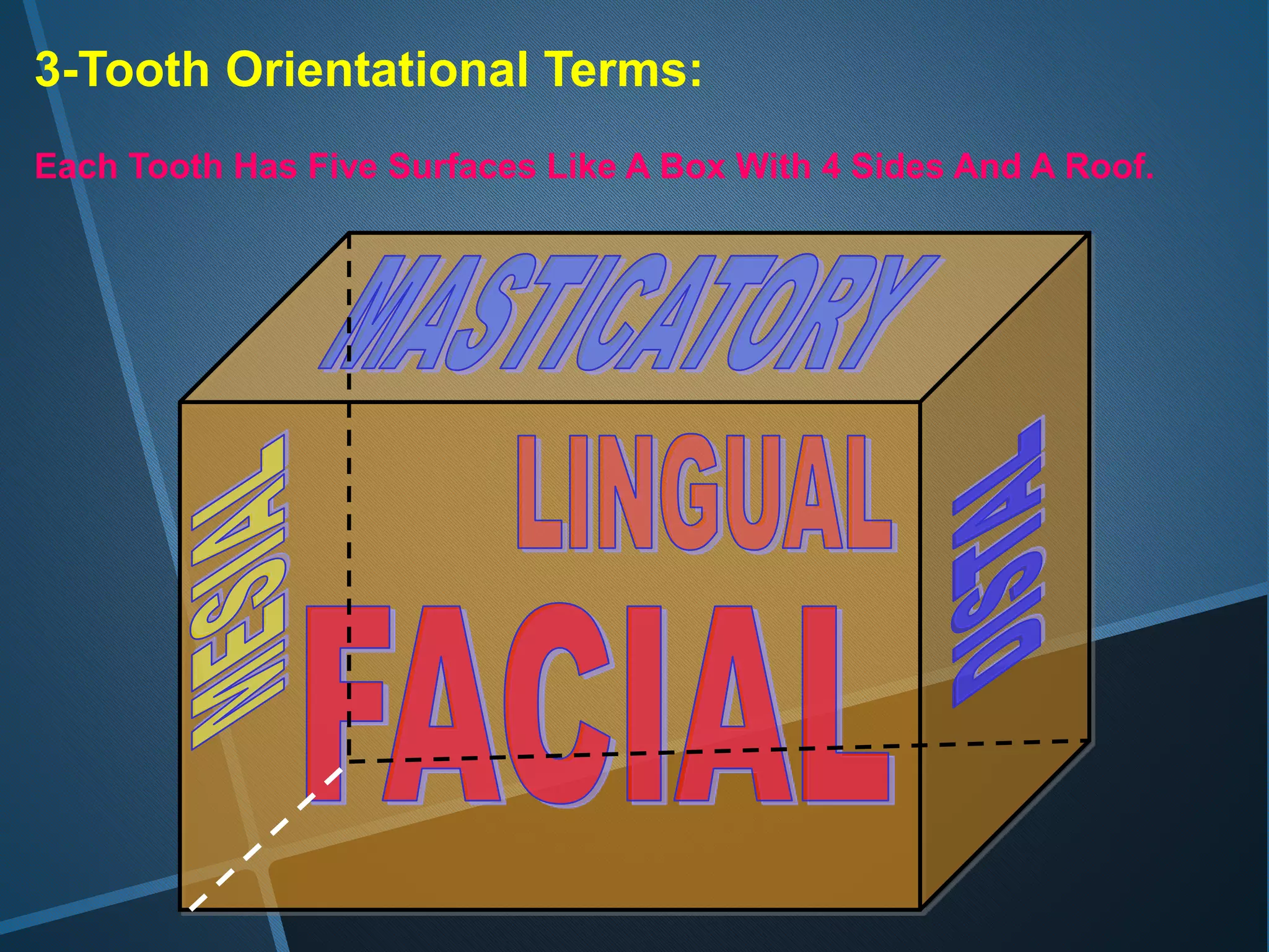
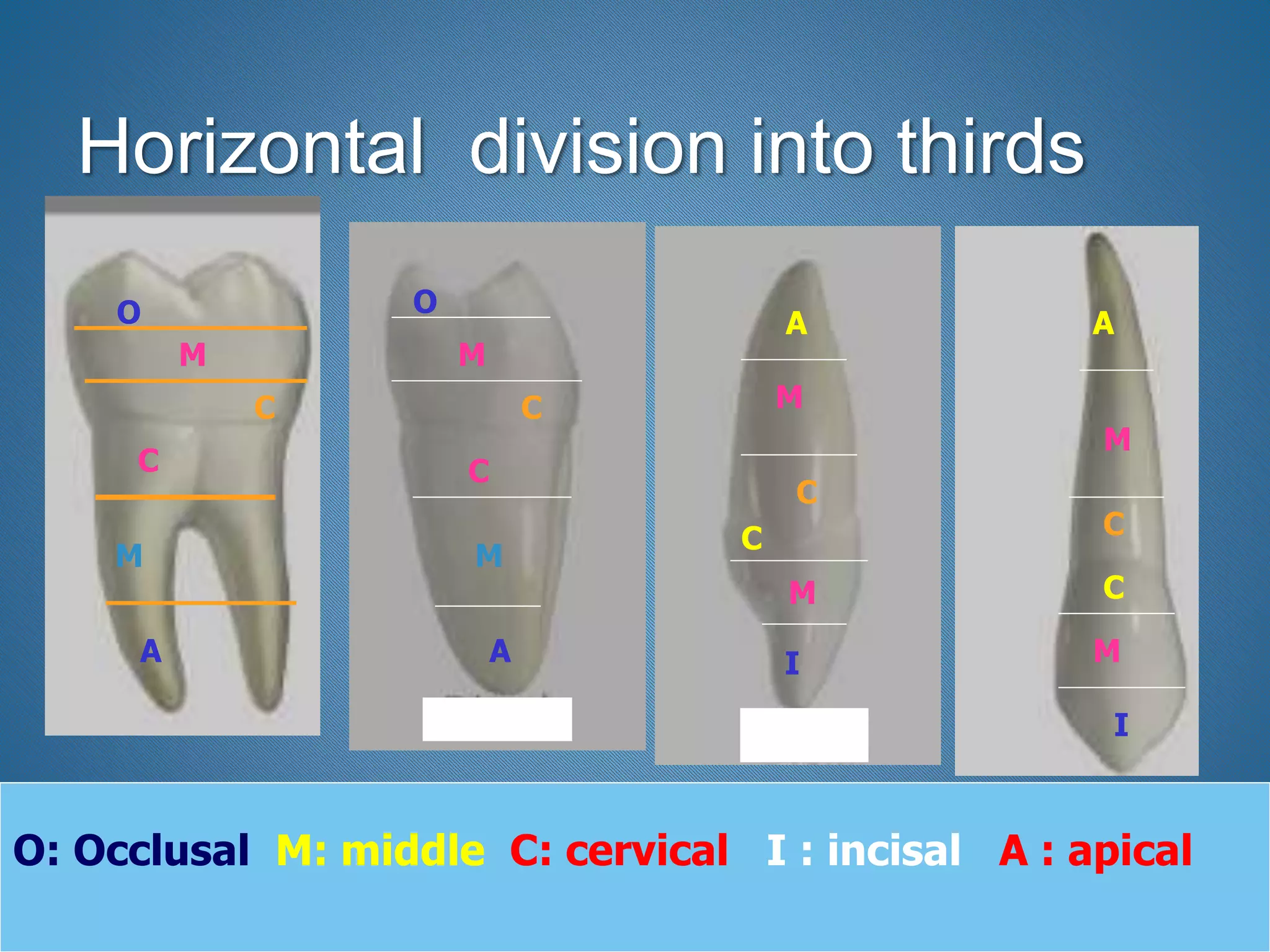
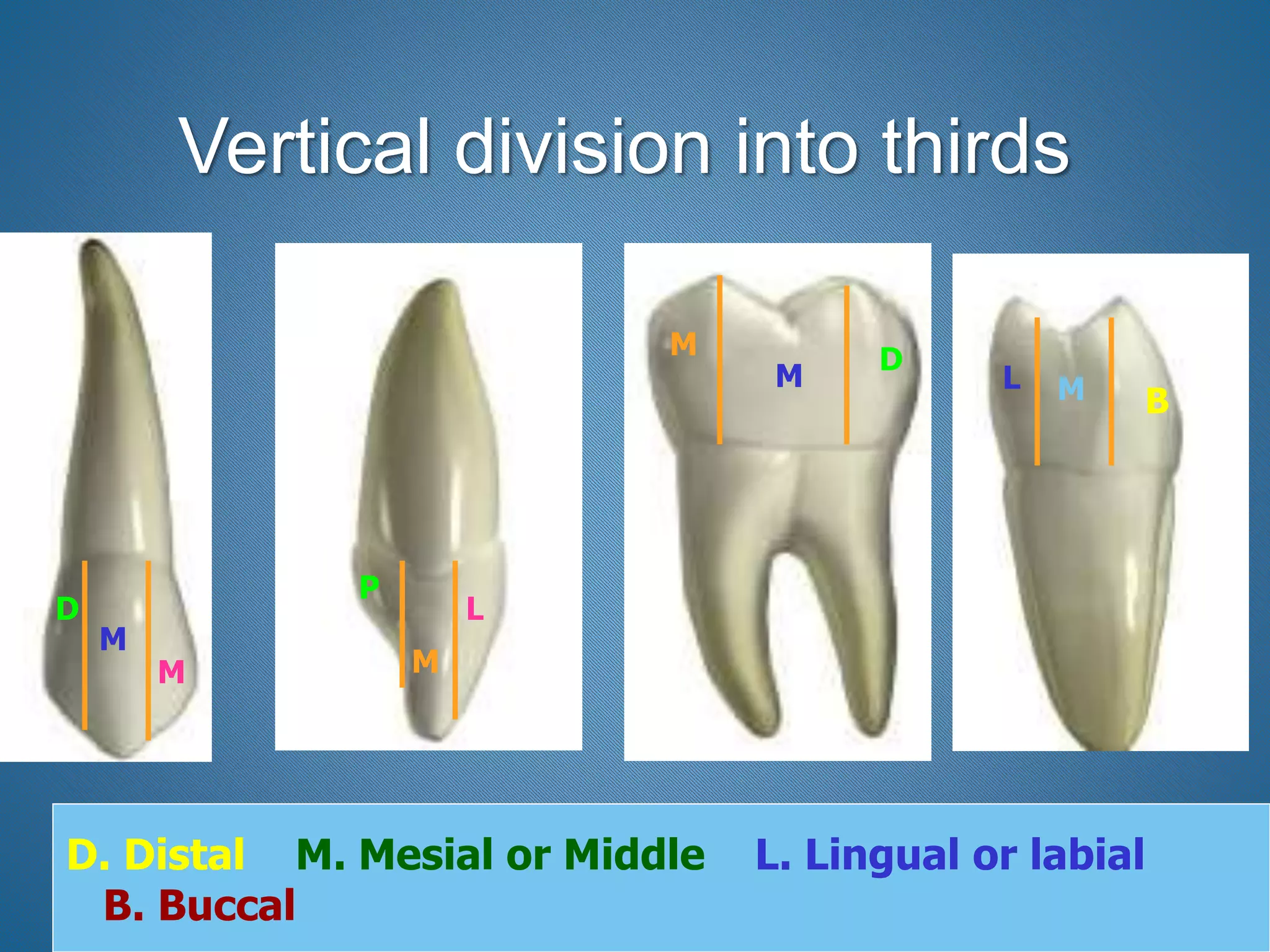
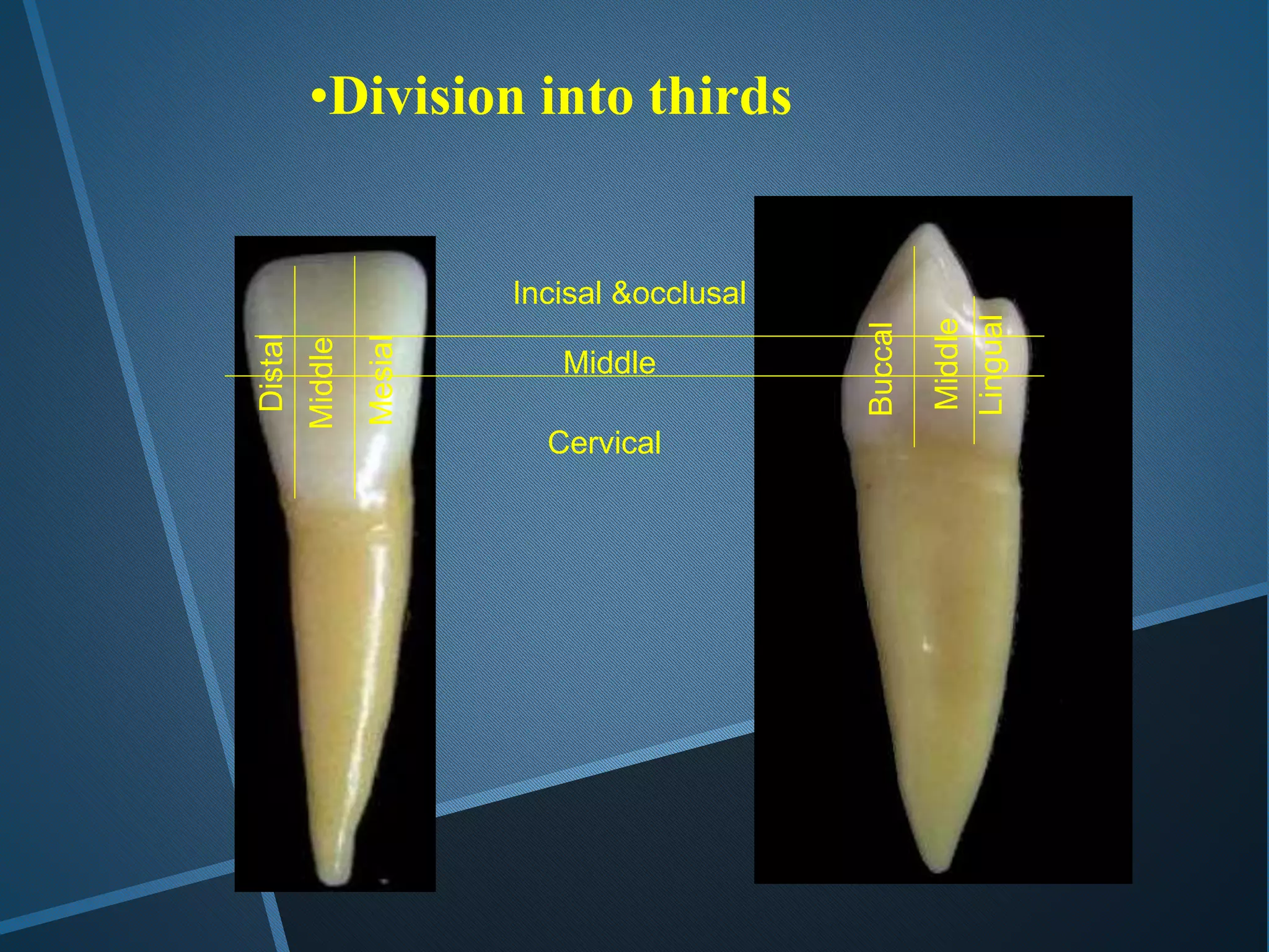
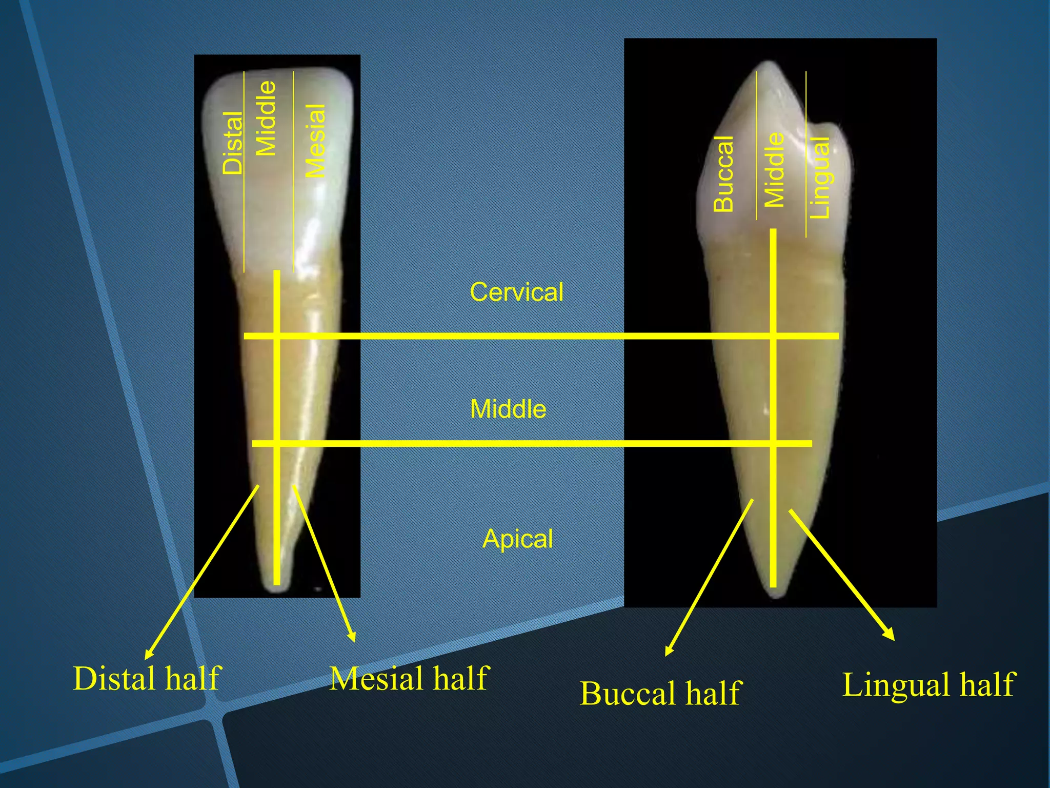
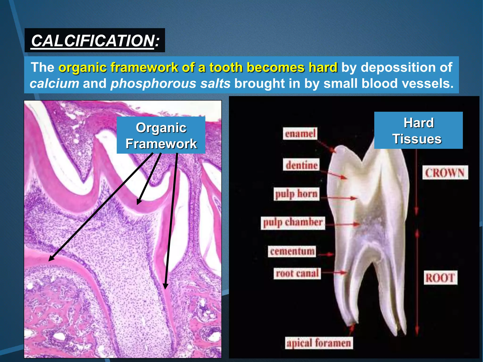
1. Humans have two dentitions - the primary dentition from 6 months to 6 years containing 20 deciduous teeth, and the permanent dentition after 12 years containing 32 teeth.
2. The primary dentition is replaced by the permanent dentition between 6-12 years of age, known as the mixed dentition period when both deciduous and permanent teeth are present.
3. Each tooth consists of a crown covered by enamel, a neck, and one or more roots. Teeth can be classified based on the number of roots as single or multi-rooted.
A Complete presentation explaining the complete morphology of Maxillary first molar, for the benefit of people like me who tried and failed to find everything in one package
some essential information about anatomy and morphology of teeth to learn ( specially dentistry students ) , collecting and presenting by Negin Aliyari
I wanna share this to all dental students and colleagues. This is a simplified and concise description of the anatomical structure of a Permanent Maxillary Central Incisor.
Centered in the maxilla, one on either side of median line, with mesial surface of each in contact with mesial surface of other
Two in number
Larger than the lateral incisor
These teeth supplement each other in function, and they are similar anatomically
Shearing or cutting teeth
Major function is to punch and cut food material during the process of mastication
These teeth have incisal ridges or edges rather than
cusps such as are found on canines & posterior teeth
First evidence of calcification
Crown completion
Eruption
Root completion
3-4 months
4-5 years
7-8 years
10-11 years
Platelet Rich Fibrin (PRF) in Dentistry, What is PRF ? , What are the difference between PRP,PRGF and PRF ?, Preparation of PRF , shapes of PRF, Role of PRF in wound healing, APPLICATIONS OF PRF, Applications of PRF In Oral and Maxillofacial Surgery, Applications of PRF In Periodontics, Applications of PRF In Endodontics, Applications of PRF In Tissue Engineering
GOALS OF SUTURING, CLASSIFICATION OF SUTURE MATERIALS According to source, CLASSIFICATION OF SUTURE MATERIALS According to Structure, CLASSIFICATION OF SURGICAL NEEDLES, IDEAL PROPERTIES OF NEEDLES, BODY OF NEEDLE, SUTURE SIZES, THE EYE OF THE NEEDLE, PRINCIPLES OF SUTURE SELECTION, Gut/ Chromic Gut, SILK, Collagen SUTURE, Vicryl (Polyglactin 910), Dexon and PGA, SURGICAL COTTON, GLYCOLIC ACID (MAXON) POLYGLYCONATE, NYLON, Polymerized Caprolactam, Polymerized Caprolactam, Polypropylene, Stainless Steel, Anesthetic Solutions, Wound Preparation, Principles And Techniques, Wound antisepsis and sterile technique, Wound antisepsis and sterile technique, Wound antisepsis and sterile technique, The interrupted suture, The full surgeon s knot, The full surgeon s knot, The simple or spiral continuous suture technique, The locked continuous suture, The locked and secured continuous suture, The external horizontal mattress suture The buried horizontal mattress suture, The buried vertical mattress suture, The simple anchored (sling) suture, The sliding anchored (sling) suture, The continuous sling suture, Suturing Tips and Approaches by Anatomic Location, How to Care for Stitches (Sutures), Removal of suture, Principle of suture removal, Reasons for failure of sutures, Possible complications of leaving sutures for many days, Other Methods of Wound Closure, Ligating Clips, Tissue Adhesives,
MTA uses, MTA types, Mineral Trioxide Aggregate, why we use mta not Portland cement, MTA Mixing, mta carrier, block matrix mta, mta map system, usage of MTA, pulp capping , pulptomy, apexification, regenerative endodontics, revitalization, revasclarization, internal & external root resorption, obturation, root perforation, root end filling, biodentine, MTA Fillapex, MM-MTA, THERACAL LC, Endosequence selar
MANAGEMENT OF ATRIOVENTRICULAR CONDUCTION BLOCK.pdfJim Jacob Roy
Cardiac conduction defects can occur due to various causes.
Atrioventricular conduction blocks ( AV blocks ) are classified into 3 types.
This document describes the acute management of AV block.
Explore natural remedies for syphilis treatment in Singapore. Discover alternative therapies, herbal remedies, and lifestyle changes that may complement conventional treatments. Learn about holistic approaches to managing syphilis symptoms and supporting overall health.
Recomendações da OMS sobre cuidados maternos e neonatais para uma experiência pós-natal positiva.
Em consonância com os ODS – Objetivos do Desenvolvimento Sustentável e a Estratégia Global para a Saúde das Mulheres, Crianças e Adolescentes, e aplicando uma abordagem baseada nos direitos humanos, os esforços de cuidados pós-natais devem expandir-se para além da cobertura e da simples sobrevivência, de modo a incluir cuidados de qualidade.
Estas diretrizes visam melhorar a qualidade dos cuidados pós-natais essenciais e de rotina prestados às mulheres e aos recém-nascidos, com o objetivo final de melhorar a saúde e o bem-estar materno e neonatal.
Uma “experiência pós-natal positiva” é um resultado importante para todas as mulheres que dão à luz e para os seus recém-nascidos, estabelecendo as bases para a melhoria da saúde e do bem-estar a curto e longo prazo. Uma experiência pós-natal positiva é definida como aquela em que as mulheres, pessoas que gestam, os recém-nascidos, os casais, os pais, os cuidadores e as famílias recebem informação consistente, garantia e apoio de profissionais de saúde motivados; e onde um sistema de saúde flexível e com recursos reconheça as necessidades das mulheres e dos bebês e respeite o seu contexto cultural.
Estas diretrizes consolidadas apresentam algumas recomendações novas e já bem fundamentadas sobre cuidados pós-natais de rotina para mulheres e neonatos que recebem cuidados no pós-parto em unidades de saúde ou na comunidade, independentemente dos recursos disponíveis.
É fornecido um conjunto abrangente de recomendações para cuidados durante o período puerperal, com ênfase nos cuidados essenciais que todas as mulheres e recém-nascidos devem receber, e com a devida atenção à qualidade dos cuidados; isto é, a entrega e a experiência do cuidado recebido. Estas diretrizes atualizam e ampliam as recomendações da OMS de 2014 sobre cuidados pós-natais da mãe e do recém-nascido e complementam as atuais diretrizes da OMS sobre a gestão de complicações pós-natais.
O estabelecimento da amamentação e o manejo das principais intercorrências é contemplada.
Recomendamos muito.
Vamos discutir essas recomendações no nosso curso de pós-graduação em Aleitamento no Instituto Ciclos.
Esta publicação só está disponível em inglês até o momento.
Prof. Marcus Renato de Carvalho
www.agostodourado.com
These simplified slides by Dr. Sidra Arshad present an overview of the non-respiratory functions of the respiratory tract.
Learning objectives:
1. Enlist the non-respiratory functions of the respiratory tract
2. Briefly explain how these functions are carried out
3. Discuss the significance of dead space
4. Differentiate between minute ventilation and alveolar ventilation
5. Describe the cough and sneeze reflexes
Study Resources:
1. Chapter 39, Guyton and Hall Textbook of Medical Physiology, 14th edition
2. Chapter 34, Ganong’s Review of Medical Physiology, 26th edition
3. Chapter 17, Human Physiology by Lauralee Sherwood, 9th edition
4. Non-respiratory functions of the lungs https://academic.oup.com/bjaed/article/13/3/98/278874
Knee anatomy and clinical tests 2024.pdfvimalpl1234
This includes all relevant anatomy and clinical tests compiled from standard textbooks, Campbell,netter etc..It is comprehensive and best suited for orthopaedicians and orthopaedic residents.
ARTIFICIAL INTELLIGENCE IN HEALTHCARE.pdfAnujkumaranit
Artificial intelligence (AI) refers to the simulation of human intelligence processes by machines, especially computer systems. It encompasses tasks such as learning, reasoning, problem-solving, perception, and language understanding. AI technologies are revolutionizing various fields, from healthcare to finance, by enabling machines to perform tasks that typically require human intelligence.
Flu Vaccine Alert in Bangalore Karnatakaaddon Scans
As flu season approaches, health officials in Bangalore, Karnataka, are urging residents to get their flu vaccinations. The seasonal flu, while common, can lead to severe health complications, particularly for vulnerable populations such as young children, the elderly, and those with underlying health conditions.
Dr. Vidisha Kumari, a leading epidemiologist in Bangalore, emphasizes the importance of getting vaccinated. "The flu vaccine is our best defense against the influenza virus. It not only protects individuals but also helps prevent the spread of the virus in our communities," he says.
This year, the flu season is expected to coincide with a potential increase in other respiratory illnesses. The Karnataka Health Department has launched an awareness campaign highlighting the significance of flu vaccinations. They have set up multiple vaccination centers across Bangalore, making it convenient for residents to receive their shots.
To encourage widespread vaccination, the government is also collaborating with local schools, workplaces, and community centers to facilitate vaccination drives. Special attention is being given to ensuring that the vaccine is accessible to all, including marginalized communities who may have limited access to healthcare.
Residents are reminded that the flu vaccine is safe and effective. Common side effects are mild and may include soreness at the injection site, mild fever, or muscle aches. These side effects are generally short-lived and far less severe than the flu itself.
Healthcare providers are also stressing the importance of continuing COVID-19 precautions. Wearing masks, practicing good hand hygiene, and maintaining social distancing are still crucial, especially in crowded places.
Protect yourself and your loved ones by getting vaccinated. Together, we can help keep Bangalore healthy and safe this flu season. For more information on vaccination centers and schedules, residents can visit the Karnataka Health Department’s official website or follow their social media pages.
Stay informed, stay safe, and get your flu shot today!
Tom Selleck Health: A Comprehensive Look at the Iconic Actor’s Wellness Journeygreendigital
Tom Selleck, an enduring figure in Hollywood. has captivated audiences for decades with his rugged charm, iconic moustache. and memorable roles in television and film. From his breakout role as Thomas Magnum in Magnum P.I. to his current portrayal of Frank Reagan in Blue Bloods. Selleck's career has spanned over 50 years. But beyond his professional achievements. fans have often been curious about Tom Selleck Health. especially as he has aged in the public eye.
Follow us on: Pinterest
Introduction
Many have been interested in Tom Selleck health. not only because of his enduring presence on screen but also because of the challenges. and lifestyle choices he has faced and made over the years. This article delves into the various aspects of Tom Selleck health. exploring his fitness regimen, diet, mental health. and the challenges he has encountered as he ages. We'll look at how he maintains his well-being. the health issues he has faced, and his approach to ageing .
Early Life and Career
Childhood and Athletic Beginnings
Tom Selleck was born on January 29, 1945, in Detroit, Michigan, and grew up in Sherman Oaks, California. From an early age, he was involved in sports, particularly basketball. which played a significant role in his physical development. His athletic pursuits continued into college. where he attended the University of Southern California (USC) on a basketball scholarship. This early involvement in sports laid a strong foundation for his physical health and disciplined lifestyle.
Transition to Acting
Selleck's transition from an athlete to an actor came with its physical demands. His first significant role in "Magnum P.I." required him to perform various stunts and maintain a fit appearance. This role, which he played from 1980 to 1988. necessitated a rigorous fitness routine to meet the show's demands. setting the stage for his long-term commitment to health and wellness.
Fitness Regimen
Workout Routine
Tom Selleck health and fitness regimen has evolved. adapting to his changing roles and age. During his "Magnum, P.I." days. Selleck's workouts were intense and focused on building and maintaining muscle mass. His routine included weightlifting, cardiovascular exercises. and specific training for the stunts he performed on the show.
Selleck adjusted his fitness routine as he aged to suit his body's needs. Today, his workouts focus on maintaining flexibility, strength, and cardiovascular health. He incorporates low-impact exercises such as swimming, walking, and light weightlifting. This balanced approach helps him stay fit without putting undue strain on his joints and muscles.
Importance of Flexibility and Mobility
In recent years, Selleck has emphasized the importance of flexibility and mobility in his fitness regimen. Understanding the natural decline in muscle mass and joint flexibility with age. he includes stretching and yoga in his routine. These practices help prevent injuries, improve posture, and maintain mobilit
HOT NEW PRODUCT! BIG SALES FAST SHIPPING NOW FROM CHINA!! EU KU DB BK substit...GL Anaacs
Contact us if you are interested:
Email / Skype : kefaya1771@gmail.com
Threema: PXHY5PDH
New BATCH Ku !!! MUCH IN DEMAND FAST SALE EVERY BATCH HAPPY GOOD EFFECT BIG BATCH !
Contact me on Threema or skype to start big business!!
Hot-sale products:
NEW HOT EUTYLONE WHITE CRYSTAL!!
5cl-adba precursor (semi finished )
5cl-adba raw materials
ADBB precursor (semi finished )
ADBB raw materials
APVP powder
5fadb/4f-adb
Jwh018 / Jwh210
Eutylone crystal
Protonitazene (hydrochloride) CAS: 119276-01-6
Flubrotizolam CAS: 57801-95-3
Metonitazene CAS: 14680-51-4
Payment terms: Western Union,MoneyGram,Bitcoin or USDT.
Deliver Time: Usually 7-15days
Shipping method: FedEx, TNT, DHL,UPS etc.Our deliveries are 100% safe, fast, reliable and discreet.
Samples will be sent for your evaluation!If you are interested in, please contact me, let's talk details.
We specializes in exporting high quality Research chemical, medical intermediate, Pharmaceutical chemicals and so on. Products are exported to USA, Canada, France, Korea, Japan,Russia, Southeast Asia and other countries.
3. *The Dentitions
1-The primary dentition: 6m to 6y
20 teeth. 10 maxillary and 10 mandibular.
2 incisors, 1 canine and 2 molars.
The dental formula is:
2 1 2
I --- C ---- M ---- = 10
2 1 2
1-Tooth types
2-Dentition periods
4. 1- primary Dentition period
( From 6 months to 6 years)
The only deciduous teeth are seen in the oral cavity
5. 2-The permanent dentition: after 12 y
(secondary, adult or succedaneous )
32 teeth, 16 maxillary and 16 mandibular.
2 incisors, 1 canine, 2 premolars and 3 molars.
The dental formula is :
2 1 2 3
I ---- C ---- PM --- M ---- = 16
2 1 2 3
6. 2- Secondary ( permanent ) Dentition
period ( after 12 years of age )
The only permanent teeth are seen in the oral cavity .
Note:
•The permanent molars are not preceded with
deciduous teeth so they may be termed as non-
succedaneous teeth.
7. 3- Mixed Dentition period
( From 6 years to 12 years of age)
Deciduous and permanent teeth are seen in the
oral cavity
8. Primary dentition
6 m - 6 y
Mixed dentition
6 y -12 y
Permanent dentition 12 y and up
Shedding: It is the
physiological loss of the
deciduous teeth and their
subsequent replacement
by permanent teeth.
Deciduous
Permanent
Mixed
9. 1- Primary dentition
6 m - 6 y
3- Permanent dentition 12 y and up
2- Mixed dentition
6 y - 12 y
X – Ray
Showing
10. Each tooth has three
anatomical parts:
1- Crown
2- The Neck: cervical line .
3- Root (s)
Tooth Anatomy
Macro-anatomy of the tooth
Enamel
Cementum
11.
12. There are three sites of Junction
Junctions of tooth tissues
1- Cemento-enamel junction
(CEJ): Cervical line
2- Dentino-enamel junction
(DEJ):
3- Dentino-cemental junction
(DCJ):
Enamel
Dentin
Cementum
Pulp
DCJ
CEJ
DEJ
14. Is the part of a tooth
that is visible in the
oral cavity above
the gum line.
Clinical
crown
Clinical
crown
Anatomical
crown
Anatomical and clinical crown
☻The part
of the tooth
covered by
enamel.
15. Part of the anatomical crown may be
covered by gingiva.
19. Teeth May Be Divided According To The Number
Of Roots
Single rooted teeth
The undivided
portion of the
root is called:
Root Trunk.
Multirooted teeth
20. •Teeth may be divided according to no. of roots into:
Single rooted teeth Multirooted teeth
21. 3-Tooth designation
1) Palmer Notation System:
The permanent teeth are numbered from 1-8 on each side
from the midline.
Upper right Upper left
8 7 6 5 4 3 2 1 1 2 3 4 5 6 7 8
8 7 6 5 4 3 2 1 1 2 3 4 5 6 7 8
Lower right Lower left
8 permanent upper right third
molar
5 Lower left second premolar
3 permanent upper left
canine
1 permanent lower right central
incisor
22. The deciduous teeth are lettered from A-E on each side from
the midline.
Upper right Upper left
E D C B A A B C D E
E D C B A A B C D E
Lower right Lower left
D Deciduous upper right first molar
Deciduous lower left canine
Deciduous upper left
lateral incisor
Deciduous lower right
second molarE
B
C
23.
24. 2- The international numbering system
(The two digit system)
The teeth are designed by using two-digit systems:
a. The first digit of the code is located at the left side of the
number and indicates the quadrant.
U.R. 1 2 U.L.
L.R. 4 3 L. L.
b. The second digit is located at the right side of the number
and indicates the number of the tooth in the quadrant.
****The two digits should be pronounced separately.
In the permanent dentition. In the deciduous dentition.
U.R. 5 6 U.L
L.R. 8 7 L.L.
26. 3)The universal numbering system
(American numbering system):
The number is always preceded by the sign # to designate that
the system is used for universal system.
Permanent teeth (1 – 32)
1 2 3 4 5 6 7 8 9 10 11 12 13 14 15 16
32 31 30 29 28 27 26 25 24 23 22 21 20 19 18 17
#22 Permanent Lower left canine #16 Permanent upper left third
molar
27. Deciduous teeth (1 – 20)
The number is preceded by d #
1 2 3 4 5 6 7 8 9 10
20 19 18 17 16 15 14 13 12 11
Or (A - T) The letter is preceded by # only
A B C D E F G H I J
T S R Q P O N M L K
d# 14 Deciduous lower left
lateral incisor
#B Deciduous upper right
first molar
37. Note: The anterior teeth have fewer line angles because the
meeting of the mesial and distal line surfaces with incisal ridge
are rounded , so the mesio-incisal and disto-incisal line
angles are practically not exist. .
Incisal ridge
Note:
38. Horizontal division into thirds
O
M
C
C
M
A
O
M
C
C
M
A I
M
C
M
C
A
I
M
C
C
M
A
O: Occlusal M: middle C: cervical I : incisal A : apical
39. Vertical division into thirds
D
D
M
M
L
M
P
M
M
BML
D. Distal M. Mesial or Middle L. Lingual or labial
B. Buccal
42. Lobe.
It is one of the primary centers of calcification and growth
formed during the crown development.
There are three, four or five
lobes
The pulp chamber has pulp
horns corresponding to
these lobes.
44. They are three small round projections of
enamel present in the incisal third of newly
erupted incisors.
After normal use, eventually the mamelones
wear down into a flat edge.
45. It is the enlargement or bulge on the cervical
third of lingual surface of the crown in anterior
teeth .
46. Elevation of various shape & size present
on
**Incisal portion of canine.
**The occlusal surface of post. Teeth
It is formed of enamel, dentin& pulp
Cusp:
47. Tubercle.
•It is a small elevation. It is produced by excessive
formation of enamel.
•Tubercle differs from cusp as it is formed of enamel
only while cusp is formed of pulp horn covered by dentin
and enamel.
48. Ridge.
It is a linear elevation on the different surfaces of the crown.
Facial Lingual Occlusal
49. A- Ridges on the facial surface (Labial and Buccal):
Labial Ridge: Buccal Ridge: Cervical Ridge:
Found on the labial
surfaces of
canines.
Usually found on the
buccal surfaces of the
premolars.
It is found on the
cervical one third of
the facial surfaces of
all the teeth.
Well developed in the
(D) followed by (E).
50. B- Ridges on the lingual surface:
1.Incisal Ridge:
3.Lingual
Ridge:
4.Marginal Ridge:
2.Cusp Ridge:
IR
51. C- Ridges on the occlusal surface of posterior teeth:
1.Marginal Ridges
2.Triangular Ridges:
1
2
1
2
57. SULCUS:
It is a broad depression or valley on the occlusal
surfaces of posterior teeth.
Its inclines meet in a developmental groove and
extend to the cusp tips.
60. True pit Faulty pit
•Present in the bottom of
central, mesial and distal
triangular fossae.
•At the end of buccal groove
Usually develops as a
result of incomplete
formation of enamel.
pit
61. *Dental anatomy terminology
Each erupted tooth is surrounded and
supported by the bone of the jaw. It’s called
socket or alveolus
The developing tooth present in a bony space
and is called crypt.
62. It is the bony space in the alveolar bone
containing the developing unerupted tooth.
It is the bony space in the alveolar bone
containing the roots of erupted tooth.
CRYPT:
SOCKET:
63. OTHER TERMS
Those surfaces of the teeth that
are cleaned by the normal
functional action of the
tongue, lips, cheeks and
lubricated by mouth fluids.
SELF CLEANSING SURFACES:
Facial
Lingual
Incisal Occlusal
64. CALCIFICATION:
The organic framework of a tooth becomes hard by depossition of
calcium and phosphorous salts brought in by small blood vessels.
Organic
Framework
Hard
Tissues