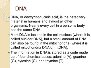
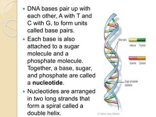
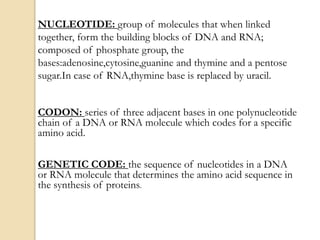
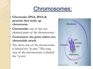
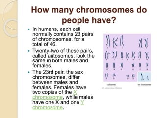
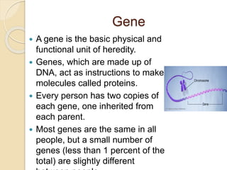
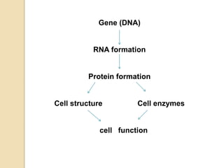
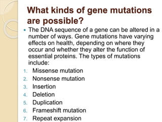
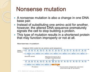
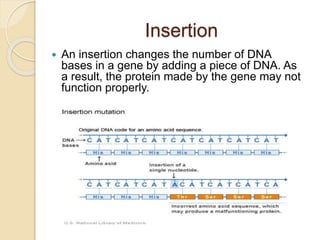
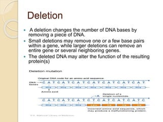
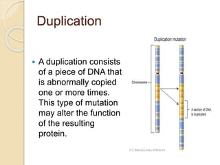
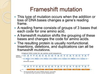
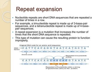
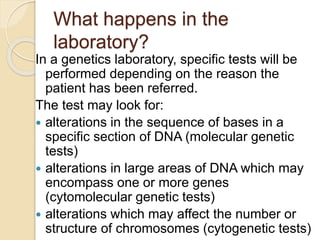
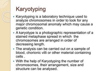
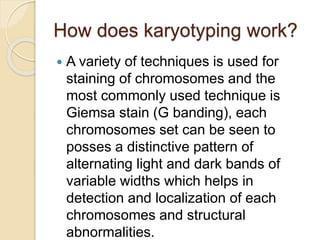
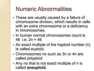
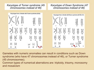
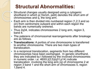
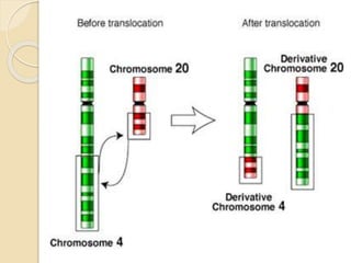
By :
Aamir Sharif
BEMS
M.phil Human Pathology
Basic of Genetics
WHAT IS
GENETICS?????
The branch of biology that deals with
heredity, especially the mechanisms of
hereditary transmission and the
variation of inherited
characteristics among
similar or related
organisms
DNA
DNA, or deoxyribonucleic acid, is the hereditary
material in humans and almost all other
organisms. Nearly every cell in a person’s body
has the same DNA.
Most DNA is located in the cell nucleus (where it is
called nuclear DNA), but a small amount of DNA
can also be found in the mitochondria (where it is
called mitochondria DNA or mtDNA).
The information in DNA is stored as a code made
up of four chemical bases: adenine (A), guanine
(G), cytosine (C), and thymine (T).
 DNA bases pair up with
each other, A with T and
C with G, to form units
called base pairs.
 Each base is also
attached to a sugar
molecule and a
phosphate molecule.
Together, a base, sugar,
and phosphate are called
a nucleotide.
 Nucleotides are arranged
in two long strands that
form a spiral called a
double helix.
NUCLEOTIDE: group of molecules that when linked
together, form the building blocks of DNA and RNA;
composed of phosphate group, the
bases:adenosine,cytosine,guanine and thymine and a pentose
sugar.In case of RNA,thymine base is replaced by uracil.
CODON: series of three adjacent bases in one polynucleotide
chain of a DNA or RNA molecule which codes for a specific
amino acid.
GENETIC CODE: the sequence of nucleotides in a DNA
or RNA molecule that determines the amino acid sequence in
the synthesis of proteins.
Chromosomes:
 Chromatin: DNA, RNA &
proteins that make up
chromosme
 Chromatids: one of the two
identical parts of the chromosome.
 Centromere: the point where two
chromatids attach
 The short arm of the chromosome
is labeled the “p arm.” The long
arm of the chromosome is labeled
the “q arm
How many chromosomes do
people have?
 In humans, each cell
normally contains 23 pairs
of chromosomes, for a
total of 46.
 Twenty-two of these pairs,
called autosomes, look the
same in both males and
females.
 The 23rd pair, the sex
chromosomes, differ
between males and
females. Females have
two copies of the X
chromosome, while males
have one X and one Y
chromosome.
Gene
 A gene is the basic physical and
functional unit of heredity.
 Genes, which are made up of
DNA, act as instructions to make
molecules called proteins.
 Every person has two copies of
each gene, one inherited from
each parent.
 Most genes are the same in all
people, but a small number of
genes (less than 1 percent of the
total) are slightly different
Gene (DNA)
RNA formation
Protein formation
Cell structure Cell enzymes
cell function
ALLELS
 Is one member of a pair or series of
different forms of a gene.
 Homozygous-an organism in which 2
copies of genes are identical i.e. have same
alleles
 Heterozygous-an organism which has
different alleles of the gene
Congential Disease.
Diseases which are present at birth.
Hereditary/Familial Disease.
Diseases which are derived from one’s
parents and trasmitted in the gametes through the
generations.
Not all congenital diseases are genetic(
congenital Syphilis) and not all genetic
diseases are congenital (Huntington disease).
Types of
Mutation
Mutation
 it is defined as a permanent changes in the
DNA.
 Mutation that affect germ cell are
transmitted to the progeny and can give rise
to inherited disease.
What kinds of gene mutations
are possible?
 The DNA sequence of a gene can be altered in a
number of ways. Gene mutations have varying
effects on health, depending on where they
occur and whether they alter the function of
essential proteins. The types of mutations
include:
1. Missense mutation
2. Nonsense mutation
3. Insertion
4. Deletion
5. Duplication
6. Frameshift mutation
7. Repeat expansion
Missense mutation
 This type of mutation is a change in one
DNA base pair that results in the
substitution of one amino acid for another in
the protein made by a gene
Nonsense mutation
 A nonsense mutation is also a change in one DNA
base pair.
 Instead of substituting one amino acid for another,
however, the altered DNA sequence prematurely
signals the cell to stop building a protein.
 This type of mutation results in a shortened protein
that may function improperly or not at all.
Insertion
 An insertion changes the number of DNA
bases in a gene by adding a piece of DNA. As
a result, the protein made by the gene may not
function properly.
Deletion
 A deletion changes the number of DNA bases by
removing a piece of DNA.
 Small deletions may remove one or a few base pairs
within a gene, while larger deletions can remove an
entire gene or several neighboring genes.
 The deleted DNA may alter the function of the resulting
protein(s)
Duplication
 A duplication consists
of a piece of DNA that
is abnormally copied
one or more times.
This type of mutation
may alter the function
of the resulting
protein.
Frameshift mutation
 This type of mutation occurs when the addition or
loss of DNA bases changes a gene’s reading
frame.
 A reading frame consists of groups of 3 bases that
each code for one amino acid.
 A frameshift mutation shifts the grouping of these
bases and changes the code for amino acids.
 The resulting protein is usually nonfunctional.
Insertions, deletions, and duplications can all be
frameshift mutations.
Repeat expansion
 Nucleotide repeats are short DNA sequences that are repeated a
number of times in a row.
 For example, a trinucleotide repeat is made up of 3-base-pair
sequences, and a tetranucleotide repeat is made up of 4-base-
pair sequences.
 A repeat expansion is a mutation that increases the number of
times that the short DNA sequence is repeated.
 This type of mutation can cause the resulting protein to function
improperly.
What happens in the
laboratory?
In a genetics laboratory, specific tests will be
performed depending on the reason the
patient has been referred.
The test may look for:
 alterations in the sequence of bases in a
specific section of DNA (molecular genetic
tests)
 alterations in large areas of DNA which may
encompass one or more genes
(cytomolecular genetic tests)
 alterations which may affect the number or
structure of chromosomes (cytogenetic tests)
Karyotyping
 Karyotyping is a laboratory technique used to
analyze chromosomes in order to look for any
major chromosomal anomaly which may cause a
genetic condition.
 A karyotype is a photographic representation of a
stained metaphase spread in which the
chromosomes are arranged in order of
decreasing length.
 The analysis can be carried out on a sample of
blood, chorionic villi or other material containing
cells .
 With the help of Karyotyping the number of
chromosomes, their arrangement, size and
structure can be analysed.
How does karyotyping work?
 A variety of techniques is used for
staining of chromosomes and the
most commonly used technique is
Giemsa stain (G banding), each
chromosomes set can be seen to
posses a distinctive pattern of
alternating light and dark bands of
variable widths which helps in
detection and localization of each
chromosomes and structural
abnormalities.
Numeric Abnormalities
 These are usually caused by a failure of
chromosome division, which results in cells
with an extra chromosome or a deficiency
in chromosomes.
 In human normal chromosomes count is
46 i.e. 2n = 46
 An exact multiple of the haploid number (n)
is called euploid.
 Chromosomes no such as 3n or 4n are
called polyploid
 Any no that is not exact multiple of n is
called aneuploid.
Gametes with numeric anomalies can result in conditions such as Down
syndrome (who have 47 chromosomes instead of 46), or Turner syndrome
(45 chromosomes).
Common types of numerical aberrations are: triploidy, trisomy, monosomy
and mosaicism
Karyotype of Turner syndrome (45
chromosomes instead of 46)
Karyotype of Down Syndrome (47
chromosomes instead of 46)
Structural Abnormalities:
 Structural changes usually designed using a cytogenic
shorthand in which p( french, petit) denotes the short arm of
chromosomes and q, the long arm.
 Each arm is then divided into numbered region (1,2,3 and so
on) from centromere outward and within each region the
bands are numerically ordrered.
 Thus 2q34, indicates chromosomes 2 long arm, region 3,
band 4.
 The paterens of chromosomal rearrangements after breakage
are as follows.
 Translocations: A portion of one chromosome is transferred
to another chromosome. There are two main types of
translocation.
 In a reciprocal translocation, segments from two different
chromosomes have been exchanged. In genetic shorthand,
they are indicated by t followed by the involved chromosomes
in numeric order. i.e. 46XX,t(2;5)(q31;p14) indicate
translocation involving the long arm (q) of chromosome 2 at
region 3 band 1 and the short arm of chromosoes 5, region
1,band 4.
 Deletions: A portion of the chromosome is
missing or deleted.
 Duplications: A portion of the chromosome
is duplicated, resulting in extra genetic
material.
 Inversions: A portion of the chromosome has
broken off, turned upside down, and
reattached. As a result, the genetic material is
inverted.
 Rings: A portion of a chromosome has
broken off and formed a circle or ring. This
can happen with or without loss of genetic
material.
 Most chromosome abnormalities occur
as an accident in the egg or sperm. In
these cases, the abnormality is present
in every cell of the body. Some
abnormalities, however, happen after
conception; then some cells have the
abnormality and some do not.
 Chromosome abnormalities can be
inherited from a parent (such as a
translocation) or be "de novo" (new to
the individual). This is why, when a child
is found to have an abnormality,
chromosome studies are often
performed on the parents.
Thank you

Basic genetics ,mutation and karyotyping

  • 1.
    By : Aamir Sharif BEMS M.philHuman Pathology Basic of Genetics
  • 2.
    WHAT IS GENETICS????? The branchof biology that deals with heredity, especially the mechanisms of hereditary transmission and the variation of inherited characteristics among similar or related organisms
  • 3.
    DNA DNA, or deoxyribonucleicacid, is the hereditary material in humans and almost all other organisms. Nearly every cell in a person’s body has the same DNA. Most DNA is located in the cell nucleus (where it is called nuclear DNA), but a small amount of DNA can also be found in the mitochondria (where it is called mitochondria DNA or mtDNA). The information in DNA is stored as a code made up of four chemical bases: adenine (A), guanine (G), cytosine (C), and thymine (T).
  • 4.
     DNA basespair up with each other, A with T and C with G, to form units called base pairs.  Each base is also attached to a sugar molecule and a phosphate molecule. Together, a base, sugar, and phosphate are called a nucleotide.  Nucleotides are arranged in two long strands that form a spiral called a double helix.
  • 5.
    NUCLEOTIDE: group ofmolecules that when linked together, form the building blocks of DNA and RNA; composed of phosphate group, the bases:adenosine,cytosine,guanine and thymine and a pentose sugar.In case of RNA,thymine base is replaced by uracil. CODON: series of three adjacent bases in one polynucleotide chain of a DNA or RNA molecule which codes for a specific amino acid. GENETIC CODE: the sequence of nucleotides in a DNA or RNA molecule that determines the amino acid sequence in the synthesis of proteins.
  • 6.
    Chromosomes:  Chromatin: DNA,RNA & proteins that make up chromosme  Chromatids: one of the two identical parts of the chromosome.  Centromere: the point where two chromatids attach  The short arm of the chromosome is labeled the “p arm.” The long arm of the chromosome is labeled the “q arm
  • 7.
    How many chromosomesdo people have?  In humans, each cell normally contains 23 pairs of chromosomes, for a total of 46.  Twenty-two of these pairs, called autosomes, look the same in both males and females.  The 23rd pair, the sex chromosomes, differ between males and females. Females have two copies of the X chromosome, while males have one X and one Y chromosome.
  • 8.
    Gene  A geneis the basic physical and functional unit of heredity.  Genes, which are made up of DNA, act as instructions to make molecules called proteins.  Every person has two copies of each gene, one inherited from each parent.  Most genes are the same in all people, but a small number of genes (less than 1 percent of the total) are slightly different
  • 9.
    Gene (DNA) RNA formation Proteinformation Cell structure Cell enzymes cell function
  • 10.
    ALLELS  Is onemember of a pair or series of different forms of a gene.  Homozygous-an organism in which 2 copies of genes are identical i.e. have same alleles  Heterozygous-an organism which has different alleles of the gene
  • 12.
    Congential Disease. Diseases whichare present at birth. Hereditary/Familial Disease. Diseases which are derived from one’s parents and trasmitted in the gametes through the generations. Not all congenital diseases are genetic( congenital Syphilis) and not all genetic diseases are congenital (Huntington disease).
  • 13.
  • 14.
    Mutation  it isdefined as a permanent changes in the DNA.  Mutation that affect germ cell are transmitted to the progeny and can give rise to inherited disease.
  • 15.
    What kinds ofgene mutations are possible?  The DNA sequence of a gene can be altered in a number of ways. Gene mutations have varying effects on health, depending on where they occur and whether they alter the function of essential proteins. The types of mutations include: 1. Missense mutation 2. Nonsense mutation 3. Insertion 4. Deletion 5. Duplication 6. Frameshift mutation 7. Repeat expansion
  • 16.
    Missense mutation  Thistype of mutation is a change in one DNA base pair that results in the substitution of one amino acid for another in the protein made by a gene
  • 17.
    Nonsense mutation  Anonsense mutation is also a change in one DNA base pair.  Instead of substituting one amino acid for another, however, the altered DNA sequence prematurely signals the cell to stop building a protein.  This type of mutation results in a shortened protein that may function improperly or not at all.
  • 18.
    Insertion  An insertionchanges the number of DNA bases in a gene by adding a piece of DNA. As a result, the protein made by the gene may not function properly.
  • 19.
    Deletion  A deletionchanges the number of DNA bases by removing a piece of DNA.  Small deletions may remove one or a few base pairs within a gene, while larger deletions can remove an entire gene or several neighboring genes.  The deleted DNA may alter the function of the resulting protein(s)
  • 20.
    Duplication  A duplicationconsists of a piece of DNA that is abnormally copied one or more times. This type of mutation may alter the function of the resulting protein.
  • 21.
    Frameshift mutation  Thistype of mutation occurs when the addition or loss of DNA bases changes a gene’s reading frame.  A reading frame consists of groups of 3 bases that each code for one amino acid.  A frameshift mutation shifts the grouping of these bases and changes the code for amino acids.  The resulting protein is usually nonfunctional. Insertions, deletions, and duplications can all be frameshift mutations.
  • 22.
    Repeat expansion  Nucleotiderepeats are short DNA sequences that are repeated a number of times in a row.  For example, a trinucleotide repeat is made up of 3-base-pair sequences, and a tetranucleotide repeat is made up of 4-base- pair sequences.  A repeat expansion is a mutation that increases the number of times that the short DNA sequence is repeated.  This type of mutation can cause the resulting protein to function improperly.
  • 23.
    What happens inthe laboratory? In a genetics laboratory, specific tests will be performed depending on the reason the patient has been referred. The test may look for:  alterations in the sequence of bases in a specific section of DNA (molecular genetic tests)  alterations in large areas of DNA which may encompass one or more genes (cytomolecular genetic tests)  alterations which may affect the number or structure of chromosomes (cytogenetic tests)
  • 24.
    Karyotyping  Karyotyping isa laboratory technique used to analyze chromosomes in order to look for any major chromosomal anomaly which may cause a genetic condition.  A karyotype is a photographic representation of a stained metaphase spread in which the chromosomes are arranged in order of decreasing length.  The analysis can be carried out on a sample of blood, chorionic villi or other material containing cells .  With the help of Karyotyping the number of chromosomes, their arrangement, size and structure can be analysed.
  • 25.
    How does karyotypingwork?  A variety of techniques is used for staining of chromosomes and the most commonly used technique is Giemsa stain (G banding), each chromosomes set can be seen to posses a distinctive pattern of alternating light and dark bands of variable widths which helps in detection and localization of each chromosomes and structural abnormalities.
  • 26.
    Numeric Abnormalities  Theseare usually caused by a failure of chromosome division, which results in cells with an extra chromosome or a deficiency in chromosomes.  In human normal chromosomes count is 46 i.e. 2n = 46  An exact multiple of the haploid number (n) is called euploid.  Chromosomes no such as 3n or 4n are called polyploid  Any no that is not exact multiple of n is called aneuploid.
  • 27.
    Gametes with numericanomalies can result in conditions such as Down syndrome (who have 47 chromosomes instead of 46), or Turner syndrome (45 chromosomes). Common types of numerical aberrations are: triploidy, trisomy, monosomy and mosaicism Karyotype of Turner syndrome (45 chromosomes instead of 46) Karyotype of Down Syndrome (47 chromosomes instead of 46)
  • 28.
    Structural Abnormalities:  Structuralchanges usually designed using a cytogenic shorthand in which p( french, petit) denotes the short arm of chromosomes and q, the long arm.  Each arm is then divided into numbered region (1,2,3 and so on) from centromere outward and within each region the bands are numerically ordrered.  Thus 2q34, indicates chromosomes 2 long arm, region 3, band 4.  The paterens of chromosomal rearrangements after breakage are as follows.  Translocations: A portion of one chromosome is transferred to another chromosome. There are two main types of translocation.  In a reciprocal translocation, segments from two different chromosomes have been exchanged. In genetic shorthand, they are indicated by t followed by the involved chromosomes in numeric order. i.e. 46XX,t(2;5)(q31;p14) indicate translocation involving the long arm (q) of chromosome 2 at region 3 band 1 and the short arm of chromosoes 5, region 1,band 4.
  • 30.
     Deletions: Aportion of the chromosome is missing or deleted.  Duplications: A portion of the chromosome is duplicated, resulting in extra genetic material.  Inversions: A portion of the chromosome has broken off, turned upside down, and reattached. As a result, the genetic material is inverted.  Rings: A portion of a chromosome has broken off and formed a circle or ring. This can happen with or without loss of genetic material.
  • 31.
     Most chromosomeabnormalities occur as an accident in the egg or sperm. In these cases, the abnormality is present in every cell of the body. Some abnormalities, however, happen after conception; then some cells have the abnormality and some do not.  Chromosome abnormalities can be inherited from a parent (such as a translocation) or be "de novo" (new to the individual). This is why, when a child is found to have an abnormality, chromosome studies are often performed on the parents.
  • 32.