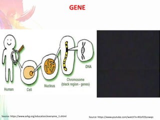
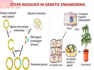
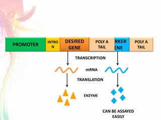
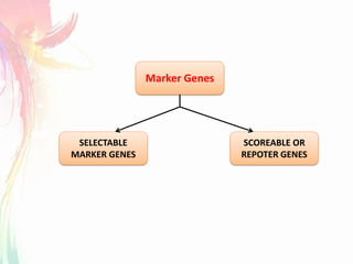
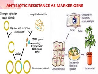
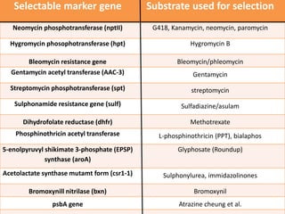
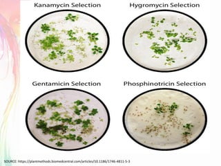
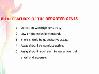
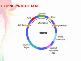
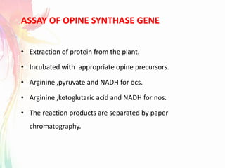
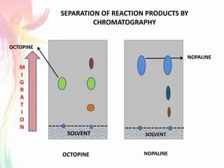
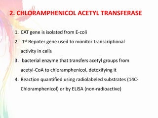
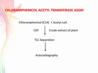
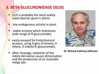
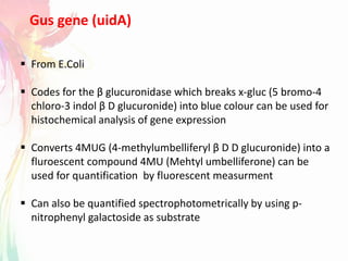
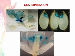
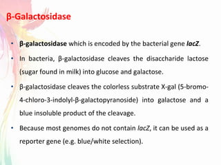
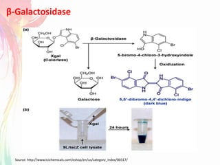
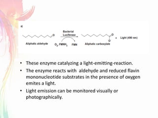
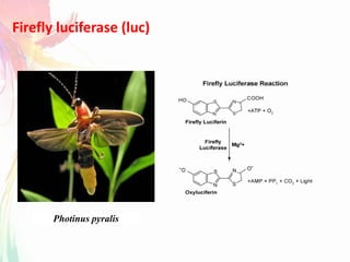
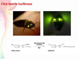
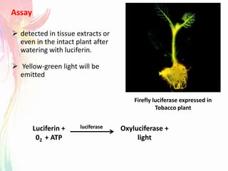
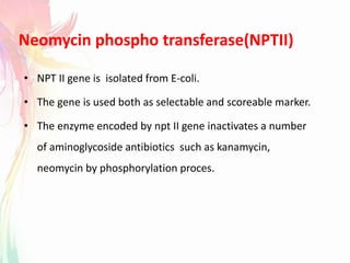
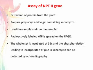
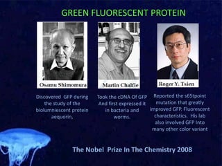
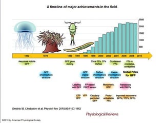
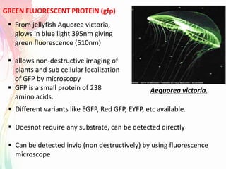
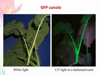
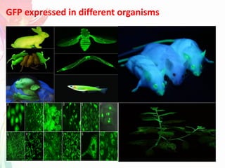
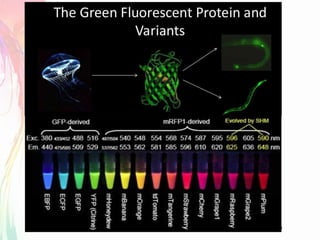
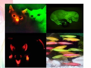
Under the guidence of
Banumathi.R.L
Lecturer
Dept. of Biotechnology
Sahyadri Science College
Shimoga
SAHYADRI SCIENCE COLLEGE
Submitted By
Submitted to
Dept. of Biotechnology
Sahyadri Science college
Shimoga
GENE
Source: https://www.ashg.org/education/everyone_1.shtml Source: https://www.youtube.com/watch?v=N5zFOScowqo
STEPS INVOLVED IN GENETIC ENGINEERING
MARKER GENES
PROMOTER
DESIRED
GENE
MARKER
GENE
TRANSCRIPTION
TRANSLATION
mRNA
ENZYME
INTRO
N
POLY A
TAIL
CAN BE ASSAYED
EASILY
PROM
OTER
POLY A
TAIL
SCOREABLE OR
REPOTER GENES
SELECTABLE
MARKER GENES
Marker Genes
SELECTABLE MARKER GENES
HOW SELECTABLE MARKER WORKS ?
TYPES OF SELECTABLE MARKER
Hygromycin
Resistant
gene
ANTIBIOTIC RESISTANCE AS MARKER GENE
Selectable marker gene Substrate used for selection
Neomycin phosphotransferase (nptII) G418, Kanamycin, neomycin, paromycin
Hygromycin phosophotransferase (hpt) Hygromycin B
Bleomycin resistance gene Bleomycin/phleomycin
Gentamycin acetyl transferase (AAC-3) Gentamycin
Streptomycin phosphotransferase (spt) streptomycin
Sulphonamide resistance gene (sulf) Sulfadiazine/asulam
Dihydrofolate reductase (dhfr) Methotrexate
Phosphinothricin acetyl transferase L-phosphinothricin (PPT), bialaphos
5-enolpyruvyl shikimate 3-phosphate (EPSP)
synthase (aroA)
Glyphosate (Roundup)
Acetolactate synthase mutamt form (csr1-1) Sulphonylurea, immidazolinones
Bromoxynill nitrilase (bxn) Bromoxynil
psbA gene Atrazine cheung et al.
SOURCE: https://plantmethods.biomedcentral.com/articles/10.1186/1746-4811-5-3
REPORTER GENESIDEAL FEATURES OF THE
1. Detection with high sensitivity
2. Low endogenous background.
3. There should be quantitative assay.
4. Assay should be nondestructive.
5. Assay should require a minimal amount of
effort and expense.
1. OPINE SYNTHASE GENE
ASSAY OF OPINE SYNTHASE GENE
• Extraction of protein from the plant.
• Incubated with appropriate opine precursors.
• Arginine ,pyruvate and NADH for ocs.
• Arginine ,ketoglutaric acid and NADH for nos.
• The reaction products are separated by paper
chromatography.
SOLVENT
M
I
G
R
A
T
I
O
N
OCTOPINE NOPALINE
SEPARATION OF REACTION PRODUCTS BY
CHROMATOGRAPHY
NOPALINE
OCTOPINE
SOLVENT
2. CHLORAMPHENICOL ACETYL TRANSFERASE
1. CAT gene is isolated from E-coli
2. 1st Repoter gene used to monitor transcriptional
activity in cells
3. bacterial enzyme that transfers acetyl groups from
acetyl-CoA to chloramphenicol, detoxifying it
4. Reaction quantified using radiolabeled substrates (14C-
Chloramphenicol) or by ELISA (non-radioactive)
CHLORAMPHENICOL ACETYL TRANSFERASE ASSAY
Chloramphenicol (C14) + Acetyl coA
CAT Crude extract of plant
TLC Separation
Autoradiography
1 2 3 4
SOLVENT
Migration
TLC Sheet
Origin
Di-acetylated
Chloramphenicol
Mono-acetylated
chloramphenicol
Unacetylated
chloramphenicol
3. BETA GLUCURONIDASE (GUS)
Dr. Richard Anthony Jefferson
 GUS is probably the most widely
used reporter gene in plants
 low endogenous activity in plant
 stable enzyme which hydrolyses
wide range of ß-glucuronides.
 easily assayed for histochemical
analysis, using X-gluc (5-bromo, 4-
chloro, 3-indolyl ß–glucuronide).
 After cleavage, oxidation of the
indole derivative causes dimerisation
and the production of an insoluble
indigo dye
 From E.Coli
 Codes for the β glucuronidase which breaks x-gluc (5 bromo-4
chloro-3 indol β D glucuronide) into blue colour can be used for
histochemical analysis of gene expression
 Converts 4MUG (4-methylumbelliferyl β D D glucuronide) into a
fluroescent compound 4MU (Mehtyl umbelliferone) can be
used for quantification by fluorescent measurment
 Can also be quantified spectrophotometrically by using p-
nitrophenyl galactoside as substrate
Gus gene (uidA)
BETA GLUCURONIDASE (GUS) ASSAY
GUS EXPRESSION
β-Galactosidase
• β-galactosidase which is encoded by the bacterial gene lacZ.
• In bacteria, β-galactosidase cleaves the disaccharide lactose
(sugar found in milk) into glucose and galactose.
• β-galactosidase cleaves the colorless substrate X-gal (5-bromo-
4-chloro-3-indolyl-β-galactopyranoside) into galactose and a
blue insoluble product of the cleavage.
• Because most genomes do not contain lacZ, it can be used as a
reporter gene (e.g. blue/white selection).
β-Galactosidase
Source: http://www.tcichemicals.com/eshop/en/us/category_index/00317/
BACTERIAL LUCIFERASE (Lux F2)
Vibrio harveyi culture
Vibrio harveyi
A. fischeri
Hawaian bobtail squid (Euprymna scolopes)
• These enzyme catalyzing a light-emitting-reaction.
• The enzyme reacts with aldehyde and reduced flavin
mononucleotide substrates in the presence of oxygen
emites a light.
• Light emission can be monitored visually or
photographically.
Firefly luciferase (luc)
Photinus pyralis
Click beetle luciferase
Assay
 detected in tissue extracts or
even in the intact plant after
watering with luciferin.
 Yellow-green light will be
emitted
Firefly luciferase expressed in
Tobacco plant
Luciferin +
02 + ATP
Oxyluciferase +
light
luciferase
Neomycin phospho transferase(NPTII)
• NPT II gene is isolated from E-coli.
• The gene is used both as selectable and scoreable marker.
• The enzyme encoded by npt II gene inactivates a number
of aminoglycoside antibiotics such as kanamycin,
neomycin by phosphorylation proces.
Assay of NPT II gene
• Extraction of protein from the plant.
• Prepare poly acryl amide gel containing kanamycin.
• Load the sample and run the sample.
• Radioactively labeled ATP is spread on the PAGE.
• The whole set is incubated at 35c and the phosphorylation
leading to incorporaton of p32 in kanamycin can be
detected by autoradiography.
The Nobel Prize In The Chemistry 2008
Discovered GFP during
the study of the
biolumniescent protein
aequorin,
Took the cDNA Of GFP
And first expressed it
in bacteria and
worms.
Reported the s65tpoint
mutation that greatly
improved GFP. Fluorescent
characteristics. His lab
also involved GFP Into
many other color variant
GREEN FLUORESCENT PROTEIN
Doug Prasher
OMISSION OF DOUG PRASHER FROM THE NOBEL PRIZE
Aequorea victoria.
 From jellyfish Aquorea victoria,
glows in blue light 395nm giving
green fluorescence (510nm)
 allows non-destructive imaging of
plants and sub cellular localization
of GFP by microscopy
 GFP is a small protein of 238
amino acids.
 Different variants like EGFP, Red GFP, EYFP, etc available.
 Doesnot require any substrate, can be detected directly
 Can be detected invio (non destructively) by using fluorescence
microscope
GREEN FLUORESCENT PROTEIN (gfp)
GFP canola
White light UV light in a darkened room
GFP expressed in different organisms
Source: https://www.newscientist.com/article/dn17003-fluorescent-puppy-is-worlds-first-transgenic-dog/
WORLDS FIRST TRANSGENIC DOG - RUPPY
CONCLUSION
Reporter genes

Reporter genes

  • 1.
    Under the guidenceof Banumathi.R.L Lecturer Dept. of Biotechnology Sahyadri Science College Shimoga SAHYADRI SCIENCE COLLEGE Submitted By Submitted to Dept. of Biotechnology Sahyadri Science college Shimoga
  • 2.
  • 3.
    STEPS INVOLVED INGENETIC ENGINEERING
  • 4.
  • 5.
  • 6.
  • 7.
    SELECTABLE MARKER GENES HOWSELECTABLE MARKER WORKS ? TYPES OF SELECTABLE MARKER
  • 8.
  • 9.
    Selectable marker geneSubstrate used for selection Neomycin phosphotransferase (nptII) G418, Kanamycin, neomycin, paromycin Hygromycin phosophotransferase (hpt) Hygromycin B Bleomycin resistance gene Bleomycin/phleomycin Gentamycin acetyl transferase (AAC-3) Gentamycin Streptomycin phosphotransferase (spt) streptomycin Sulphonamide resistance gene (sulf) Sulfadiazine/asulam Dihydrofolate reductase (dhfr) Methotrexate Phosphinothricin acetyl transferase L-phosphinothricin (PPT), bialaphos 5-enolpyruvyl shikimate 3-phosphate (EPSP) synthase (aroA) Glyphosate (Roundup) Acetolactate synthase mutamt form (csr1-1) Sulphonylurea, immidazolinones Bromoxynill nitrilase (bxn) Bromoxynil psbA gene Atrazine cheung et al.
  • 10.
  • 11.
    REPORTER GENESIDEAL FEATURESOF THE 1. Detection with high sensitivity 2. Low endogenous background. 3. There should be quantitative assay. 4. Assay should be nondestructive. 5. Assay should require a minimal amount of effort and expense.
  • 12.
  • 13.
    ASSAY OF OPINESYNTHASE GENE • Extraction of protein from the plant. • Incubated with appropriate opine precursors. • Arginine ,pyruvate and NADH for ocs. • Arginine ,ketoglutaric acid and NADH for nos. • The reaction products are separated by paper chromatography.
  • 14.
    SOLVENT M I G R A T I O N OCTOPINE NOPALINE SEPARATION OFREACTION PRODUCTS BY CHROMATOGRAPHY NOPALINE OCTOPINE SOLVENT
  • 15.
    2. CHLORAMPHENICOL ACETYLTRANSFERASE 1. CAT gene is isolated from E-coli 2. 1st Repoter gene used to monitor transcriptional activity in cells 3. bacterial enzyme that transfers acetyl groups from acetyl-CoA to chloramphenicol, detoxifying it 4. Reaction quantified using radiolabeled substrates (14C- Chloramphenicol) or by ELISA (non-radioactive)
  • 17.
    CHLORAMPHENICOL ACETYL TRANSFERASEASSAY Chloramphenicol (C14) + Acetyl coA CAT Crude extract of plant TLC Separation Autoradiography
  • 18.
    1 2 34 SOLVENT Migration TLC Sheet Origin Di-acetylated Chloramphenicol Mono-acetylated chloramphenicol Unacetylated chloramphenicol
  • 19.
    3. BETA GLUCURONIDASE(GUS) Dr. Richard Anthony Jefferson  GUS is probably the most widely used reporter gene in plants  low endogenous activity in plant  stable enzyme which hydrolyses wide range of ß-glucuronides.  easily assayed for histochemical analysis, using X-gluc (5-bromo, 4- chloro, 3-indolyl ß–glucuronide).  After cleavage, oxidation of the indole derivative causes dimerisation and the production of an insoluble indigo dye
  • 20.
     From E.Coli Codes for the β glucuronidase which breaks x-gluc (5 bromo-4 chloro-3 indol β D glucuronide) into blue colour can be used for histochemical analysis of gene expression  Converts 4MUG (4-methylumbelliferyl β D D glucuronide) into a fluroescent compound 4MU (Mehtyl umbelliferone) can be used for quantification by fluorescent measurment  Can also be quantified spectrophotometrically by using p- nitrophenyl galactoside as substrate Gus gene (uidA)
  • 21.
  • 22.
  • 23.
    β-Galactosidase • β-galactosidase whichis encoded by the bacterial gene lacZ. • In bacteria, β-galactosidase cleaves the disaccharide lactose (sugar found in milk) into glucose and galactose. • β-galactosidase cleaves the colorless substrate X-gal (5-bromo- 4-chloro-3-indolyl-β-galactopyranoside) into galactose and a blue insoluble product of the cleavage. • Because most genomes do not contain lacZ, it can be used as a reporter gene (e.g. blue/white selection).
  • 24.
  • 25.
    BACTERIAL LUCIFERASE (LuxF2) Vibrio harveyi culture Vibrio harveyi
  • 26.
    A. fischeri Hawaian bobtailsquid (Euprymna scolopes)
  • 27.
    • These enzymecatalyzing a light-emitting-reaction. • The enzyme reacts with aldehyde and reduced flavin mononucleotide substrates in the presence of oxygen emites a light. • Light emission can be monitored visually or photographically.
  • 28.
  • 29.
  • 30.
    Assay  detected intissue extracts or even in the intact plant after watering with luciferin.  Yellow-green light will be emitted Firefly luciferase expressed in Tobacco plant Luciferin + 02 + ATP Oxyluciferase + light luciferase
  • 31.
    Neomycin phospho transferase(NPTII) •NPT II gene is isolated from E-coli. • The gene is used both as selectable and scoreable marker. • The enzyme encoded by npt II gene inactivates a number of aminoglycoside antibiotics such as kanamycin, neomycin by phosphorylation proces.
  • 32.
    Assay of NPTII gene • Extraction of protein from the plant. • Prepare poly acryl amide gel containing kanamycin. • Load the sample and run the sample. • Radioactively labeled ATP is spread on the PAGE. • The whole set is incubated at 35c and the phosphorylation leading to incorporaton of p32 in kanamycin can be detected by autoradiography.
  • 33.
    The Nobel PrizeIn The Chemistry 2008 Discovered GFP during the study of the biolumniescent protein aequorin, Took the cDNA Of GFP And first expressed it in bacteria and worms. Reported the s65tpoint mutation that greatly improved GFP. Fluorescent characteristics. His lab also involved GFP Into many other color variant GREEN FLUORESCENT PROTEIN
  • 34.
    Doug Prasher OMISSION OFDOUG PRASHER FROM THE NOBEL PRIZE
  • 36.
    Aequorea victoria.  Fromjellyfish Aquorea victoria, glows in blue light 395nm giving green fluorescence (510nm)  allows non-destructive imaging of plants and sub cellular localization of GFP by microscopy  GFP is a small protein of 238 amino acids.  Different variants like EGFP, Red GFP, EYFP, etc available.  Doesnot require any substrate, can be detected directly  Can be detected invio (non destructively) by using fluorescence microscope GREEN FLUORESCENT PROTEIN (gfp)
  • 37.
    GFP canola White lightUV light in a darkened room
  • 38.
    GFP expressed indifferent organisms
  • 41.
  • 42.