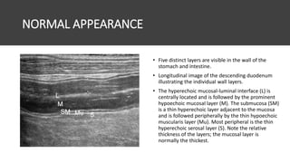
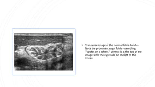
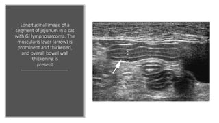
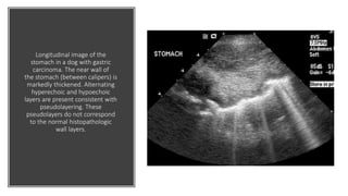
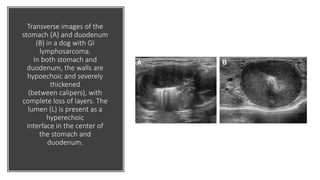
Ultrasound of the
Gastrointestinal Tract
Significance
• Understanding with the normal and abnormal ultrasound appearance of
the canine and feline gastrointestinal (GI) tract provides a marked
advantage in the diagnosis of GI disease.
• Although gas may inhibit complete visualization, in many cases, ultrasound
is able to confirm or rule out suspected disease.
• It should be noted that ultrasound of the GI tract does not eliminate the
need for abdominal radiographs.
• Ultrasound evaluation of the GI tract provides information about
• bowel wall thickness and layers,
• assessment of motility, and visualization of important adjacent structures such as
lymph nodes and peritoneum.
• abdominal ultrasound may replace the need for GI contrast studies in many cases,
saving time, money, radiation exposure, and stress to the patient.
Technique
• Techniques and approaches vary somewhat on patient conformation and
position.
• Scanning the patient in dorsal recumbency allows relatively complete
evaluation of the GI tract,
• Although right and left lateral recumbency may be necessary to
redistribute gas and fluid within individual portions of the stomach and
bowel.
• Right lateral intercostal windows are often helpful in visualizing the pylorus
and proximal duodenum in deep-chested dogs.
• Scanning the ventral abdomen of the standing dog or cat may allow
improved visualization of the dependent pylorus and gastric body.
• In most patients, a high-frequency transducer (7.5 MHz or higher) is used
for better visualization of stomach, small intestines, and colonic wall
thickness and layers.
Technique
• A sector or curvilinear transducer is best for intercostal windows due
to the smaller contact area of these probes.
• The canine stomach can be accessed immediately caudal to the liver,
at the level of the xiphoid.
• The feline gastric fundus and body are left-sided structures, with the
pylorus located in a more midline location.
• In both species, the left-sided dorsal fundus should be followed
ventrally and to the right, toward the pylorus, using both longitudinal
and transverse imaging planes.
(A)Ultrasound
image of the long
axis view of the
normal canine
stomach.
(B) Ultrasound
image of the long
axis view of the
normal feline
stomach
• F = fundus
• B= body of stomach
• L = liver
• S= stomach
• P= pancreas
• Ultrasound image of the longitudinal
view of the normal canine descending
duodenum. The duodenum is located
immediately ventral to the right
kidney (RK) as it extends from cranial
to caudal along the right lateral
abdomen. Ventral is at the top of the
image, with cranial on the left side of
the image.
• Transverse image of the
proximal duodenum at
the level of the duodenal
papilla. The bile duct
(arrow) is visible entering
the papilla. Ventral is at
the top of the image,
with the right on the left
side of the image.
• (A) Longitudinal image of the normal
feline ileo-colic junction.
• The terminal ileum (I) is seen entering the
gas-filled colon (C). Ventral is at the top of
the image, and cranial is to the left.
• (B) Transverse image of the normal feline
terminal ileum just before it enters the
gas-filled colon (C).
• Note the ‘‘wagon-wheel’’ appearance of
the layers in the ileum.
• Ventral is at the top of the image, with
the right on the left of the image.
NORMAL APPEARANCE
• Five distinct layers are visible in the wall of the
stomach and intestine.
• Longitudinal image of the descending duodenum
illustrating the individual wall layers.
• The hyperechoic mucosal-luminal interface (L) is
centrally located and is followed by the prominent
hypoechoic mucosal layer (M). The submucosa (SM)
is a thin hyperechoic layer adjacent to the mucosa
and is followed peripherally by the thin hypoechoic
muscularis layer (Mu). Most peripheral is the thin
hyperechoic serosal layer (S). Note the relative
thickness of the layers; the mucosal layer is
normally the thickest.
• Transverse image of the normal feline fundus.
Note the prominent rugal folds resembling
‘‘spokes on a wheel.’’ Ventral is at the top of the
image, with the right side on the left of the
image.
ABNORMAL APPEARANCE
• The stomach and bowel should be evaluated for wall thickness,
appearance of wall layers, peristaltic activity, and luminal contents
and diameter.
• Adjacent lymph nodes should be evaluated for enlargement, and the
presence of free gas or fluid should be noted.
Image (A): Longitudinal image of the
descending duodenum. The duodenal
lumen (D) is distended
with fluid. An echogenic structure with
round, well-defined margins (B) is
obstructing
the duodenum.
Images: (B) Longitudinal and (C) cross-
sectional images of the
jejunum of a cat presented for vomiting.
A well-defined linear echogenic
structure (arrows)
with acoustic shadowing is present
within the jejunal lumen. This structure
did not change
in appearance or move with peristaltic
activity.
Longitudinal image of a
segment of jejunum in a cat
with GI lymphosarcoma. The
muscularis layer (arrow) is
prominent and thickened,
and overall bowel wall
thickening is
present
Longitudinal image of the
stomach in a dog with gastric
carcinoma. The near wall of
the stomach (between calipers) is
markedly thickened. Alternating
hyperechoic and hypoechoic
layers are present consistent with
pseudolayering. These
pseudolayers do not correspond
to the normal histopathologic
wall layers.
Transverse images of the
stomach (A) and duodenum
(B) in a dog with GI
lymphosarcoma.
In both stomach and
duodenum, the walls are
hypoechoic and severely
thickened
(between calipers), with
complete loss of layers. The
lumen (L) is present as a
hyperechoic
interface in the center of
the stomach and
duodenum.
For Further
Reading
• http://www.sonopath.com/sites/default/files/download
s/article_casey_GASTRO_Ultrasound_The_GI_Tract.pdf
• Vet Clin Small Anim 39 (2009) 747–759
• doi:10.1016/j.cvsm.2009.04.010

Ultrasonography of GIT.pptx

  • 1.
  • 2.
    Significance • Understanding withthe normal and abnormal ultrasound appearance of the canine and feline gastrointestinal (GI) tract provides a marked advantage in the diagnosis of GI disease. • Although gas may inhibit complete visualization, in many cases, ultrasound is able to confirm or rule out suspected disease. • It should be noted that ultrasound of the GI tract does not eliminate the need for abdominal radiographs. • Ultrasound evaluation of the GI tract provides information about • bowel wall thickness and layers, • assessment of motility, and visualization of important adjacent structures such as lymph nodes and peritoneum. • abdominal ultrasound may replace the need for GI contrast studies in many cases, saving time, money, radiation exposure, and stress to the patient.
  • 3.
    Technique • Techniques andapproaches vary somewhat on patient conformation and position. • Scanning the patient in dorsal recumbency allows relatively complete evaluation of the GI tract, • Although right and left lateral recumbency may be necessary to redistribute gas and fluid within individual portions of the stomach and bowel. • Right lateral intercostal windows are often helpful in visualizing the pylorus and proximal duodenum in deep-chested dogs. • Scanning the ventral abdomen of the standing dog or cat may allow improved visualization of the dependent pylorus and gastric body. • In most patients, a high-frequency transducer (7.5 MHz or higher) is used for better visualization of stomach, small intestines, and colonic wall thickness and layers.
  • 4.
    Technique • A sectoror curvilinear transducer is best for intercostal windows due to the smaller contact area of these probes. • The canine stomach can be accessed immediately caudal to the liver, at the level of the xiphoid. • The feline gastric fundus and body are left-sided structures, with the pylorus located in a more midline location. • In both species, the left-sided dorsal fundus should be followed ventrally and to the right, toward the pylorus, using both longitudinal and transverse imaging planes.
  • 5.
    (A)Ultrasound image of thelong axis view of the normal canine stomach. (B) Ultrasound image of the long axis view of the normal feline stomach • F = fundus • B= body of stomach • L = liver • S= stomach • P= pancreas
  • 6.
    • Ultrasound imageof the longitudinal view of the normal canine descending duodenum. The duodenum is located immediately ventral to the right kidney (RK) as it extends from cranial to caudal along the right lateral abdomen. Ventral is at the top of the image, with cranial on the left side of the image.
  • 7.
    • Transverse imageof the proximal duodenum at the level of the duodenal papilla. The bile duct (arrow) is visible entering the papilla. Ventral is at the top of the image, with the right on the left side of the image.
  • 8.
    • (A) Longitudinalimage of the normal feline ileo-colic junction. • The terminal ileum (I) is seen entering the gas-filled colon (C). Ventral is at the top of the image, and cranial is to the left. • (B) Transverse image of the normal feline terminal ileum just before it enters the gas-filled colon (C). • Note the ‘‘wagon-wheel’’ appearance of the layers in the ileum. • Ventral is at the top of the image, with the right on the left of the image.
  • 9.
    NORMAL APPEARANCE • Fivedistinct layers are visible in the wall of the stomach and intestine. • Longitudinal image of the descending duodenum illustrating the individual wall layers. • The hyperechoic mucosal-luminal interface (L) is centrally located and is followed by the prominent hypoechoic mucosal layer (M). The submucosa (SM) is a thin hyperechoic layer adjacent to the mucosa and is followed peripherally by the thin hypoechoic muscularis layer (Mu). Most peripheral is the thin hyperechoic serosal layer (S). Note the relative thickness of the layers; the mucosal layer is normally the thickest.
  • 10.
    • Transverse imageof the normal feline fundus. Note the prominent rugal folds resembling ‘‘spokes on a wheel.’’ Ventral is at the top of the image, with the right side on the left of the image.
  • 11.
    ABNORMAL APPEARANCE • Thestomach and bowel should be evaluated for wall thickness, appearance of wall layers, peristaltic activity, and luminal contents and diameter. • Adjacent lymph nodes should be evaluated for enlargement, and the presence of free gas or fluid should be noted.
  • 12.
    Image (A): Longitudinalimage of the descending duodenum. The duodenal lumen (D) is distended with fluid. An echogenic structure with round, well-defined margins (B) is obstructing the duodenum. Images: (B) Longitudinal and (C) cross- sectional images of the jejunum of a cat presented for vomiting. A well-defined linear echogenic structure (arrows) with acoustic shadowing is present within the jejunal lumen. This structure did not change in appearance or move with peristaltic activity.
  • 13.
    Longitudinal image ofa segment of jejunum in a cat with GI lymphosarcoma. The muscularis layer (arrow) is prominent and thickened, and overall bowel wall thickening is present
  • 14.
    Longitudinal image ofthe stomach in a dog with gastric carcinoma. The near wall of the stomach (between calipers) is markedly thickened. Alternating hyperechoic and hypoechoic layers are present consistent with pseudolayering. These pseudolayers do not correspond to the normal histopathologic wall layers.
  • 15.
    Transverse images ofthe stomach (A) and duodenum (B) in a dog with GI lymphosarcoma. In both stomach and duodenum, the walls are hypoechoic and severely thickened (between calipers), with complete loss of layers. The lumen (L) is present as a hyperechoic interface in the center of the stomach and duodenum.
  • 16.