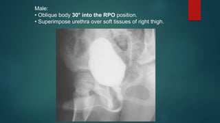
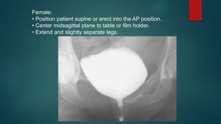
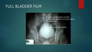
Cystourethrography
NTAMBI MUSA
AP PROJECTION
• LPO AND RPO POSITIONS
• LATERAL POSITION (OPTIONAL):
CYSTOGRAPHY
Pathology Demonstrated
Signs of cystitis, obstruction, vesicoureteral reflux, and
bladder calculi are visualized. Lateral demonstrates
possible fistulas between bladder and uterus or rectum.
Cystography
BASIC
• AP (CR 10° to 15° caudad)
• Both oblique positions (45° to 60°)
AP (10° to 15° caudad)
Posterior Oblique Positions:
• 45° to 60° body rotation. (Steep oblique positions are used to visualize
posterolateral aspect of bladder, especially UV junction.)
RPO (45° to 60°).
45°
- RPO (30°) POSITION – MALE
- AP PROJECTION - FEMALE: VOIDING
CYSTOURETHROGRAPHY
Pathology Demonstrated
Functional study of the urinary bladder and urethra determines
cause of urinary retention and evaluates for possible
vesicoureteral reflux.
Voiding Cystourethrography
BASIC
• Male—RPO (30°)
• Female—AP
Male:
• Oblique body 30° into the RPO position.
• Superimpose urethra over soft tissues of right thigh.
Female:
• Position patient supine or erect into the AP position.
• Center midsagittal plane to table or film holder.
• Extend and slightly separate legs.
COMPRESSION
Compression
Distention
Release
Flow  Full
Visualization of length
 Normal peristalsis may leave portions of
ureters empty of contrast
 To inhibit ureteric drainage and promote
distension of pelvicalyceal systems,
optimising visualization
 Proximal ureters and intrarenal
collecting system optimally distended
 With contraindications
15-MINUTE
 After compression is released, there is
transient increase in flow down the
ureters  TAKE RADIOGRAPH
 Peristalsis makes visualization of
entire length uncommon
 Kidneys and ureters still visualized,
although less clearly
 Bladder begins to light up
FULL BLADDER FILM
 Bladder should appear smooth,
balloon-like, globular structure above
the pelvic rim
 Kidneys, ureters less visible
FULL BLADDER FILM
 Bladder should appear smooth,
balloon-like, globular structure above
the pelvic rim
 Kidneys, ureters less visible
POST-VOID FILM
 Visualize residual urine
 < 50cc of fluid
POST-VOID FILM
 Visualize residual urine
 < 50cc of fluid

Cystourethrography - Lecture.pptx

  • 1.
  • 2.
    AP PROJECTION • LPOAND RPO POSITIONS • LATERAL POSITION (OPTIONAL): CYSTOGRAPHY Pathology Demonstrated Signs of cystitis, obstruction, vesicoureteral reflux, and bladder calculi are visualized. Lateral demonstrates possible fistulas between bladder and uterus or rectum. Cystography BASIC • AP (CR 10° to 15° caudad) • Both oblique positions (45° to 60°)
  • 3.
    AP (10° to15° caudad)
  • 5.
    Posterior Oblique Positions: •45° to 60° body rotation. (Steep oblique positions are used to visualize posterolateral aspect of bladder, especially UV junction.) RPO (45° to 60°).
  • 6.
  • 7.
    - RPO (30°)POSITION – MALE - AP PROJECTION - FEMALE: VOIDING CYSTOURETHROGRAPHY Pathology Demonstrated Functional study of the urinary bladder and urethra determines cause of urinary retention and evaluates for possible vesicoureteral reflux. Voiding Cystourethrography BASIC • Male—RPO (30°) • Female—AP
  • 8.
    Male: • Oblique body30° into the RPO position. • Superimpose urethra over soft tissues of right thigh.
  • 9.
    Female: • Position patientsupine or erect into the AP position. • Center midsagittal plane to table or film holder. • Extend and slightly separate legs.
  • 10.
    COMPRESSION Compression Distention Release Flow  Full Visualizationof length  Normal peristalsis may leave portions of ureters empty of contrast  To inhibit ureteric drainage and promote distension of pelvicalyceal systems, optimising visualization  Proximal ureters and intrarenal collecting system optimally distended  With contraindications
  • 11.
    15-MINUTE  After compressionis released, there is transient increase in flow down the ureters  TAKE RADIOGRAPH  Peristalsis makes visualization of entire length uncommon  Kidneys and ureters still visualized, although less clearly  Bladder begins to light up
  • 12.
    FULL BLADDER FILM Bladder should appear smooth, balloon-like, globular structure above the pelvic rim  Kidneys, ureters less visible
  • 13.
    FULL BLADDER FILM Bladder should appear smooth, balloon-like, globular structure above the pelvic rim  Kidneys, ureters less visible
  • 14.
    POST-VOID FILM  Visualizeresidual urine  < 50cc of fluid
  • 15.
    POST-VOID FILM  Visualizeresidual urine  < 50cc of fluid

Editor's Notes

  • #11 Ureters are compressed against pelvis Place belt and pneumatic balloons at upper edge anterior superior iliac spine Paddles should nearly meet at the midline Contraindications: Omitted in children Aortic aneurysm Ureteral obstruction Acute abdominal/flank pain Tender abdomen Recent abdominal surgery Abdominal stomas Colostomy, ileostomy, ileal conduit
  • #12 Obstruction lower down (ie pelvic portion of the ureters) may now be visualized Ureters: A full-length radiograph at this stage will best demonstrate the ureters. If there is any hold-up of contrast medium in a ureter, further views are indicated prone view  contrast medium will collect in a more dependent portion of the ureter oblique view  relationship of the ureter to a possible phlebolith will be seen Bladder: Bladder will be well filled at this stage Oblique views may help to show any irregularity of the wall of the bladder or demonstrate the relationship of a pelvic mass to the bladder As the bladder becomes progressively distended, the intraluminal contrast material should be spheric and smoothly marginated and the wall progressively less evident NOTE: contrast material–opacified urine is heavier than nonopacified urine. Gravity maneuvers such as imaging with the patient in the prone or dependent oblique position often assist with visualization of unopacified portions of the ureters, especially in cases of obstruction. Supine: ureters are sometimes not seen because of inherent peristalsis Prone: ureters are much seen because abdominal contents pushes the ureters anteriorly and blocks peristalsis
  • #13 - If bladder is not found above pelvic rim, or appears to descend upon filling, a cystocoele should be considered
  • #14 - If bladder is not found above pelvic rim, or appears to descend upon filling, a cystocoele should be considered
  • #15 If bladder is not found above pelvic rim, or appears to descend upon filling, a cystocoele should be considered Summary of Findings: Renal papilla cup shaped, no abnormal filling defects, pelvocalyces not dilated, ureters 3-5mm diameter, urinary bladder located above pelvic brim, moderately dilated with no filling defects
  • #16 If bladder is not found above pelvic rim, or appears to descend upon filling, a cystocoele should be considered Summary of Findings: Renal papilla cup shaped, no abnormal filling defects, pelvocalyces not dilated, ureters 3-5mm diameter, urinary bladder located above pelvic brim, moderately dilated with no filling defects