2. Definition
Cervical ectopy is a condition where the squamous epithelium of the ectocervix is
replaced by columnar epithelium, which is continuous with the endocervix. It is
not an ulcer.
Etiology
1. Congenital
2. Acquired
3. Congenital
At birth, in about one-third of cases, the columnar epithelium of the
endocervix extends beyond the external os. This condition persists only for a
few days until the level of estrogen derived from the mother falls. Thus, the
congenital ectopy heals spontaneously.
Acquired
Hormonal:
The squamocolumnar junction is not static and its movement, either inwards
or outwards is dependent on estrogen. When the estrogen level is high, it
moves out so that the columnar epithelium extends onto the vaginal portion
of the cervix replacing the squamous epithelium. This state is observed during
during pregnancy and amongst ‘pill users’. The squamocolumnar junction
returns back to its normal position after 3 months following delivery and little
earlier following withdrawal of ‘pill’.
4. Infection:
The role of infection as the primary cause of ectopy has been discarded. However,
chronic cervicitis may be associated or else the infection may supervene on an
ectopy because of the delicate columnar epithelium which is more vulnerable to
trauma and infection.
5. Pathogenesis
In the active phase of ectopy, the squamocolumnar junction
moves out from the os. The columnar epithelium of the
endocervix maintains its continuity while covering the ectocervix
replacing the squamous epithelium. The replaced epithelium is
usually arranged in a single layer (flat type) or may be so
hyperplastic as to fold inwards to accommodate in the increased
area—a follicular ectopy. At times, it becomes heaped up to fold
inwards and outwards—
a papillary ectopy. Underneath the epithelium, there are
evidences of round cell infiltration and glandular proliferation.
The features of infection are probably secondary rather than
primary. The columnar epithelium is less resistant to infection
than the squamous epithelium.
6. During the process of healing, the squamocolumnar junction gradually moves
up towards the external os. The squamous epithelium grows beneath the
columnar epithelium until it reaches at or near to its original position at the
external os. Alternatively, the replacement is probably by squamous
metaplasia of the columnar cells. The possibility of squamous metaplasia of
the reserve cells
During the process, the squamous epithelium may obstruct the mouth of the
underlying glands (normally not present in ectocervix) → pent up secretion →
retention cyst → Nabothian follicle. Alternatively, the epithelium may burrow
inside the gland lumina. This process of replacement by the squamous
epithelium is called epidermidization
7. Clinical Features
Symptoms:
The lesion may be asymptomatic. However, the following symptoms may be
present. ™
Vaginal discharge—The discharge may be excessively mucoid. It may be
mucopurulent, offensive and irritant in presence of infection; may be even
blood-stained due to premenstrual congestion.
™
Contact bleeding especially during pregnancy and ‘pill use’ either following
coitus or defecation may be associated. ™
Associated cervicitis may produce backache, pelvic pain and at times,
infertility.
8. Signs:
Internal examination reveals : •
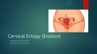
Per speculum—There is a bright red area
surrounding and extending beyond the external os in the ectocervix. The outer
edge is clearly demarcated. The lesion may be smooth or having small papillary
folds. It is neither tender nor bleeds to touch. On rubbing with a gauze piece,
there may be multiple oozing spots (sharp bleeding in isolated spots in
carcinoma). The feel is soft and granular giving rise to a grating sensation
9. Diagnosis—The diagnosis is confused with:
Ectropion: The lips of the cervix are curled back to expose the endocervix.
This may be apparent when the lips of the cervix are stretched by the bivalve
speculum.
Early carcinoma: It is indurated, friable and usually ulcerated which bleeds to
touch. Confirmation is by biopsy.
Primary lesion (chancre): The ulcer has a punched out appearance.
Tubercular ulcer: There is indurated ulcer with caseation at the base. Biopsy
confirms the diagnosis.
10. Management Guidelines: All cases should be subjected to cytological
examination from the cervical smear to exclude dysplasia or malignancy.
Symptomatic cases ™
Detected during pregnancy and early puerperium, the treatment should be
withheld for at least 12 weeks postpartum. In pill users, the ‘pill’ should be
stopped and barrier method is advised. ™
Persistent ectopy with troublesome discharge should be treated surgically by—
(i) thermal cauterization, (ii) cryosurgery and (iii) laser vaporization. All the
methods employed are based on the principle of destruction of the columnar
epithelium to be followed by its healing by the squamous epithelium.