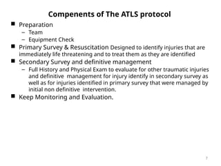
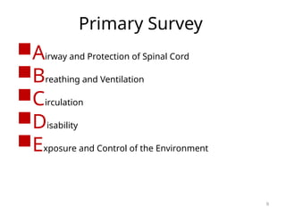
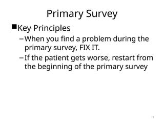
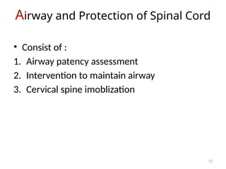
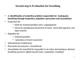
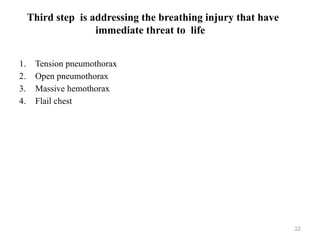
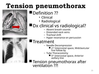
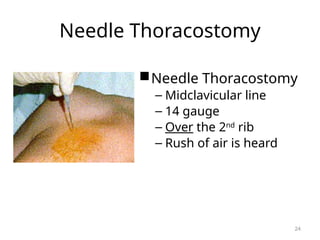
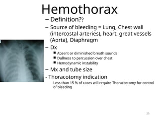
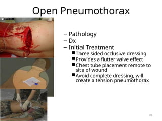
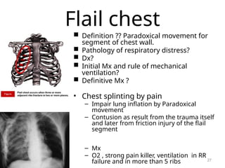
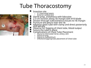
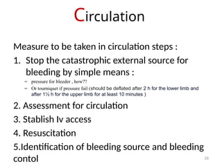
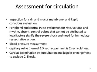
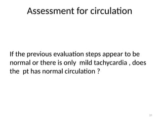
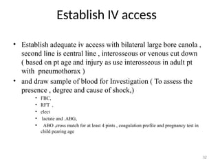
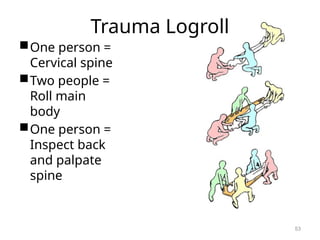
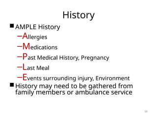
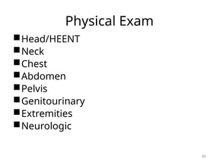
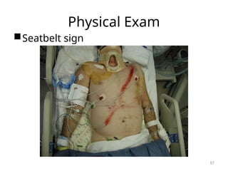
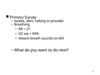
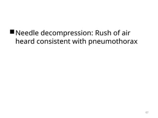
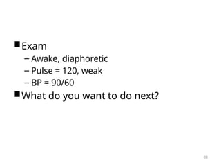
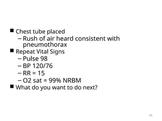
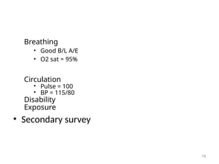
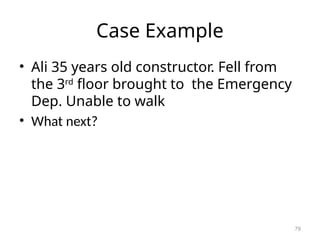
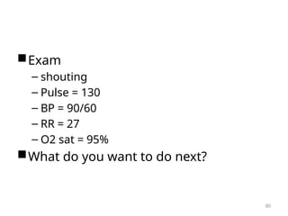
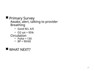
ATLS
1
Dr. Mohammed Abdullrahman ,
"Arab Board - General Surgery
Residency and Membership of
the Royal College of Surgeons
(MRCS)"
2
Goal of ATLS protocol
1. What is the goal of ATLS protocol?
2. What is the parts of ATLS protocol?
3. What is the first simple rapid method to assess the
patency of airway ?
4. Mention 4 signs of respiratory distress?
5. How to know the pt is in shock in 10 seconds
evaluation?
6. What type of iv access provide the most rapid
infusion rate ?
3
Goal of ATLS protocol
• To provides a organized approach for trauma care to
improve survival and outcomes, through rapidly
identify and address life-threatening injuries in a
structured sequence.
• Origins: Developed by the American College of
Surgeons in the 1970s.
Injury: Scale of the Global
Problem
• 5.8 million deaths/year
• 10% of worlds deaths
• 32% more deaths than HIV, TB
and
Malaria combined
Source:
Global
Burden
of
Disease,
WHO,
2004
4
Injury: Scale of the Global
Problem
Source: World Report on Road Traffic Injury Prevention 2004
5
World Health Organization, who.int
Epidemiology
 Golden Hour = 80% of trauma
deaths in first hour after injury
 Rapid trauma care has greatest
level of impact in these patients
Immediately Hours Days/Weeks
50%
35%
15%
Trimodal Distribution of Trauma Deaths
6
Compenents of The ATLS protocol
 Preparation
– Team
– Equipment Check
 Primary Survey & Resuscitation Designed to identify injuries that are
immediately life threatening and to treat them as they are identified
 Secondary Survey and definitive management
– Full History and Physical Exam to evaluate for other traumatic injuries
and definitive management for injury identify in secondary survey as
well as for injuries identified in primary survey that were managed by
initial non definitive intervention.
 Keep Monitoring and Evaluation.
7
Preparation for Patient Arrival
Organize Trauma
Response Team
8
Primary Survey
Airway and Protection of Spinal Cord
Breathing and Ventilation
Circulation
Disability
Exposure and Control of the Environment
9
Why the primary survey had been
organized in this order ABCDE ?
10
Primary Survey
Primary Survey
Key Principles
–When you find a problem during the
primary survey, FIX IT.
–If the patient gets worse, restart from
the beginning of the primary survey
11
Airway and Protection of Spinal Cord
12
• Consist of :
1. Airway patency assessment
2. Intervention to maintain airway
3. Cervical spine imoblization
Cusses and risks of airway obstruction in
trauma ?
Airway Assessment
• Ask pt his name or what happens , if pt can talk clearly indicate his airway is
patent currently and no immediate hazards to his airway, but does not indicate
secure airway, if the pt can talk clearly generally do not have a need for
immediate airway management , so go to the second step ( breathing- B )
adter C spine fixation, if not do further assessment for airway patency by
LOOK ,LISETN & FEEL
14
Airway Assessment
• Look for
– The level of conscious Agitation, drawsy, Coma
– For central cyanosis( late sign)
– Fascial fracture, laryngeal or tracheal injury
– Open the pt mouth and look for foreign body or nasopharyngeal bleeding ,
– For neck emphysema or hematoma compressing larynx or trachea
• Listen for abnormal sound as
– strider , indicated severe airway narrowing or compression
– or gurgling (indicate liquid or semiliquid material in upper airway),
– snoring (indicated partial laryngeal obstruction by tongue)
– expiratory wheezing (indicate lower airway obstruction)
• Feel for breathing if pt has no clear breathing effort by your check or cotton piece
and look for chest movement ( for 10 second)
15
Airway
Interventions
 Initial measures to maintain Airway Patency
– Finger swap and Suction of Secretions
– Chin Lift/Jaw thrust
– Nasopharyngeal Airway
– Needle cricothyroidtomy
 Definitive measures : indicated if initial measures
fail to solve the problem , GCS <8 and in CPR
– Endotracheal Intubation
– Surgical Cricothyroidotomy
– Surgical tracheostomy
16
Protection of Spinal Cord
 General Principle:
• All pt with blunt trauma or gun shot to neck, should presume
to have C- spine injury
• Immobilization of C – spine is priority in primary survey to
protect the spinal cord from disruption during mobilization if
the C- spine has instability, until injury has been excluded by
radiography or clinical physical exam
• Clearance of spinal cord injury isn't priority and should
not precede resuscitation
17
Protection of Spinal Cord
 C- Spine immobilization maneuver
Return head to neutral position and Do not apply traction and do
tribal immobilization by :
1. Rigid Cervical Spinal Collar
2. On table immobilization by tabe
3. and sand bag or block
18
Breathing and
Ventilation
19
Steps should be taken in Breathing are :
• First step is high flow oxygen supply to all pt via non re-breathable mask 15 L per
M
• Second step is evaluation for adequacy breathing
• Third step is addressing the breathing injury that have immediate threat to life
Second step is Evaluation for breathing
20
1. Evaluation for adequacy of breathing and oxygen delivery by
exclusion for signs of respiratory distress via:
– RR counting ,
– Oxygen saturation by pulse oximeter
– cyanosis
– and accessory muscle ,
Second step is Evaluation for breathing
21
• 2. Identification of underline problem responsible for inadequate
breathing through inspection, palpation ,percussion and auscultation:
• Inspection for
• Neck for tracheal position and J. engorgement
• Chest for paradoxical movement of chest, chest flail segment, and
open wound,
• Palpation for
• tracheal position ,
• symmetry of chest movement
• Subcutaneous emphysema
• Percussion for pneumo / hemothorax
• Auscultation for lung filed for inequality in air entry, decreasing or absence
breathing sound or added sound( weez, crepitation, crackling)
Third step is addressing the breathing injury that have
immediate threat to life
22
1. Tension pneumothorax
2. Open pneumothorax
3. Massive hemothorax
4. Flail chest
Definition ??
• Clinical
• Radiological
Dx clinical vs radiological?
– Absent breath sounds
– Distended neck veins
– Tracheal shift
– Hyper resonance on percussion
Treatment
– Needle Decompression
 2nd
Intercostal space, Midclavicular
line follow by
– Tube Thoracostomy
 5th
Intercostal space, Anterior
axillary line
 Tension pneumothorax after
ventilation ??!
Tension pneumothorax
23
Needle Thoracostomy
Needle Thoracostomy
– Midclavicular line
– 14 gauge
– Over the 2nd
rib
– Rush of air is heard
24
Hemothorax
– Definition??
– Source of bleeding = Lung, Chest wall
(intercostal arteries), heart, great vessels
(Aorta), Diaphragm
– Dx
 Absent or diminished breath sounds
 Dullness to percussion over chest
 Hemodynamic instability
– Mx and tube size
- Thoracotomy indication
Less than 15 % of cases will require Thoracostomy for control
of bleeding
25
Open Pneumothorax
– Pathology
– Dx
– Initial Treatment
Three sided occlusive dressing
Provides a flutter valve effect
Chest tube placement remote to
site of wound
Avoid complete dressing, will
create a tension pneumothorax
26
Flail chest
 Definition ?? Paradoxical movement for
segment of chest wall.
 Pathology of respiratory distress?
 Dx?
 Initial Mx and rule of mechanical
ventilation?
 Definitive Mx ?
• Chest splinting by pain
– Impair lung inflation by Paradoxical
movement
– Contusion as result from the trauma itself
and later from friction injury of the flail
segment
– Mx
– O2 , strong pain killer, ventilation in RR
failure and in more than 5 ribs 27
Tube Thoracostomy
 Insertion site
– 5th
intercostal space,
– Anterior axillary line
 Sterile prep, anesthesia with lidocaine
 2-3 cm incision along rib margin with #10 blade
 Dissect through subcutaneous tissues to rib margin
 Puncture the pleura over the rib
 Advance chest tube with clamp and direct posteriorly
and apically
 Observe for fogging of chest tube, blood output
 Suture the tube in place
 Complications of Chest Tube Placement
– Injury to intercostal nerve, artery, vein
– Injury to lung
– Injury to mediastinum
– InfectionInappropriate placement of chest tube
28
Circulation
29
Measure to be taken in circulation steps :
1. Stop the catastrophic external source for
bleeding by simple means :
– pressure for bleeder , how??
– Or tourniquet if pressure fail (should be deflated after 2 h for the lower limb and
after 1½ h for the upper limb for at least 10 minutes )
2. Assessment for circulation
3. Stablish Iv access
4. Resuscitation
5.Identification of bleeding source and bleeding
contol
Assessment for circulation
30
• Inspection for skin and mucus membrane, and Rapid
conscious evaluation,
• Peripheral and central Pulse evaluation for rate, volume and
rhythm, absent central pulses that cannot be attributed to
local factors signify the severe shock and need for immediate
resuscitative action.
• Blood pressure measurment,
• capillary refile (normal 1.5 sec , upper limit is 2 sec, coldness,
• Cardiac examination by auscultation and jugular engorgement
to exclude C. Shock .
Assessment for circulation
31
If the previous evaluation steps appear to be
normal or there is only mild tachycardia , does
the pt has normal circulation ?
Establish IV access
32
• Establish adequate iv access with bilateral large bore canola ,
second line is central line , interosseous or venous cut down
( based on pt age and injury as use interosseous in adult pt
with pneumothorax )
• and draw sample of blood for Investigation ( To assess the
presence , degree and cause of shock,)
• FBC,
• RFT ,
• elect
• lactate and .ABG,
• ABO ,cross match for at least 4 pints , coagulation profile and pregnancy test in
child pearing age
Resuscitation
33
Is based on class of shock ??
Circulation
 Shock
– Impaired tissue perfusion
– Tissue oxygenation is inadequate to meet metabolic demand
– Prolonged shock state leads to multi-organ system failure
and cell death
 Clinical Signs of Shock
– Altered mental status
– Tachycardia (HR > 100) = Most common sign
– Arterial Hypotension (SBP < 120)
– Inadequate Tissue Perfusion
 Pale skin color
 Cool clammy skin
 Delayed cap refill (> 3 seconds)
 Altered LOC
 Decreased Urine Output (UOP < 0.5 mL/kg/hr)
34
Resuscitation
35
Resuscitation based on class of hemorrhagic shock ;
Class 1by monitoring ,
Class 2 by iv fluid , 10 to 20 ml of crystalloid fluid as bolus in
15 minutes
Class 3, iv fluid vs blood transfusion
Class 4 iv fluid until group o negative blood is ready follow by
crosshatched blood transfusion under massive transfusion
protocol
Resuscitation
36
• Type of fluid given :
– Crystalloid vs colloid?
– RL vs NS vs Dex 5% ?
– Blood vs Crystalloid ?
• Type of blood poduct in transfusion 1:1:1 ??
• What is Massive blood transfusion protocol , and when
need to be activated?
• Rule of transexamic acid ?
Resuscitation
37
How to monitor response?
Target of transfusion ? Effective circulation and later on end
organ perfusion during secondary survey .
Types of response to resuscitation?
• Adequate Response : mean no active bleeding , continue monitoring and identify
source of bleeding and it usually managed conservative . Except if there is other
indication for surgery.
• unsustain response : because of redistribution of iv fluid or active bleeding , give
blood transfusion if not given before and surgical intervention if after 3 units pt
condition not improved
• In adequate response : pt need further resuscitation , so start or add blood
transfusion and measure CVP, if still in adequate response after 3 uints, the cause
is likely the active bleeding , so pt need surgical intervention .
• No response : mean sever active bleeding , so pt need surgical intervention .
Resuscitation
38
Types of response to resuscitation?
• Adequate Response : mean no active bleeding , continue monitoring and
identify source of bleeding and it usually managed conservative . Except
if there is other indication for surgery.
• unsustain response : because of redistribution of iv fluid or active
bleeding , give blood transfusion if not given before and surgical
intervention if after 3 units pt condition not improved
• In adequate response : pt need further resuscitation , so start or add blood
transfusion and measure CVP, if still in adequate response after 3 uints,
the cause is likely the active bleeding , so pt need surgical intervention .
• No response : mean sever active bleeding , so pt need surgical
intervention .
Identify source of bleeding and control
What is the source of internal bleeding in
pt with shock in blunt trauma?
– Chest
– Abdomen
– Pelvis
– Multiple bone fractures !
39
Identify source of bleeding and control
How to identify the source of bleeding and
when ?
In pt with unresponsive or unsustain response
shock the source should be identify during primary
survey , the other is identify after completing the
primary survey :
– Examination In all pt with shock
– FAST in pt with unresponsive or unsustain
response
– Aspiration in pt with unresponsive or unsustain
response with negative FAST
– Intraoperative !!
40
Identify source of bleeding and control
Pelvic fracture
Pelvic fracture Dx?
Source of bleeding ?
Mx?
Splenic injury Mx?
Liver laceration Mx ?
41
Identify source of bleeding and control
 Types of Shock in Trauma
– Hemorrhagic
 Assume hemorrhagic shock in all trauma patients until
proven otherwise
 Results from Internal or External Bleeding
– Obstructive
 Cardiac Tamponade
 Tension Pneumothorax
– Neurogenic
 Spinal Cord injury
42
Circulation
Pericardial Tamponade
– Pericardium or sac around heart fills
with blood due to penetrating or blunt
injury to chest
– Beck’s Triad
Distended jugular veins
Hypotension
Muffled heart sounds
– Treatment
Rapid evacuation of pericardial space
Performed through a pericardiocentesis
(temporizing measure)
Open thoracotomy
H
e
a
r
t
Blood
Pericardium
Epicardium
Aceofhearts1968(Wikimedia)
43
Pericardiocentesis
 Puncture the skin 1-2 cm inferior to xiphoid
process
 45/45/45 degree angle
 Advance needle to tip of left scapula
 Withdraw on needle during advance of needle
 Preferable under ultrasound guidance or EKG
lead V attachment
44
Adjuncts to Primary Survey
• Radiology
– C-spine, CXR, Pelvis
– FAST
• Folly catheter , way, C/I?
• Repeat investigation
45
Disability
 Baseline Neurologic Exam
– Pupillary Exam
 Dilated pupil – suggests transtentorial herniation on ipsilateral side
– AVPU Scale
 Alert
 Responds to verbal stimulation
 Responds to pain
 Unresponsive
– Glasgow Coma Scale: 3-15
– Rectal Exam
 Normal Rectal Tone
 Note: If intubation prior to neuro assessment, consider
quick neuro assessment to determine degree of injury
46
Disability
 Glasgow Coma Scale
– Eye
 Spontaneously opens 4
 To verbal command 3
 To pain 2
 No response 1
– Best Motor Response
 Obeys verbal commands 6
 Localizes to pain 5
 Withdraws from pain 4
 Flexion to pain (Decorticate Posturing) 3
 Extension to pain (Decerebrate Posturing) 2
 No response 1
– Verbal Response
 Oriented/Conversant 5
 Disoriented/Confused 4
 Inappropriate words 3
 Incomprehensible words 2
 No response 1
GCS ≤ 8
Intubate
47
Disability
Key Principles
– Precise diagnosis is not necessary at this point
in evaluation
– Prevention of further injury and identification
of neurologic injury is the goal
– Decreased level of consciousness = Head injury
until proven otherwise
– Maintenance of adequate cerebral perfusion is
key to prevention of further brain injury
Adequate oxygenation
Avoid hypotension
– Involve neurosurgeon early for clear
intracranial lesions
48
Disability
Cervical Spinal Clearance
– Patients must be alert and oriented to
person, place and time
– No neurological deficits
– Not clinically intoxicated with alcohol or
drugs
– Non-tender at all spinous processes
– No distracting injuries
– Painless range of motion of neck
49
Exposure
 Remove all clothing
– Examine for other signs of injury
– Injuries cannot be diagnosed until seen by
provider
 Logroll the patient to examine patient’s back
– Maintain cervical spinal immobilization
– Palpate along thoracic and lumbar spine
– Minimum of 3 people, often more providers
required
 Avoid hypothermia
– Apply warm blankets after removing clothes
– Hypothermia = Coagulopathy
Increases risk of hemorrhage
50
Exposure
51
Exposure
52
Trauma Logroll
One person =
Cervical spine
Two people =
Roll main
body
One person =
Inspect back
and palpate
spine
53
Secondary Survey
Secondary Survey is completed after
primary survey is completed and
patient has been adequately
resuscitated.
No patient with abnormal vital signs
should proceed through a secondary
survey
Secondary Survey includes a brief
history and complete physical exam
54
History
AMPLE History
–Allergies
–Medications
–Past Medical History, Pregnancy
–Last Meal
–Events surrounding injury, Environment
History may need to be gathered from
family members or ambulance service
55
Physical Exam
Head/HEENT
Neck
Chest
Abdomen
Pelvis
Genitourinary
Extremities
Neurologic
56
Physical Exam
Seatbelt sign
57
Adjuncts to Secondary Survey
– Focused Abdominal Sonography in Trauma
(FAST)
– Additional films
 Cat scan imaging
 Angiography
 Foley Catheter
– Blood at urethral meatus = No Foley
catheter
58
FAST Exam
• Focused Abdominal Sonography in
Trauma
• Has largely replaced deep peritoneal
lavage (DPL)
• Bedside ultrasound looking for blood
collection in an unstable patient.
• If the patient is unstable and a blood
collection is found, proceed emergently
to the operating theater.
59
FAST
• Sensitivity of 94.6%
• Specificity of 95.1%
• Overall accuracy of 94.9% in identifying
the presence of intra-abdominal
injuries.
– Yoshil: J Trauma 1998; 45
60
FAST
Right Upper Quadrant - Morrison’s
Pouch
• Between the liver and kidney in RUQ.
• First place that fluid collects in supine
patient.
61
FAST Exam - RUQ
62
Trauma in Special Populations
Pediatric
 Broselow Tape
Pregnancy
– Supine Hypotensive Syndrome
Enlarged uterus inferior vena cava
Decreases venous return, decrease C.O.
Left lateral decubitus position to avoid
excessive hypotension
63
Definitive Care
 Secondary Survey followed by radiographic
evaluation
– CatScan
– Consultation
Neurosurgery
Orthopedic Surgery
Vascular Surgery
 Transfer to Definitive Care
– Operating Room
– ICU
– Higher level facility
64
Case Example
 Ahmed 45 y/o male involved in
a rollover MVA and was ejected
from the vehicle. Patient was
unrestrained. Patient was not
ambulatory on scene of
accident and is brought into
trauma bay for evaluation.
– What concerns you about story?
– First steps of evaluation and
management
65
 Primary Survey
– Awake, alert, talking to provider
– Breathing
• RR = 27
• O2 sat = 93%
• Absent breath sounds on left
– What do you want to do next?
66
Needle decompression: Rush of air
heard consistent with pneumothorax
67
Exam
– Awake, diaphoretic
– Pulse = 120, weak
– BP = 90/60
What do you want to do next?
68
 Chest tube placed
– Rush of air heard consistent with
pneumothorax
 Repeat Vital Signs
– Pulse 98
– BP 120/76
– RR = 15
– O2 sat = 99% NRBM
 What do you want to do next?
69
– Circulation
– Disability
– Exposure
• Secondary survey
70
71
Case Example
• Omar had an accident while driving 100km/h
on motorcycle with out helmet. Broght to ED
agitated.
• What to do next?
72
Exam
– Agitated
– Pulse = 125
– BP = 110/75
– RR = 35
– O2 sat = 85%
What do you want to do next?
73
 Primary Survey
– Awake, blood on face and deformity, not talking to
provider
What do you want to do next?
74
Physical Exam
Difficult airway
75
– Good B/L A/E
– O2 sat = 95%
– What next?
76
– Pulse = 100
– BP = 115/80
– RR = 25
– O2 sat = 95%
What do you want to do next?
77
Breathing
• Good B/L A/E
• O2 sat = 95%
Circulation
• Pulse = 100
• BP = 115/80
Disability
Exposure
• Secondary survey
78
Case Example
• Ali 35 years old constructor. Fell from
the 3rd
floor brought to the Emergency
Dep. Unable to walk
• What next?
79
Exam
– shouting
– Pulse = 130
– BP = 90/60
– RR = 27
– O2 sat = 95%
What do you want to do next?
80
 Primary Survey
Awake, alert, talking to provider
Breathing
• Good B/L A/E
• O2 sat = 95%
Circulation
• Pulse = 130
• BP = 90/60
 WHAT NEXT?
81
• 2 IV CANULA
• 2 L RL
• BP went down
• HR went up
• WHAT NEXT?
82
83
84
85
86
Disability
Unable to move right leg
Exposure
Open wound with bone exposed
• Secondary survey
– Pulseless limb
• How do you manage?
87
• Control bleeding
• Control pain
• Reduce
• splint
• Check NV
• Tetanus
• Abx
88
89
90
Conclusion
 Assessment of the trauma patient is a standard
algorithm designed to ensure life threatening
injuries do not get missed
 Primary Survey + Resuscitation
– Airway
– Breathing
– Circulation
– Disability
– Exposure
 Secondary Survey
 Definitive Care
91
Questions?
Dkscully (flickr)
92
References
 American College of Surgeons. Advanced Trauma
Life Support. 6th
Edition. 1997.
 Feliciano, David et al. Trauma. 6th
Edition. McGraw
Hill. New York. 2008.
 Hockberger, Robert et al. Rosen’s Emergency
Medicine: Concepts and Clinical Practice. 6th
Edition. Mosby. 2006.
 Tintinalli et al. Tintinalli’s Emergency Medicine: A
Comprehensive Study Guide. 6th
Edition. McGraw
Hill. 2003.
93

ATLS ,advanced trauma life support .pptx

  • 1.
    ATLS 1 Dr. Mohammed Abdullrahman, "Arab Board - General Surgery Residency and Membership of the Royal College of Surgeons (MRCS)"
  • 2.
    2 Goal of ATLSprotocol 1. What is the goal of ATLS protocol? 2. What is the parts of ATLS protocol? 3. What is the first simple rapid method to assess the patency of airway ? 4. Mention 4 signs of respiratory distress? 5. How to know the pt is in shock in 10 seconds evaluation? 6. What type of iv access provide the most rapid infusion rate ?
  • 3.
    3 Goal of ATLSprotocol • To provides a organized approach for trauma care to improve survival and outcomes, through rapidly identify and address life-threatening injuries in a structured sequence. • Origins: Developed by the American College of Surgeons in the 1970s.
  • 4.
    Injury: Scale ofthe Global Problem • 5.8 million deaths/year • 10% of worlds deaths • 32% more deaths than HIV, TB and Malaria combined Source: Global Burden of Disease, WHO, 2004 4
  • 5.
    Injury: Scale ofthe Global Problem Source: World Report on Road Traffic Injury Prevention 2004 5 World Health Organization, who.int
  • 6.
    Epidemiology  Golden Hour= 80% of trauma deaths in first hour after injury  Rapid trauma care has greatest level of impact in these patients Immediately Hours Days/Weeks 50% 35% 15% Trimodal Distribution of Trauma Deaths 6
  • 7.
    Compenents of TheATLS protocol  Preparation – Team – Equipment Check  Primary Survey & Resuscitation Designed to identify injuries that are immediately life threatening and to treat them as they are identified  Secondary Survey and definitive management – Full History and Physical Exam to evaluate for other traumatic injuries and definitive management for injury identify in secondary survey as well as for injuries identified in primary survey that were managed by initial non definitive intervention.  Keep Monitoring and Evaluation. 7
  • 8.
    Preparation for PatientArrival Organize Trauma Response Team 8
  • 9.
    Primary Survey Airway andProtection of Spinal Cord Breathing and Ventilation Circulation Disability Exposure and Control of the Environment 9
  • 10.
    Why the primarysurvey had been organized in this order ABCDE ? 10 Primary Survey
  • 11.
    Primary Survey Key Principles –Whenyou find a problem during the primary survey, FIX IT. –If the patient gets worse, restart from the beginning of the primary survey 11
  • 12.
    Airway and Protectionof Spinal Cord 12 • Consist of : 1. Airway patency assessment 2. Intervention to maintain airway 3. Cervical spine imoblization
  • 13.
    Cusses and risksof airway obstruction in trauma ?
  • 14.
    Airway Assessment • Askpt his name or what happens , if pt can talk clearly indicate his airway is patent currently and no immediate hazards to his airway, but does not indicate secure airway, if the pt can talk clearly generally do not have a need for immediate airway management , so go to the second step ( breathing- B ) adter C spine fixation, if not do further assessment for airway patency by LOOK ,LISETN & FEEL 14
  • 15.
    Airway Assessment • Lookfor – The level of conscious Agitation, drawsy, Coma – For central cyanosis( late sign) – Fascial fracture, laryngeal or tracheal injury – Open the pt mouth and look for foreign body or nasopharyngeal bleeding , – For neck emphysema or hematoma compressing larynx or trachea • Listen for abnormal sound as – strider , indicated severe airway narrowing or compression – or gurgling (indicate liquid or semiliquid material in upper airway), – snoring (indicated partial laryngeal obstruction by tongue) – expiratory wheezing (indicate lower airway obstruction) • Feel for breathing if pt has no clear breathing effort by your check or cotton piece and look for chest movement ( for 10 second) 15
  • 16.
    Airway Interventions  Initial measuresto maintain Airway Patency – Finger swap and Suction of Secretions – Chin Lift/Jaw thrust – Nasopharyngeal Airway – Needle cricothyroidtomy  Definitive measures : indicated if initial measures fail to solve the problem , GCS <8 and in CPR – Endotracheal Intubation – Surgical Cricothyroidotomy – Surgical tracheostomy 16
  • 17.
    Protection of SpinalCord  General Principle: • All pt with blunt trauma or gun shot to neck, should presume to have C- spine injury • Immobilization of C – spine is priority in primary survey to protect the spinal cord from disruption during mobilization if the C- spine has instability, until injury has been excluded by radiography or clinical physical exam • Clearance of spinal cord injury isn't priority and should not precede resuscitation 17
  • 18.
    Protection of SpinalCord  C- Spine immobilization maneuver Return head to neutral position and Do not apply traction and do tribal immobilization by : 1. Rigid Cervical Spinal Collar 2. On table immobilization by tabe 3. and sand bag or block 18
  • 19.
    Breathing and Ventilation 19 Steps shouldbe taken in Breathing are : • First step is high flow oxygen supply to all pt via non re-breathable mask 15 L per M • Second step is evaluation for adequacy breathing • Third step is addressing the breathing injury that have immediate threat to life
  • 20.
    Second step isEvaluation for breathing 20 1. Evaluation for adequacy of breathing and oxygen delivery by exclusion for signs of respiratory distress via: – RR counting , – Oxygen saturation by pulse oximeter – cyanosis – and accessory muscle ,
  • 21.
    Second step isEvaluation for breathing 21 • 2. Identification of underline problem responsible for inadequate breathing through inspection, palpation ,percussion and auscultation: • Inspection for • Neck for tracheal position and J. engorgement • Chest for paradoxical movement of chest, chest flail segment, and open wound, • Palpation for • tracheal position , • symmetry of chest movement • Subcutaneous emphysema • Percussion for pneumo / hemothorax • Auscultation for lung filed for inequality in air entry, decreasing or absence breathing sound or added sound( weez, crepitation, crackling)
  • 22.
    Third step isaddressing the breathing injury that have immediate threat to life 22 1. Tension pneumothorax 2. Open pneumothorax 3. Massive hemothorax 4. Flail chest
  • 23.
    Definition ?? • Clinical •Radiological Dx clinical vs radiological? – Absent breath sounds – Distended neck veins – Tracheal shift – Hyper resonance on percussion Treatment – Needle Decompression  2nd Intercostal space, Midclavicular line follow by – Tube Thoracostomy  5th Intercostal space, Anterior axillary line  Tension pneumothorax after ventilation ??! Tension pneumothorax 23
  • 24.
    Needle Thoracostomy Needle Thoracostomy –Midclavicular line – 14 gauge – Over the 2nd rib – Rush of air is heard 24
  • 25.
    Hemothorax – Definition?? – Sourceof bleeding = Lung, Chest wall (intercostal arteries), heart, great vessels (Aorta), Diaphragm – Dx  Absent or diminished breath sounds  Dullness to percussion over chest  Hemodynamic instability – Mx and tube size - Thoracotomy indication Less than 15 % of cases will require Thoracostomy for control of bleeding 25
  • 26.
    Open Pneumothorax – Pathology –Dx – Initial Treatment Three sided occlusive dressing Provides a flutter valve effect Chest tube placement remote to site of wound Avoid complete dressing, will create a tension pneumothorax 26
  • 27.
    Flail chest  Definition?? Paradoxical movement for segment of chest wall.  Pathology of respiratory distress?  Dx?  Initial Mx and rule of mechanical ventilation?  Definitive Mx ? • Chest splinting by pain – Impair lung inflation by Paradoxical movement – Contusion as result from the trauma itself and later from friction injury of the flail segment – Mx – O2 , strong pain killer, ventilation in RR failure and in more than 5 ribs 27
  • 28.
    Tube Thoracostomy  Insertionsite – 5th intercostal space, – Anterior axillary line  Sterile prep, anesthesia with lidocaine  2-3 cm incision along rib margin with #10 blade  Dissect through subcutaneous tissues to rib margin  Puncture the pleura over the rib  Advance chest tube with clamp and direct posteriorly and apically  Observe for fogging of chest tube, blood output  Suture the tube in place  Complications of Chest Tube Placement – Injury to intercostal nerve, artery, vein – Injury to lung – Injury to mediastinum – InfectionInappropriate placement of chest tube 28
  • 29.
    Circulation 29 Measure to betaken in circulation steps : 1. Stop the catastrophic external source for bleeding by simple means : – pressure for bleeder , how?? – Or tourniquet if pressure fail (should be deflated after 2 h for the lower limb and after 1½ h for the upper limb for at least 10 minutes ) 2. Assessment for circulation 3. Stablish Iv access 4. Resuscitation 5.Identification of bleeding source and bleeding contol
  • 30.
    Assessment for circulation 30 •Inspection for skin and mucus membrane, and Rapid conscious evaluation, • Peripheral and central Pulse evaluation for rate, volume and rhythm, absent central pulses that cannot be attributed to local factors signify the severe shock and need for immediate resuscitative action. • Blood pressure measurment, • capillary refile (normal 1.5 sec , upper limit is 2 sec, coldness, • Cardiac examination by auscultation and jugular engorgement to exclude C. Shock .
  • 31.
    Assessment for circulation 31 Ifthe previous evaluation steps appear to be normal or there is only mild tachycardia , does the pt has normal circulation ?
  • 32.
    Establish IV access 32 •Establish adequate iv access with bilateral large bore canola , second line is central line , interosseous or venous cut down ( based on pt age and injury as use interosseous in adult pt with pneumothorax ) • and draw sample of blood for Investigation ( To assess the presence , degree and cause of shock,) • FBC, • RFT , • elect • lactate and .ABG, • ABO ,cross match for at least 4 pints , coagulation profile and pregnancy test in child pearing age
  • 33.
  • 34.
    Circulation  Shock – Impairedtissue perfusion – Tissue oxygenation is inadequate to meet metabolic demand – Prolonged shock state leads to multi-organ system failure and cell death  Clinical Signs of Shock – Altered mental status – Tachycardia (HR > 100) = Most common sign – Arterial Hypotension (SBP < 120) – Inadequate Tissue Perfusion  Pale skin color  Cool clammy skin  Delayed cap refill (> 3 seconds)  Altered LOC  Decreased Urine Output (UOP < 0.5 mL/kg/hr) 34
  • 35.
    Resuscitation 35 Resuscitation based onclass of hemorrhagic shock ; Class 1by monitoring , Class 2 by iv fluid , 10 to 20 ml of crystalloid fluid as bolus in 15 minutes Class 3, iv fluid vs blood transfusion Class 4 iv fluid until group o negative blood is ready follow by crosshatched blood transfusion under massive transfusion protocol
  • 36.
    Resuscitation 36 • Type offluid given : – Crystalloid vs colloid? – RL vs NS vs Dex 5% ? – Blood vs Crystalloid ? • Type of blood poduct in transfusion 1:1:1 ?? • What is Massive blood transfusion protocol , and when need to be activated? • Rule of transexamic acid ?
  • 37.
    Resuscitation 37 How to monitorresponse? Target of transfusion ? Effective circulation and later on end organ perfusion during secondary survey . Types of response to resuscitation? • Adequate Response : mean no active bleeding , continue monitoring and identify source of bleeding and it usually managed conservative . Except if there is other indication for surgery. • unsustain response : because of redistribution of iv fluid or active bleeding , give blood transfusion if not given before and surgical intervention if after 3 units pt condition not improved • In adequate response : pt need further resuscitation , so start or add blood transfusion and measure CVP, if still in adequate response after 3 uints, the cause is likely the active bleeding , so pt need surgical intervention . • No response : mean sever active bleeding , so pt need surgical intervention .
  • 38.
    Resuscitation 38 Types of responseto resuscitation? • Adequate Response : mean no active bleeding , continue monitoring and identify source of bleeding and it usually managed conservative . Except if there is other indication for surgery. • unsustain response : because of redistribution of iv fluid or active bleeding , give blood transfusion if not given before and surgical intervention if after 3 units pt condition not improved • In adequate response : pt need further resuscitation , so start or add blood transfusion and measure CVP, if still in adequate response after 3 uints, the cause is likely the active bleeding , so pt need surgical intervention . • No response : mean sever active bleeding , so pt need surgical intervention .
  • 39.
    Identify source ofbleeding and control What is the source of internal bleeding in pt with shock in blunt trauma? – Chest – Abdomen – Pelvis – Multiple bone fractures ! 39
  • 40.
    Identify source ofbleeding and control How to identify the source of bleeding and when ? In pt with unresponsive or unsustain response shock the source should be identify during primary survey , the other is identify after completing the primary survey : – Examination In all pt with shock – FAST in pt with unresponsive or unsustain response – Aspiration in pt with unresponsive or unsustain response with negative FAST – Intraoperative !! 40
  • 41.
    Identify source ofbleeding and control Pelvic fracture Pelvic fracture Dx? Source of bleeding ? Mx? Splenic injury Mx? Liver laceration Mx ? 41
  • 42.
    Identify source ofbleeding and control  Types of Shock in Trauma – Hemorrhagic  Assume hemorrhagic shock in all trauma patients until proven otherwise  Results from Internal or External Bleeding – Obstructive  Cardiac Tamponade  Tension Pneumothorax – Neurogenic  Spinal Cord injury 42
  • 43.
    Circulation Pericardial Tamponade – Pericardiumor sac around heart fills with blood due to penetrating or blunt injury to chest – Beck’s Triad Distended jugular veins Hypotension Muffled heart sounds – Treatment Rapid evacuation of pericardial space Performed through a pericardiocentesis (temporizing measure) Open thoracotomy H e a r t Blood Pericardium Epicardium Aceofhearts1968(Wikimedia) 43
  • 44.
    Pericardiocentesis  Puncture theskin 1-2 cm inferior to xiphoid process  45/45/45 degree angle  Advance needle to tip of left scapula  Withdraw on needle during advance of needle  Preferable under ultrasound guidance or EKG lead V attachment 44
  • 45.
    Adjuncts to PrimarySurvey • Radiology – C-spine, CXR, Pelvis – FAST • Folly catheter , way, C/I? • Repeat investigation 45
  • 46.
    Disability  Baseline NeurologicExam – Pupillary Exam  Dilated pupil – suggests transtentorial herniation on ipsilateral side – AVPU Scale  Alert  Responds to verbal stimulation  Responds to pain  Unresponsive – Glasgow Coma Scale: 3-15 – Rectal Exam  Normal Rectal Tone  Note: If intubation prior to neuro assessment, consider quick neuro assessment to determine degree of injury 46
  • 47.
    Disability  Glasgow ComaScale – Eye  Spontaneously opens 4  To verbal command 3  To pain 2  No response 1 – Best Motor Response  Obeys verbal commands 6  Localizes to pain 5  Withdraws from pain 4  Flexion to pain (Decorticate Posturing) 3  Extension to pain (Decerebrate Posturing) 2  No response 1 – Verbal Response  Oriented/Conversant 5  Disoriented/Confused 4  Inappropriate words 3  Incomprehensible words 2  No response 1 GCS ≤ 8 Intubate 47
  • 48.
    Disability Key Principles – Precisediagnosis is not necessary at this point in evaluation – Prevention of further injury and identification of neurologic injury is the goal – Decreased level of consciousness = Head injury until proven otherwise – Maintenance of adequate cerebral perfusion is key to prevention of further brain injury Adequate oxygenation Avoid hypotension – Involve neurosurgeon early for clear intracranial lesions 48
  • 49.
    Disability Cervical Spinal Clearance –Patients must be alert and oriented to person, place and time – No neurological deficits – Not clinically intoxicated with alcohol or drugs – Non-tender at all spinous processes – No distracting injuries – Painless range of motion of neck 49
  • 50.
    Exposure  Remove allclothing – Examine for other signs of injury – Injuries cannot be diagnosed until seen by provider  Logroll the patient to examine patient’s back – Maintain cervical spinal immobilization – Palpate along thoracic and lumbar spine – Minimum of 3 people, often more providers required  Avoid hypothermia – Apply warm blankets after removing clothes – Hypothermia = Coagulopathy Increases risk of hemorrhage 50
  • 51.
  • 52.
  • 53.
    Trauma Logroll One person= Cervical spine Two people = Roll main body One person = Inspect back and palpate spine 53
  • 54.
    Secondary Survey Secondary Surveyis completed after primary survey is completed and patient has been adequately resuscitated. No patient with abnormal vital signs should proceed through a secondary survey Secondary Survey includes a brief history and complete physical exam 54
  • 55.
    History AMPLE History –Allergies –Medications –Past MedicalHistory, Pregnancy –Last Meal –Events surrounding injury, Environment History may need to be gathered from family members or ambulance service 55
  • 56.
  • 57.
  • 58.
    Adjuncts to SecondarySurvey – Focused Abdominal Sonography in Trauma (FAST) – Additional films  Cat scan imaging  Angiography  Foley Catheter – Blood at urethral meatus = No Foley catheter 58
  • 59.
    FAST Exam • FocusedAbdominal Sonography in Trauma • Has largely replaced deep peritoneal lavage (DPL) • Bedside ultrasound looking for blood collection in an unstable patient. • If the patient is unstable and a blood collection is found, proceed emergently to the operating theater. 59
  • 60.
    FAST • Sensitivity of94.6% • Specificity of 95.1% • Overall accuracy of 94.9% in identifying the presence of intra-abdominal injuries. – Yoshil: J Trauma 1998; 45 60
  • 61.
    FAST Right Upper Quadrant- Morrison’s Pouch • Between the liver and kidney in RUQ. • First place that fluid collects in supine patient. 61
  • 62.
  • 63.
    Trauma in SpecialPopulations Pediatric  Broselow Tape Pregnancy – Supine Hypotensive Syndrome Enlarged uterus inferior vena cava Decreases venous return, decrease C.O. Left lateral decubitus position to avoid excessive hypotension 63
  • 64.
    Definitive Care  SecondarySurvey followed by radiographic evaluation – CatScan – Consultation Neurosurgery Orthopedic Surgery Vascular Surgery  Transfer to Definitive Care – Operating Room – ICU – Higher level facility 64
  • 65.
    Case Example  Ahmed45 y/o male involved in a rollover MVA and was ejected from the vehicle. Patient was unrestrained. Patient was not ambulatory on scene of accident and is brought into trauma bay for evaluation. – What concerns you about story? – First steps of evaluation and management 65
  • 66.
     Primary Survey –Awake, alert, talking to provider – Breathing • RR = 27 • O2 sat = 93% • Absent breath sounds on left – What do you want to do next? 66
  • 67.
    Needle decompression: Rushof air heard consistent with pneumothorax 67
  • 68.
    Exam – Awake, diaphoretic –Pulse = 120, weak – BP = 90/60 What do you want to do next? 68
  • 69.
     Chest tubeplaced – Rush of air heard consistent with pneumothorax  Repeat Vital Signs – Pulse 98 – BP 120/76 – RR = 15 – O2 sat = 99% NRBM  What do you want to do next? 69
  • 70.
    – Circulation – Disability –Exposure • Secondary survey 70
  • 71.
  • 72.
    Case Example • Omarhad an accident while driving 100km/h on motorcycle with out helmet. Broght to ED agitated. • What to do next? 72
  • 73.
    Exam – Agitated – Pulse= 125 – BP = 110/75 – RR = 35 – O2 sat = 85% What do you want to do next? 73
  • 74.
     Primary Survey –Awake, blood on face and deformity, not talking to provider What do you want to do next? 74
  • 75.
  • 76.
    – Good B/LA/E – O2 sat = 95% – What next? 76
  • 77.
    – Pulse =100 – BP = 115/80 – RR = 25 – O2 sat = 95% What do you want to do next? 77
  • 78.
    Breathing • Good B/LA/E • O2 sat = 95% Circulation • Pulse = 100 • BP = 115/80 Disability Exposure • Secondary survey 78
  • 79.
    Case Example • Ali35 years old constructor. Fell from the 3rd floor brought to the Emergency Dep. Unable to walk • What next? 79
  • 80.
    Exam – shouting – Pulse= 130 – BP = 90/60 – RR = 27 – O2 sat = 95% What do you want to do next? 80
  • 81.
     Primary Survey Awake,alert, talking to provider Breathing • Good B/L A/E • O2 sat = 95% Circulation • Pulse = 130 • BP = 90/60  WHAT NEXT? 81
  • 82.
    • 2 IVCANULA • 2 L RL • BP went down • HR went up • WHAT NEXT? 82
  • 83.
  • 84.
  • 85.
  • 86.
  • 87.
    Disability Unable to moveright leg Exposure Open wound with bone exposed • Secondary survey – Pulseless limb • How do you manage? 87
  • 88.
    • Control bleeding •Control pain • Reduce • splint • Check NV • Tetanus • Abx 88
  • 89.
  • 90.
  • 91.
    Conclusion  Assessment ofthe trauma patient is a standard algorithm designed to ensure life threatening injuries do not get missed  Primary Survey + Resuscitation – Airway – Breathing – Circulation – Disability – Exposure  Secondary Survey  Definitive Care 91
  • 92.
  • 93.
    References  American Collegeof Surgeons. Advanced Trauma Life Support. 6th Edition. 1997.  Feliciano, David et al. Trauma. 6th Edition. McGraw Hill. New York. 2008.  Hockberger, Robert et al. Rosen’s Emergency Medicine: Concepts and Clinical Practice. 6th Edition. Mosby. 2006.  Tintinalli et al. Tintinalli’s Emergency Medicine: A Comprehensive Study Guide. 6th Edition. McGraw Hill. 2003. 93

Editor's Notes

  • #4  WHO
  • #5  WHO
  • #7 Basics of Trauma Assessment The trauma assessment is comprised of several steps designed to construct a standardized method for trauma assessment to ultimately decrease trauma morbidity and mortality. It is comprised of several steps: Preparation Triage Primary Survey Resuscitation Secondary Survey Monitoring and Evaluation Transfer to definitive care saf
  • #9 Primary Survey Designed to identify injuries that may be immediately life threatening and to treat them as they are identified. The order of evaluation corresponds to the severity of the injury to cause bad outcomes. Airway and Protection of spinal cord Breathing and Ventilation Circulation Disability Exposure and control of the environment
  • #10 Airway and Protection of Spinal Cord First Question: Does the patient have a secure airway? Loss of airway can kill the patient in 3 minutes asfa
  • #12 Airway and Protection of Spinal Cord First Question: Does the patient have a secure airway? Loss of airway can kill the patient in 3 minutes asfa
  • #14 Airway and Protection of Spinal Cord First Question: Does the patient have a secure airway? Loss of airway can kill the patient in 3 minutes asfa
  • #15 Airway and Protection of Spinal Cord First Question: Does the patient have a secure airway? Loss of airway can kill the patient in 3 minutes asfa