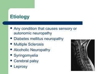
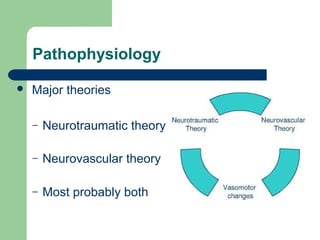
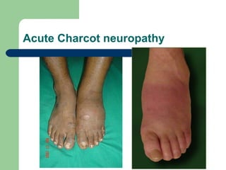
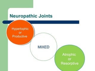
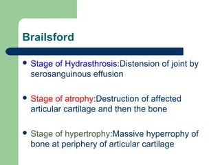
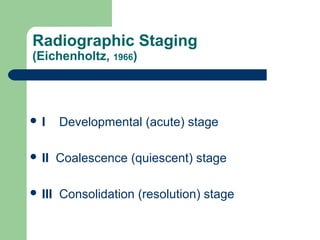
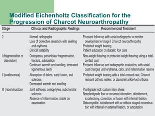
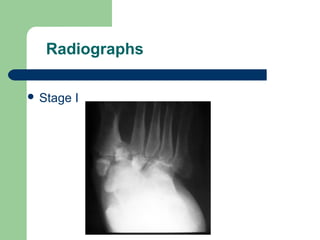
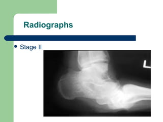
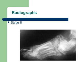
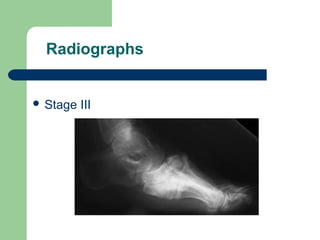
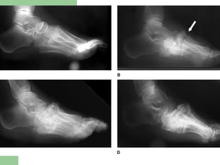
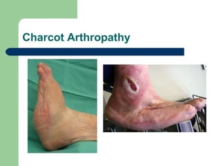
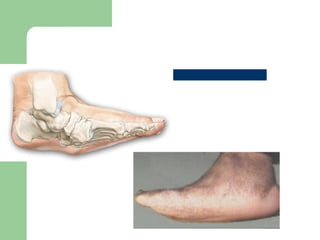
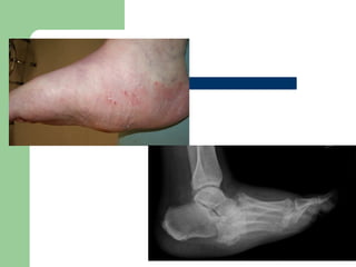
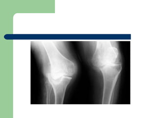
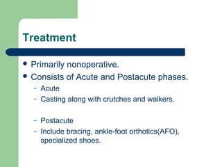
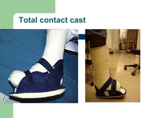
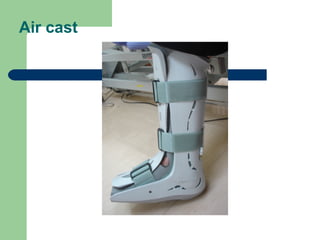
Neuropathic Arthropathy
Dr.ARAIB LAIGA
Definition
 Neuropathic arthropathy , neuropathic
osteoarthropathy, Charcot joint refers to
progressive condition of the musculoskeletal
system that is characterized by joint
dislocations, pathologic fractures, and
debilitating deformities.
 Charcot Arthropathy :James K DeOrio, MD Associate Professor of Orthopedic
Surgery, Duke University School of Medicine .
 Walter Panis, MD Clinical Instructor, Department of Physical Medicine and
Rehabilitation, Spaulding Rehabilitation Hospital, Harvard Medical School
Etiology
 Any condition that causes sensory or
autonomic neuropathy
 Diabetes mellitus neuropathy
 Multiple Sclerosis
 Alcoholic Neuropathy
 Syringomyelia
 Cerebral palsy
 Leprosy
 Tabes Dorsalis
 Spinal cord injury
 Myelomeningocele
 Intra-articular steroid injections
 Congenital insensitivity to pain
 CMTD
 Familial interstial Polyneuropathy
 Amyloidosis
 Pernicious Anemia
Pathophysiology
 Major theories
– Neurotraumatic theory
– Neurovascular theory
– Most probably both
Neurotraumatic Theory
 Loss of peripheral sensation and proprioception
leads to repetitive micro trauma to the joint in
question
 This damage goes unnoticed by the neuropathic
patient, and the resultant inflammatory resorption
of traumatized bone renders that region weak and
susceptible to further trauma.
 Poor fine motor control generates unnatural
pressure on certain joints, leading to additional
microtrauma.
Neurovascular theory
 A.C.Brower theory
 Postulates that neurologic changes produced by
an underlying medical disorder create a
hypervascular region in the subchondral bone
that is characterized by increased osteoclastic
resorption and osteoporosis.
 More recent theories implicate the role of
inflammatory cytokines such as TNF-α and
IL-1 in the pathogenesis of Charcot
neuroarthropathy.
 On the molecular level, these factors lead to
increased expression of nuclear transcription
factor-κB, which in turn stimulates osteoclast
formation.
Clinical History
 A careful history may reveal an unrecognized
traumatic event.
 Charcot neuroarthropathy most frequently
presents in the fifth decade, after an average
duration of diabetes of 20 to 24 years; in
those with type 2 diabetes.
Presentation
 DEPENDS OF DURATION OF DISEASE
 Mild swelling w/o deformity-Moderate
deformity with extreme swelling.
 Signs of inflammation.
 Profound unilateral swelling. WBC and
ESR may
be normal
 Increase in localized
temp
 Erythema,
 Joint effusion.
 75% pt. have pain.
 The deep tendon
reflexes at the knee are
absent in a majority of
patients.
Acute Charcot neuropathy
On Examination
Marked Irregularities identified as bony
projections.
Bone formation in soft tissues.
Bag of Bones:
Joint can be passively and painlessly moved in
all Directions
Diagnosis
 Xrays.
 Indium-111 WBC scan.
 Gallium scan.
 USG
 MRI
 Radionuclide scans
Lab Studies
 Inflammatory markers
ESR and WBC
– elevated in both infection and Charcot arthropathy
 Serum albumin >3.0g/dL
IMAGING
 Early Changes similar to OA
 Nontraumatic dislocations may be an early
sign.
 LaterRadiographic evidence of joint
distention caused by fluid, hypertrophic
synovitis, osteophytes, and subluxation.
Atrophic Stage:
 Rapid joint destruction
 Loose bodies
 Subchondral bone erosions
 Subluxation
 Pathological#
Hypertrophic Stage
 Reduced jt space.
 Subchondral bone sclerosis
 Pathological # healing with callus
 Multiple osteophyte formation with exoxtosis
formation.
 Dislocations of joints
Radiographic features
6D’s Yochum and Rowe
 Dense bones (subchondral sclerosis)
 Degeneration
 Destruction of articular cartilage
 Deformity (pencil-point deformity of
metatarsal heads)
 Debris (loose bodies)
 Dislocation
Commonly Affected Joints
 Foot Involvment
 Knee involvement
 Hip involvement
 Shoulder
 Elbow
Anatomic Classification
(Sanders and Frykberg, 1991)
 I - forefoot, 10-30%
 II - Lisfranc’s joint, most
common
 III - midtarsal joint, often
including naviculocuneiform
joint
 IV - ankle and subtalar joints,
8-10%
 V - (“posterior pillar”) fractures
Neuropathic Joints
Hypertophic
or
Productive
Hypertophic
or
Productive
MIXED
Atrophic
or
Resorptive
Brailsford
 Stage of Hydrasthrosis:Distension of joint by
serosanguinous effusion
 Stage of atrophy:Destruction of affected
articular cartilage and then the bone
 Stage of hypertrophy:Massive hyperrophy of
bone at periphery of articular cartilage
Radiographic Staging
(Eichenholtz, 1966)
 I Developmental (acute) stage
 II Coalescence (quiescent) stage
 III Consolidation (resolution) stage
Modified Eichenholtz Classification for the
Progression of Charcot Neuroarthropathy
Radiographs
 Stage I
Radiographs
 Stage II
Radiographs
 Stage II
Radiographs
 Stage III
Charcot Arthropathy
HIP
 Charcot neuroarthropathy in the hip is rare.
 Painless and Functional: no treatment
 Try conservative management
 50% of fractures of the femoral neck in
diabetics developed Charcot's joints.
KNEE
 Most Commonly secondary to Syphilis.
 Results in Gross Instability
 If only one knee is involved and destruction
is severe, fusion is indicated.
 Total knee arthroplasty ???
Treatment
 Primarily nonoperative.
 Consists of Acute and Postacute phases.
– Acute
– Casting along with crutches and walkers.
– Postacute
– Include bracing, ankle-foot orthotics(AFO),
specialized shoes.
Treatment
 Casting- changed every 1-2weeks, if
ulcerations are present changed every week
for wound care, duration from 3-6 months.
 Shoes, bracing, and orthotics- duration
from 6-24 months.
 Typical total healing time 1-2 years.
Early stage
 Total Contact cast.
Total contact cast
Air cast
CROW boots
Treatment
2. Pharmacological Treatment.
 Pilot study first using pamidronate,1994.
Other Bisphosphonates were used to
decrease disease activity and bone
turnover markers.
 Calcitonin were also used.
 Given for 12 weeks or till temp gradient is
less than 2 on 2 consecutive visits.
Surgical options
 Arthrodesis
 Exostosectomy of bony prominences
 Osteotomies
 Reconstructive Surgeries
 Autologous bone Grafting
 Amputations
Surgical treatment
Ankle:
 Arthrodesis of ankle to place the foot
Plantigrade.
 IM nail/Charnley/Ilizarov External Fixators
 Average time for Fusion:20 months(IM nail).
 Talus -- fragmented and avascular--talectomy and
tibiocalcaneal arthrodesis.
Internal or External Fixation??
Hindfoot neuroarthropathy
 Mainstay of Treatment is NONSURGICAL.
 Arthrodesis indicated for…
 Hindfoot valgus with subluxation of the
subtalar joint or midtarsals to prevent
ulceration and infection.
Complication
 Ulcers
 Osteomyelitis
 Gross Deformity of the foot
 Gangrene.
THANK
YOU

Ghega

  • 2.
  • 3.
    Definition  Neuropathic arthropathy, neuropathic osteoarthropathy, Charcot joint refers to progressive condition of the musculoskeletal system that is characterized by joint dislocations, pathologic fractures, and debilitating deformities.  Charcot Arthropathy :James K DeOrio, MD Associate Professor of Orthopedic Surgery, Duke University School of Medicine .  Walter Panis, MD Clinical Instructor, Department of Physical Medicine and Rehabilitation, Spaulding Rehabilitation Hospital, Harvard Medical School
  • 4.
    Etiology  Any conditionthat causes sensory or autonomic neuropathy  Diabetes mellitus neuropathy  Multiple Sclerosis  Alcoholic Neuropathy  Syringomyelia  Cerebral palsy  Leprosy
  • 5.
     Tabes Dorsalis Spinal cord injury  Myelomeningocele  Intra-articular steroid injections  Congenital insensitivity to pain  CMTD  Familial interstial Polyneuropathy  Amyloidosis  Pernicious Anemia
  • 6.
    Pathophysiology  Major theories –Neurotraumatic theory – Neurovascular theory – Most probably both
  • 7.
    Neurotraumatic Theory  Lossof peripheral sensation and proprioception leads to repetitive micro trauma to the joint in question  This damage goes unnoticed by the neuropathic patient, and the resultant inflammatory resorption of traumatized bone renders that region weak and susceptible to further trauma.  Poor fine motor control generates unnatural pressure on certain joints, leading to additional microtrauma.
  • 8.
    Neurovascular theory  A.C.Browertheory  Postulates that neurologic changes produced by an underlying medical disorder create a hypervascular region in the subchondral bone that is characterized by increased osteoclastic resorption and osteoporosis.
  • 9.
     More recenttheories implicate the role of inflammatory cytokines such as TNF-α and IL-1 in the pathogenesis of Charcot neuroarthropathy.  On the molecular level, these factors lead to increased expression of nuclear transcription factor-κB, which in turn stimulates osteoclast formation.
  • 10.
    Clinical History  Acareful history may reveal an unrecognized traumatic event.  Charcot neuroarthropathy most frequently presents in the fifth decade, after an average duration of diabetes of 20 to 24 years; in those with type 2 diabetes.
  • 11.
    Presentation  DEPENDS OFDURATION OF DISEASE  Mild swelling w/o deformity-Moderate deformity with extreme swelling.  Signs of inflammation.  Profound unilateral swelling. WBC and ESR may be normal
  • 12.
     Increase inlocalized temp  Erythema,  Joint effusion.  75% pt. have pain.  The deep tendon reflexes at the knee are absent in a majority of patients.
  • 13.
  • 14.
    On Examination Marked Irregularitiesidentified as bony projections. Bone formation in soft tissues. Bag of Bones: Joint can be passively and painlessly moved in all Directions
  • 15.
    Diagnosis  Xrays.  Indium-111WBC scan.  Gallium scan.  USG  MRI  Radionuclide scans
  • 16.
    Lab Studies  Inflammatorymarkers ESR and WBC – elevated in both infection and Charcot arthropathy  Serum albumin >3.0g/dL
  • 17.
    IMAGING  Early Changessimilar to OA  Nontraumatic dislocations may be an early sign.  LaterRadiographic evidence of joint distention caused by fluid, hypertrophic synovitis, osteophytes, and subluxation.
  • 18.
    Atrophic Stage:  Rapidjoint destruction  Loose bodies  Subchondral bone erosions  Subluxation  Pathological#
  • 19.
    Hypertrophic Stage  Reducedjt space.  Subchondral bone sclerosis  Pathological # healing with callus  Multiple osteophyte formation with exoxtosis formation.  Dislocations of joints
  • 20.
    Radiographic features 6D’s Yochumand Rowe  Dense bones (subchondral sclerosis)  Degeneration  Destruction of articular cartilage  Deformity (pencil-point deformity of metatarsal heads)  Debris (loose bodies)  Dislocation
  • 22.
    Commonly Affected Joints Foot Involvment  Knee involvement  Hip involvement  Shoulder  Elbow
  • 23.
    Anatomic Classification (Sanders andFrykberg, 1991)  I - forefoot, 10-30%  II - Lisfranc’s joint, most common  III - midtarsal joint, often including naviculocuneiform joint  IV - ankle and subtalar joints, 8-10%  V - (“posterior pillar”) fractures
  • 24.
  • 25.
    Brailsford  Stage ofHydrasthrosis:Distension of joint by serosanguinous effusion  Stage of atrophy:Destruction of affected articular cartilage and then the bone  Stage of hypertrophy:Massive hyperrophy of bone at periphery of articular cartilage
  • 26.
    Radiographic Staging (Eichenholtz, 1966) I Developmental (acute) stage  II Coalescence (quiescent) stage  III Consolidation (resolution) stage
  • 27.
    Modified Eichenholtz Classificationfor the Progression of Charcot Neuroarthropathy
  • 28.
  • 29.
  • 30.
  • 31.
  • 33.
  • 36.
    HIP  Charcot neuroarthropathyin the hip is rare.  Painless and Functional: no treatment  Try conservative management  50% of fractures of the femoral neck in diabetics developed Charcot's joints.
  • 39.
    KNEE  Most Commonlysecondary to Syphilis.  Results in Gross Instability  If only one knee is involved and destruction is severe, fusion is indicated.  Total knee arthroplasty ???
  • 41.
    Treatment  Primarily nonoperative. Consists of Acute and Postacute phases. – Acute – Casting along with crutches and walkers. – Postacute – Include bracing, ankle-foot orthotics(AFO), specialized shoes.
  • 42.
    Treatment  Casting- changedevery 1-2weeks, if ulcerations are present changed every week for wound care, duration from 3-6 months.  Shoes, bracing, and orthotics- duration from 6-24 months.  Typical total healing time 1-2 years.
  • 44.
    Early stage  TotalContact cast.
  • 45.
  • 46.
  • 47.
  • 48.
    Treatment 2. Pharmacological Treatment. Pilot study first using pamidronate,1994. Other Bisphosphonates were used to decrease disease activity and bone turnover markers.  Calcitonin were also used.  Given for 12 weeks or till temp gradient is less than 2 on 2 consecutive visits.
  • 49.
    Surgical options  Arthrodesis Exostosectomy of bony prominences  Osteotomies  Reconstructive Surgeries  Autologous bone Grafting  Amputations
  • 50.
    Surgical treatment Ankle:  Arthrodesisof ankle to place the foot Plantigrade.  IM nail/Charnley/Ilizarov External Fixators  Average time for Fusion:20 months(IM nail).  Talus -- fragmented and avascular--talectomy and tibiocalcaneal arthrodesis.
  • 51.
  • 56.
    Hindfoot neuroarthropathy  Mainstayof Treatment is NONSURGICAL.  Arthrodesis indicated for…  Hindfoot valgus with subluxation of the subtalar joint or midtarsals to prevent ulceration and infection.
  • 57.
    Complication  Ulcers  Osteomyelitis Gross Deformity of the foot  Gangrene.
  • 58.

Editor's Notes

  • #36 ROCKERS BOTTOM DEFORMITY:-a convex deformity of the foot’s plantar aspect caused by the collapse of metatarsal bones