Download to read offline

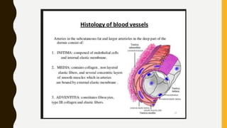


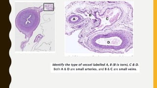
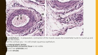

The document summarizes the key components of arteries, capillaries, and venules. It describes how the intima of small arteries appears as a "dotted line" of endothelial cell nuclei due to post-mortem contraction wrinkling the endothelium. It notes that the media is the thickest and most conspicuous layer, containing clearly visible smooth muscle cell nuclei. The adventitia does not form a distinct layer and instead merges with surrounding connective tissue. Letters in the image denote specific structures, with E labeling the endothelium, F the internal elastic lamina, and G, H, I, and J labeling arterioles and venules.