NEONATAL DISEASES RESPIRATORY  DISDRESS SYNDROME PATHOLOGIC JAUNDISE SCIEREDERMA NEONATOUM
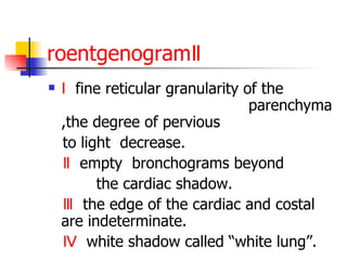
Neonatal respiratory distress syndrome  ( NRDS ) Infant respiratory distress syndrome  ( IRDS ) Respiratory distress syndrome of newborn Hyaline membrane disease
Introduction A  syndrome  caused in  premature   infants  by  developmental insufficiency of  surfactant  production  and structural immaturity in the  lungs . It can also  result from a genetic problem with the production of surfactant associated proteins. RDS affects about  1% of newborn infants and is the leading cause of  death in  preterm  infants.The incidence decreases with advancing  gestational  age .The syndrome is  more frequent in infants of diabetic mothers and in premature twins.
DEFINITION NRDS is a condition caused by insufficient pulmonary development or alveolar stability.It is a major cause of death in the neonatal period, with characteristic  radiographic ,clinical, and physiologic signs  that show difficult initiating normal respiration,progressive dyspnea, cyanosis,and respiratory failure developed within hours of life.
DEFINITION MAJOR  CAUSE  OF  DEATH OCCURS  PRIMARILY  IN  PREMATURE  INFANTS CHARACTERISTIC
About incidence  An estimated  30%  of all neonatal deaths result from RDS or/and its complications. RDS occurs primarily in premature infants; incidence is inversely proportional to the gestational age and birthweight.It occurs in  60-80%  of infants less than  28wk  of gestational age, in  15-30%  of those between  32-36wk  , in about  5%  beyond  37wk ,and rarely at term.
Table 1 A  <28wk(60-80%)   B  32-36wk(15-30%) C  >37wk(5%)
Associated factors
Associated factors An increased frequency is associated with infant of  diabetic mothers Prematurity Multifetal cesarean   section Asphyxia cold stress prior affected and etc.
ETIOLOGY AND PATHOPHYSIOLOGY
ETIOLOGY AND PATHOPHYSIOLOGY The absence of surfactant The failure to develop a functional residual capacity  The tendency of affected lungs to become  atelectatic   correlate   with high surface tensions
about Surfactant The lungs  of  infants  with  respiratory distress syndrome are developmentally deficient in a material called  surfactant , which helps  prevent  collapse  of  the terminal  air- spaces  throughout  the normal cycle of inhalation and exhalati- on.
about Surfactant Surfactant is a complex system of  lipids ,  proteins  and  glycoproteins  which are produced in specialized lung cells called Type II cells or Type II pneumocytes. The surfactant is packaged by the cell in structures called  lamellar bodies , and extruded into the air-spaces. The lamellar bodies then unfold into a complex lining of the air-space. This layer reduces the surface tension .
about Surfactant With progressive gestational age ,increasing amounts of Surfactant are synthesized and stored in  type Ⅱ alveolar cells .  These active agents are released into the alveoli,reducing the surface tension and maintain alveolar stability by preventing the collapse of small air spaces at end-expiration.
about Surfactant
about Surfactant By reducing surface tension, surfactant prevents the air-spaces from completely collapsing on exhalation. In addition, the decreased surface tension allows re-opening of the air-space with a lower amount of force. Therefore, without adequate amounts of surfactant, the air-spaces collapse and are very difficult to expand.
Table 2  (composition)
about Surfactant Surfactant is present in high concentrations  in fetal lung homogenates by  20 wk  of gestation but does not reach the surface of the lung until later. It appears in the amniotic fluid between  28  and 32  wk. Mature levels of pulmonary surfactant are usually present after  35  wk. So the amounts produced or released may be insufficient to meet postnatal demands because of immaturity.
about Surfactant Surfactant synthesis depends in part on normal PH, temperature, and pulmonary perfusion. Asphyxia, hypoxemia, and pulmonary ischemia, particularly in association with hypovolemia, hypotention, and cold stress,may supress surfactant synthesis. The epithelial lining of the lung may also be injured by high oxygen concentrations , resulting in further reduction in surfactant .
lungs less compliant Alveolar atelectasis Hyaline membrane formation Interstitial edema  make the lungs less compliant, requiring greater pressure to expend the small alveoli and airways.
about hyaline membranes Microscopically, a surfactant deficient lung is characterized by collapsed air-spaces alternating with hyper-expanded areas, vascular congestion and, in time,  hyaline  membranes. Hyaline membranes are composed of  fibrin , cellular debris,  red blood cells , rare  neutrophils  and  macrophages . They appear as an eosinophilic, amorphous material, lining or filling the air spaces and blocking gas exchange .
about hyaline membranes As a result, blood passing through the lungs is unable to pick up oxygen and unload carbon dioxide. Blood oxygen levels fall and carbon dioxide rises, resulting in rising blood acid levels and  hypoxia ..
Structural immaturity Structural immaturity, as manifest by decreased number of gas-exchange units and thicker walls, also contributes to the disease process.
highly compliant chest wall In these infants, the lower chest wall is pulled in as the diaphragm descends and the intrathoracic pressure becomes negative, thus limiting the amount of intrathoracic pressure that can be produced; the result is a tendency for atelectasis to develop. The highly compliant chest wall of the premature infant ,  offers less resistance than that of the mature infant against the natural tendency of the lungs  to collapse.
Table  3
Atelectasis Atelectasis resulting in perfused but not ventilated alveoli, which causes hypoxia, decreased lung compliance, small tidal volumes, increased  physiologic dead space ,increased work of breathing, and insufficient alveolar ventilation eventually resulting in hypercarbia.
Table 4
Pathology  The characteristic  pathology  seen in babies who die from RDS was the source of the name &quot;hyaline membrane disease&quot;. These waxy-appearing layers line the collapsed tiny air sacs (&quot; alveoli &quot;) of the lung. In addition, the lungs show bleeding, over-distention of airways and damage to the lining cells.
Clinical manifestations This condition usually occurs in premature  infants ,with normal crying at birth ,in  6-12  hours, progressive dyspnea can be found .
Clinical manifestations This condition  is  self-limited  ,if the baby can live for three days ,improvement will set in .But many baby with complications such as phneumonia ,the condition will depravation,until infection is restrained.
Clinical manifestations Signs of HMD usually appear within minutes of birth, although they may not be recognized for several hours until rapid, shallow respirations have increased to  ≧60/min .  Some patients require resuscitation at birth because of intrapartum asphyxia or initial severe respiratory distress .
Clinical manifestations Characteristically,  tachypnea , prominent(often audible) grunting,  intercostal and subcostal  retractions,   nasal flaring , and  duskiness  are seen. There is increasing  cyanosis,  which is often relatively unresponsive to oxygen administration.  Breath sounds  may be normal or diminished with a harsh tubular quality, and on deep inspiration, fine  rales  may be heard, especially over the posteriorly lung bases.
.
Clinical manifestations The  natural course  is characterized by progressive worsening of cyanosis and dyspnea. If inadequately treated,  blood pressure  and body  temperature  may fall; fatigue,   cyanosis , and  pallor  increase, and  grunting  decreases or disappears as the condition worsens.  Apnea  and irregular respirations occur as infants tire. These  are ominous signs requiring immediate intervention. There may also be a  mixed respiratory-metabolic acidosis, edema, ileus, and oliguria.
Clinical manifestations As the disease progresses, the baby may develop ventilatory failure (rising carbon dioxide concentrations in the blood), and prolonged cessations of breathing (&quot; apnea &quot;).
The clinical course Whether treated or not, the clinical course for the acute disease lasts about 2 to 3 days.  the first , the patient worsens and requires more support.  the second  the baby may be remarkably stable on adequate support and resolution is noted  the third  day, heralded by a prompt diuresis
Clinical manifestations Signs of asphyxia secondary to apnea or partial respiratory failure occur when there is rapid progression of the disease.In many cases the symptoms and signs may reach a peak within  3 days, after which gradual improvement sets in .
Clinical manifestations Improvement is often heralded by a spontaneous  diuresis  and the ablilty to  oxygenate the infant with lower inspired oxygen levels  . Death is rare on the 1 st  day of illness,but usually occurs between days 2 and 7.
 
Diagnosis clinical course   roentgenogram   blood gas   and acid-base
roentgenogramⅠ There may be considerable variation among films, depending on the phase of respiration and the use of CPAP, often resulting in poor correlation between the roentgenograms and clinical course.
roentgenogramⅡ Ⅰ   fine reticular granularity of the  parenchyma ,the degree of pervious  to light  decrease. Ⅱ  empty  bronchograms beyond  the cardiac shadow. Ⅲ  the edge of the cardiac and costal are indeterminate. Ⅳ  white shadow called “white lung”.
blood gas   and acid-base The laboratory findings are characterized initially by hypoxemia and later by progressive hypoxemia, hypercarbia, and variable metabolic acidosis.we can see PH , BE ,CO2CP decline,and sometime Na,K,Cl increase.
DiagnosisⅡ- Foam test Extract amnoitic fluid or bronchial secretion ,mixed with equivalent 95% alcohol,shake for 15 second and stay for  15 minutes.Then observe the surface of the liquid. If we can see foam we can exclude  this condition.
Prevention Most cases of hyaline membrane disease can be ameliorated or prevented if mothers who are about to deliver prematurely can be given one of a group of hormones  glucocorticoids . This speeds the production of surfactant.
Prevention For very premature deliveries, a glucocorticoid is given without testing the fetal lung maturity. In pregnancies of greater than 30 weeks, the fetal lung maturity may be tested by sampling the amount of surfactant in the amniotic fluid, obtained by inserting a needle through the mother's abdomen and uterus .
 
Several tests are available lecithin-sphingomyelin ratio For assessing fetal lung maturity The presence of Phosphatidol glycerol ( PG  ) usually indicates fetal lung maturity The S/A ratio the result is given as mg of surfactant per gm of protein. An S/A ratio <35 indicates immature lungs, between 35-55 is indeterminate, and >55 indicates mature surfactant production (correlates with an L/S ratio of 2.2 or greater).
 
lecithin-sphingomyelin ratio Lungs require  surfactant , a soapy sort of substance, to lower the surface pressure of the  alveoli  in the lungs. This is especially important for  trying to expand their lungs for that first critical breath after birth. Surfactant is a mixture of lipids, proteins, and  gly n coproteins .  Lecithin  and  sphingomyeli  being two of   them.
lecithin-sphingomyelin ratio Lecithin makes the surfactant mixture more effective. An L/S ratio of 2 indicates a relatively low risk of  infant respiratory distress syndrome , and less than 1.5 is associated with a high risk of infant respiratory distress syndrome.  If  preterm delivery  is necessary and the L/S ratio is low, the mother may need to receive steroids to hasten the fetus's surfactant production .
Prevention prevention of prematurity glucocorticoid therapy Surfactant therapy
Prevention Most important is the  prevention of prematurity , including avoidance of unnecessary poorly-timed cesarean section, appropriate management of the high-risk pregnancy and labor. In timing cesarean sections or inducing labor, estimation of the fetal head circumference by ultrasound and determination of the lecithin concentration in the amniotic fluid should be considered.
prevention of prematurity Preventing prematurity is the most important way to prevent neonatal RDS. Ideally, this effort begins with the first prenatal visit, which should be scheduled as soon as a mother discovers that she is pregnant.  Good prenatal care results in larger, healthier babies and fewer premature births.
management of the high-risk pregnancy and labor If a mother does go into labor early, a lab test will be done to determine the maturity of the infant's lungs. When possible, labor is usually halted until the test shows that the baby's lungs have matured. This decreases the chances of developing RDS.
treatment of pulmonary immaturity In some cases, medicines called corticosteroids may be given to help . It is not clear if additional doses of corticosteroids are safe or effective.
Prevention The administration of  dexamethasone or betamethasone  to women  48-72  hr before delivery of fetuses at   24 and 34 weeks  of gestation significantly reduces the incidence and the mortality and morbidity from HMD.  It is appropriate to administer these  corticosteroids  intramuscularly to pregnant women whose lecithin in amniotic fluid indicates fetal lung immaturity and who are likely to deliver in 1 wk or whose labor may be delayed 48 hr or more.
Prevention Prenatal  glucocorticoid therapy Decreases the severity of RDS Reduces the incidence of other complications of prematurity  (intraventricular,hemorrhage,patent ductus arteriosus, and pneumothorax).
Prevention Administration of one dose of  surfactant  into the trachea of premature infants immediately after birth during the first  24 hr  of life reduces the mortality from HMD .
Treatment The basic defect requiring treatment is inadequate pulmonary exchange of oxygen and carbon dioxide; metabolic acidosis and circulatory insufficiency are secondary manifestations.
Treatment Basic  inadequate pulmonary exchange of oxygen and carbon dioxide second  metabolic acidosis and circulatory insufficiency
Treatment Despite greatly improved RDS treatment, in recent years, many controversies still exist.
 
Treatment Early  supportive care  of the LBW infant,especially in the treatment of acidosis ,hypoxia, hypotension, and hypothermia, appears to lessen the severity of HMD.Therapy requires careful and frequent monitoring of heart and respiratory rates, arterial PO 2 , PCO 2 , PH, bicarbonate, electrolytes,blood glucose,hematocrit, blood pressure,and temperature.
Treatment The goal of treatment is to minimize abnormal physiologic variations and superimposed iatrogenic problems. The management of these infants is best carried out in a specially in stalled and equipped hospital unit,the  neonatal intensive care nursery .
Treatment (1)Supportive care (2)Oxygenation and artificial ventilation (3)Surfactant replacement  (4) Correction of metabolic acidosis (5) Symptomatic treatment (6) Antibacterial therapy for preventing infections.
Treatment (1)Supportive care The general principles for supportive care of any LBW infant shoud be adhered to ,including gentle handling and minimal disturbance consistent with management.To avoid chilling and to educe the metabolic rate, infants should be placed in an  Isolette  and core temperature maintained between  36.5 to 37℃ . Calories and fluids  should be provided intravenously.
Treatment (2)Oxygenation and artificial ventilation   Warm humidified oxygen should be provided at a concentration sufficient initially to keep atrerial levels between  55 and 70mm Hg  with stable vital signs to maintain normal tissue oxygenation while minimizing the risk of oxygen toxicity. If the atrerial oxygen tension cannot be maintained above  50mm  Hg at inspired oxygen concentrations of  70% ,applying  CPAP .
oxygen toxicity Infants will be given warm, moist oxygen. This is critically important, but needs to be given carefully to reduce the side effects associated with too much oxygen . Injury retina,lead to blindness. Lead to bronchopulmonary dysplasia .
CPAP A treatment called continuous positive airway pressure (CPAP) that delivers slightly pressurized air through the nose can help keep the airways open and may prevent the need for a breathing machine for many babies. Even with CPAP, oxygen and pressure will be reduced as soon as possible to prevent side effects associated with excessive oxygen or pressure.
assisted mechanical ventilation Infants  with  severe  HMD  or those who develop complications resulting in persistent apnea  require assisted mechanical  ventilation.
  assisted mechanical ventilation  A breathing machine can be lifesaving, especially for babies with the following: High levels of carbon dioxide in the arteries  Low blood oxygen in the arteries  Low blood pH (acidity)
assisted mechanical ventilation There are a number of different types of breathing machines available. However, the devices can damage fragile lung tissues, and breathing machines should be avoided or limited when possible.
Treatment
assisted mechanical ventilation (1)arterial blood pH of less than 7.20; (2)arterial blood Pco2 of 60mm Hg or more; (3)arterial blood Po2 of 50mm Hg or less at oxygen concentrations of 70-100%; (4)persistent apnea. These are reasonable indications
assisted mechanical ventilation . Assisted ventilation by pressure or flow limited conventional respirators through an endotracheal tube including  positive end-expiratory pressure(PEEP).
Treatment (3)Surfactant replacement   Partially synthesized or subtraction from calf lung or amniotic fluids . Surfactant therapy can improve the oxygenation dramatically.
Surfactant replacement Delivering artificial surfactant directly to the infant's lungs can be enormously important, but how much should be given and who should receive it and when is still under investigation
Treatment (4)Correction of metabolic acidosis The  dosage  of sodium bicarbonate should be calculated as follows: 5% NaHCO3(ml)= CO2CP X BW(kg)X 1.2
Treatment (5)Symptomatic treatment 20% manitol  in a dosage of 5-10ml/Kg/dose IV to relieve cerebra edema.  Furosemide  in a dosage of 1-2 mg/Kg/dose to increase urine output. Sod. Luminal  in a dosage of 5-10mg/Kg/dose to sedate the patient. Digoxin ,0.025mg/Kg/dose to correct heart failure. Corticotoid  to promote the production of the surfactant. Ⅻ
Treatment (6)Antibacterial therapy for preventing infections.
Treatment A variety of other treatments may be used, including: Extracorporeal membrane oxygenation  (ECMO) to directly put oxygen in the blood if a breathing machine can't be used  Inhaled nitric oxide  to improve oxygen levels
Reduce the infant's oxygen needs It is important that all babies with RDS receive excellent supportive care, including  Few disturbances  Gentle handling  Maintaining ideal body temperature
Nonphysilogic jaundice Pathological jaundice
Introduction A yellowing of the  skin  and other tissues of a newborn  infant .  In newborns jaundice is detected by blanching the skin with digital pressure so that it reveals underlying skin and subcutaneous tissue .
Incidence Jaundice is observed during the 1 st  wk of life in approximately  60% of term  infants and  80% of preterm  infants.The color usually results from the accumulation in the skin of unconjugated, nonpolar, lipid-soluble bilirubin pigment (indirect-reacting)
bruise A  bruise , also called a  contusion , is an  injury  to  biological tissue  in which the  capillaries  are damaged, allowing  blood  to seep into the surrounding tissue .
bruise Minor bruises may be easily recognized  by their characteristic  blue  or  purple  appearance (idiomatically described as &quot;black and blue&quot;) in the days following the injury.
bruise Sometimes bruises can be serious, leading to other more life threatening forms of  hematoma , or can be associated with serious injuries, including  fractures  and  internal bleeding ..
Bilirubin The yellow breakdown product of normal  heme   catabolism . Heme is formed from hemoglobin, a principal component of  red blood cells . Bilirubin is excreted in  bile , and its levels are elevated in certain diseases. It is responsible for the yellow color of  bruises  and the yellow color in  jaundice .
Bilirubin  Metabolism Erythrocytes  (red blood cells) generated in the  bone marrow  are disposed of in the  spleen  when they get old or damaged. This releases  hemoglobin , which is broken down to  heme , as the globin parts are turned into  amino acids .
Bilirubin  Metabolism The heme is then turned into unconjugated bilirubin in the  macrophages  of the spleen.  This unconjugated bilirubin is not soluble in water. It is then bound to  albumin  and sent to the  liver .
Bilirubin  Metabolism In the liver it is conjugated  with  glucuronic acid , making it soluble in water ,bocome  conjugated bilirubin .
Bilirubin  Metabolism Much of it goes into the bile and thus  out into the intestine. Some of the conjugated bilirubin is metabolised by  colonic bacteria to  urobilinogen  .
Bilirubin  Metabolism urobilinogen , which is further metabolized to  stercobilinogen , and finally oxidised to  stercobilin . This stercobilin gives feces its brown color. Some of the urobilinogen is reabsorbed and excreted in the urine along with an oxidized form,  urobilin .
Enterohepatic circulation Refers to the circulation of  bile  from the  liver , where it is produced, to the  intestine ,  then back to the liver Endogenous bacteria play an important role in enterohepatic circulation.
  Metabolic features of neonatal bilirubin The newborn infant ’ s metabolism of bilirubin is in transition from the  fetal stage,  during which the placenta is the principal route of elimination of the lipid-soluble bilirubin,to the  adult stage , during which the water-soluble conjugated form is excreted from the hepatic cell into the biliary system and then into the gastrointestinal tract .
    Metabolic features of neonatal bilirubin (1)  Increased  production of bilirubin ( A )Increased blood cell  number ( B )Shortened blood cell life ( C )Increased blood cell destructions
Metabolic features of neonatal bilirubin (2)  The ability for Bilirubin and albumin's to  link and delivery  decrease. ( A ) acidosis ( B ) Lower  serum albumin
Metabolic features of neonatal bilirubin (3)  Poor   ability for   hepatocyte to  uptake non-conjugated bilirubin  because of inadequate ligadin (Y and  Z protein )function (5 ~ 20% ) uptake non-conjugated bilirubin and transport  to smooth endoplasmic reticulum
Metabolic features of neonatal bilirubin (4) Enzyme deficiency  ( A )Glucuronosyltransferase  ( B )Uridine diphosphate  glucose dehydrogenase (UDPG dehydrogenase)
Metabolic features of neonatal bilirubin (5) Increased entero  hepatic circulation of bilirubin ( A ) Decreased Endogenous  bacteria ( B ) Increased  β -glucuronidase
Some explaining The haemoglobin concentration falls rapidly in the first days after birth from heamolysis (1g of heamoglobin yields  35mg of bilirubin) The red cell life span of newborn infant(70 days),is markedly shorter than that of adults(120 days). Hepatic bilirubin metabolism is less efficient in the first days of life.
  Associated factors Unconjugated hyperbilirubinemia may be increased by lots of  factors (1) increases the load of bilirubin to be metabolized by the liver (hemolytic anemias, shortened red cell life due to immaturity or to transfused cells, increased enterohepatic circulation, infection
Associated factors (2) may damage or reduce the activity of the transferase enzyme(hypoxia, infection, possibly hypothermia and thyroid deficiency)
Associated factors (3) may compete for or block the transferase enzyme (drugs and other substances requiring glucuronic acid conjugation for excretion)
Associated factors (4)  leads to an absence of or decreased amounts of the enzyme or to reduction of bilirubin uptake by the liver cell (genetic defect,prematurity)
Associated factors The risk of toxic effects from elevated levels of unconjugated bilirubin in the serum is increased by factors that  reduce the retention  of bilirubin in the circulation , or by factors that  increase the permeability  of the blood-brain barrier or nerve cell membanes to bilirubin or the susceptibility of brain cells to its toxicity such as asphyxia, prematurity,  and infection.
Clinical Manifestation Jaundice may be present  at birth or may appear at any time during the neonatal period , depending on the condition responsible for it. Jaundice usually begins on the face and, as the serum level increases, progresses to the abdomen and then the feet.
Clinical Manifestation A serum bilirubin level is determined for those patients with progressing jaundice, symptoms, or a risk for hemolysis or sepsis.
Clinical Manifestation In neonates the dermal  icterus  is first noted in the face and as the bilirubin level rises proceeds caudal to the trunk and then to the extremities . Notoriously inaccurate rules of thumb have been applied to the physical exam of the jaundiced infant
Rule of thumb Infants whose jaundice is restricted to the face and part of the trunk above the  umbilicus , have the bilirubin less than 12 mg/dL (less dangerous level).  Infants whose palms and soles are yellow, have serum bilirubin level over 15 mg/dL (more serious level).
examine However,  even trained examiners (physicians and nurses) make poor estimations based on physical appearance . In infants jaundice can be measured using  invasive  or non-invasive methods. In non invasive method Transcutaneous bilirubinometer are used
Transcutaneous bilirubinometer Hand held, portable and rechargable but expensive .
Transcutaneous bilirubinometer When pressure is applied to the photoprobe, a xenon tube generates a strobe light; And this light passes through the subcutaneous tissue. The reflected light returns through the second fiber optic bundle to the spectrophotometric module. The intensity of the yellow color in this light, after correcting for the hemoglobin, is measured and instantly displayed in arbitrary units .
Physiological jaundice Most  infants  develop visible jaundice due to elevation of unconjugated  bilirubin  concentration during their first week.  This common condition is called physiological jaundice. This pattern of  hyperbilirubinemia  has been classified into two functionally distinct periods.
Physiological jaundice Phase one  Term infants : jaundice lasts for about 5 days with a rapid rise of serum bilirubin up to 12 mg/dL.  Preterm infants:  jaundice lasts for about a week, with a rapid rise of serum bilirubin up to 15 mg/dL.  Phase two - bilirubin levels decline about 2 mg/dL for 2 weeks, eventually mimicking adult values .  Preterm infants - phase two can last more than 1 month.  In babies who receive exclusive breast feedings, phase two can last more than 1 month
Physiological jaundice The  diagnosis  of physiologic jaundice in term or preterm infants can be established only by excluding known causes of jaundice on the basis of the  history  and  clinical  and  laboratory findings .
definition Jaundice with peak serum  bilirubin of more than  12 mg/dl for full-term infant or 15 mg/dl for premature   is named as nonphysiologic jaundice
Pathological Jaundice of Neonates  Increased production   Fetomaternal blood group incompatibility:  Rh ,  ABO   Hereditary spherocytosis.  Non-spherocytic hemolytic anemia:  G-6-PD deficiency Sepsis.  Increased enterohepatic circulation: Pyloris stenosis, or large bowel obstruction.
Pathological Jaundice of Neonates Decreased clearance   Inborn errors of metabolism: Criggler-Najjar syndrome type I and II  Drugs and Hormones: Hypothryoidism, breast milk jaundice
Pathological Jaundice of Neonates In general,a search to determine the cause of jaundce should be made if (1) it appears in the first  24 hr  of life; (2) serum bilirubin is rising at a rate greater than  5 mg/dL ;  (3)  serum bilirubin is greater than  12 mg/dl  in full-term  or  15  mg/dL  in preterm infants; (4) jaundice persists after the  2   nd  wk of life;  (5) direct-reacting bilirubin is greater than  2 mg/dL ;  at any time. (6) appears again after disappeared.
Differentiation  Neonatal jaundice                                              Unconjugated   bilirubin         Conjugated  bilirubin                                                   Pathologic     Physiological jaundice of Neonates   Hepatic    Post-hepatic                                  Hemolytic    Non-hemolytic                                         Intrinsic causes   Extrinsic causes   
      Extrinsic causes       Intrinsic causes                                                                             Non-hemolytic     Hemolytic                                                           Post-hepatic     Hepatic Physiological jaundice of Neonates     Pathologic                                                                         Conjugated  bilirubin             Unconjugated   bilirubin                                                                               Neonatal jaundice
Color Jaundice resulting from deposition of indirect bilirubin in the skin tends to appear bright yellow or orange jaundice of the obstructive type(direct bilirubin), a greenish or muddy yellow This difference is usually apparent only in severe jaundice. The infant may be lethargic and may feed poorly.
JAUNDICE ASSOCIATED WITH BREAST-FEEDING An estamated 1 of 200 breast-fed term infants develops significant elevations in unconjugated bilirubin .The term applies to jaundice in a newborn baby who is exclusively breastfed and in whom other causes of jaundice have been ruled out. The jaundice appears at the end of the first week of life and hence overlaps physiological jaundice. It can last for up to two months. Several factors are thought to be responsible for this condition.
JAUNDICE ASSOCIATED WITH BREAST-FEEDING If breast-feeding is discontinued,the serum bilirubin level falls rapidly,usually reaching the normal levels within a few days. Cessation of breast-feeding for 1-2 days and substitutions of formula  for breast milk results in a rapid decline in serum bilirubin,after which nursing can be resumed without a return of the hyperbilirubinimia to  its previously high levels.
factors responsible First, in exclusively breastfed babies the establishment of normal gut flora is delayed. The bacteria in the adult gut convert conjugated  bilirubin  to  stercobilinogen  which is then oxidized to  stercobilin  and excreted in the stool. In the absence of sufficient bacteria the bilirubin is de-conjugated and reabsorbed. This process of re-absorption is called entero-hepatic circulation
factors responsible Second, the breast-milk of some women contains a metabolite of progesterone called . This substance inhibits the action of the enzyme uridine diphosphoglucuronic acid (UDPGA)  glucuronyl   transferase  responsible for conjugation and subsequent excretion of bilirubin. Reduced conjugation of bilirubin leads to increased level of bilirubin in the blood .
factors responsible Third, an enzyme in breast milk called  lipoprotein lipase  produces increased concentration of nonesterified free fatty acids that inhibit hepatic glucuronyl transferase which again leads to decreased conjugation and subsequent excretion of bilirubin.
JAUNDICE ASSOCIATED WITH BREAST-FEEDING Breast-milk jaundice does not usually cause any complication (like kernicterus) if the baby is otherwise healthy. The serum bilirubin level rarely goes above 20 mg /dL. It is usually not necessary to discontinue breast-feeding as the condition resolves spontaneously. Adequate hydration should be maintained by giving extra fluids if necessary
  OBSTRUCTIVE JAUNDICE  COLOUR SKIN: greenish or muddy yellow  URINE:  deep yellow  STOOL:  pale for an example: congenital biliary atresia
Biliary atresia A  rare condition  in  newborn infants  in which the common  bile duct  between the  liver  and the  small intestine  is blocked or absent. If unrecognised, the condition leads to  liver failure  but not to  kernicterus . This is because the liver is still able to conjugate bilirubin, and conjugated bilirubin is unable to cross the blood-brain barrier.
Biliary atresia The cause of the condition is unknown. The only effective treatments are certain surgeries, or liver transplantation .
Biliary atresia Initially, the symptoms are indistinguishable from  neonatal jaundice . Symptoms are usually evident between one and six weeks after birth. Besides jaundice, other symptoms include clay colored stools, dark urine, swollen abdominal region and large hardened liver .Prolonged jaundice that is resistant to phototherapy.
Kernicterus ⅰ Definition Kernicterus is a neurologic syndrome resulting from the deposition of uncojugated bilirubin in brain cells.
Kernicterusⅱ Uncojugated bilirubin may cross the  blood-brain  barrier and enter the brain by diffusion if the bilirubin-binding capacity of albumin and other plasma proteins is exceeded and plasma free bilirubin levels increase.  Alternatively, bilirubin may enter the brain following damage to the blood-brain barrier by asphyxia or hyperosmolatity.
Kernicterus  ⅲ The precise blood level above which uncojugated bilirubin will be toxic for an individual infant is unpredictable, but kernicterus is rare in healthy term infants and in the absence of hemolysis if the serum level is under 25mg/dL.  There is little evidence to suggest that the level of uncojugated bilirubin affects the IQ of healthy term infants without hemolytic disease.
Kernicterusⅳ The less mature the infant, the greater the susceptibility to kernicterus . In exceptional circumstances, kernicterus in VLBW infants with serum bilirubin concentrations as low as 8-12 mg/dL has been associated with an apparentlly cumulative effect of a number of risk  factors.
Kernicterus  ⅴ CLINICAL MANIFESTATIONS Signs and symptoms of kernicterus usually appear 2-5 days after birth in term infants and as late as the 7 th  day in premature ones, but hyperbilirubinemia may lead to the syndrome at any time during the neonatal period .
Kernicterus  ⅵ The early signs may be subtle and indistinguishable from those of sepsis, asphyxia, hypoglycemia, intracranial hemorrhage, and other acute systemic illnesses in the neonatal infant. Lethargy, poor feeding, and loss of the Moro reflex are common initial signs.
Kernicterusⅶ Subsequently, the infant may appear gravely ill and prostrated with diminished tendon reflexes and respiratory distress. Opisthotonos, with bulging fontanel, twitching of face or limbs, and a shrill high-pitched cry may follow. In advanced cases convulsions and spasm occur, with the infant stiffly extending his or her arms in inward rotation with fists clenched. Rigidity is rare at this late stage.
Kernicterusⅷ Many infants who progress to these severe neurologic signs die; the survivors are usually seriously damaged but may appear to recover and for 2-3 mo manifest few abnormalities. Later in the  1 st   yr of life opisthotonos, muscular rigidity, irregular movements, and convulsions tend to recur. In the  2 nd  yr opisthotonos and seizures abate but irregular, involuntary movements, muscular rigidty, or, in some infants, hypotonia increase steadily.  ⅷ
Kernicterus  ⅸ By 2 yr of age the complete neurologic syndrome is often apparent, consisting of bilateral choreoathetosis with involuntary muscle spasm, extrapyramidal signs, seizures, mental deficiency, dysarthric speech, high-frequency hearing loss, squints, and defective upward movement of the eyes. Pyramidal signs, hypotonia, and ataxia occur in a few infants.
Kernicteruⅹ In mildly affected infants the syndrome may be characterized only by mild to moderate neuromuscular incoordination, partial deafness, or “minimal brain dysfunction,” occurring singly or in combination; these problems may be inapparent until the child enters school.
Kernicterⅹⅰ PATHOLOGY.  The surface of the brain is usually pale yellow. On cutting, certain regions are characteristically stained yellow by unconjugated bilirubin, particularly the corpus subthalamicum, hippocampus and adjacent olfactory areas, striate bodies, thalamus, globus pallidus, putamen, inferior clivus, cerebellar nuclei, and cranial nerve nuclei.
Kernicterⅹⅱ INCIDENCE AND PROGNOSES . Using pathologic criteria,  one third of  infants (all gestational ages) with untreated hemolytic disease and bilirubin levels in excess of 20 mg/dL will develop kernicterus. The incidence at autopsy in hyperbilirubinemic premature infants is  2-16% .  Reliable estimates of the freqency of the clinical syndrome are not available because of the wide spectrum of manifestations.
Kernicterⅹⅲ Overt neurologic signs have a grave prognosis; 75%  or more of such infants die,and  80%  of affected survivors have bilateral choreoathetosis with involuntary muscle spasm. Mental retardation, deafness, and spastic quadriplegia are common.
Treatment Treatment Feeding Correct acidosis and replenish glucose Phototherapy Chinese herbal medicine Enzyme inducer Adrenl cortica hormone  Blood plasma or albumin Exchange transfusions
Feeding  It can reduce the amount of unconjugated bilirubin  produced by enterohepatic circulation . Increased feedings help move bilirubin through the neonate’s metabolic system
Correct acidosis and replenish glucose They can help  bilirubin  transporting and combining  in the liver.
Phototherapy Indication:TB>12 to 15 mg/dl Colour:  blue Wave length:420to 470 nm Distance: 50cm
  Phototherapy Infants with neonatal jaundice are treated with colored light called phototherapy.  Exposing infants to high levels of colored light breaks down the bilirubin.
Phototherapy works through a process of isomerization (same molecule but with a different arrangement of the atoms) that changes the bilirubin into water-soluble isomers that can be passed without getting stuck in the liver.
Phototherapy In phototherapy, blue light is typically used because it is more effective at breaking down bilirubin
Phototherapy The efficiency of the treatment was measured by the rate of decline of serum bilirubin
Phototherapy The light can be applied with overhead lamps, which means that the baby's eyes need to be covered
Chinese herbal medicine Yinchen 1.5g Gancao 1.5g Zhidahuang 3g Huangqin 9g One dose daily,continue to 3-5 days .
Enzyme inducer Both phenobarbital and nikethamide can induce the activity of glucuronly transferase in the smooth endoplasmic reticulum of hepatocyte,speeding its combining with unconjugated bilirubin.
Adrenl cortica hormone  It can restrain  the antigenantibody  reaction ,reduce hemolisis, and promote the cell enzyme system.
Blood plasma or albumin Offering the albumin to combine bilirubin to reduce the free unconjugated bilirubin.
Exchange transfusions Much like with phototherapy the level at which exchange transfusions should occur depends on the health status and age of the newborn. It should however be used for any newborn with a total serum bilirubin of greater then 428 umol/l  ( 25 mg/dL )
about hypothermia  The body tempreture of small neonate falls below 35°C, lassitude can be noted; severe derangements of cardiovascular, renal, hepatic, immunological, and hematological systems may also occur depending in part on the duration and severity of hypothermia.
about hypothermia We hypothesize that excess neonatal mortality during winter months, especially due to pneumonia and sepsis or sudden infant death syndrome (SIDS) is an indicator of missed cold injury syndrome.
Scleredema neonatorum Scleredema is a syndrom  ,caused primarily by cold injury,usually occurs in cold season, so we sometime call it cold injury syndrom.on the other hand ,it is associated with agents such as prematurity,axphysia,infection and so on.For an example, it can occur in the durition of severe septicaemia.
Defition Scleredema neonatorum is a disorder of adipose tissue that occurs primarily in preterm.Infections,asphysia and cold injury may also be the etiologic agents.it is one of the major cause of death in neonatal period in china . It’s  clinical character:adipose tissue sclerosis and edema.
Etiology External agent Internal agent
External agents  Cold injury Intake absence disease
Cold injury
Sympathetic excitation
Intake absence
Disease Pneumonia Septicaemia Asphysia heart failure shock
Explaining  Certain diseases increasing consumption of energy, insufficient caloric intake, combined with hypoxic, the lack of heat production, then even in the case of the normal circumstance, there would be a low body temperature and skin scleredema.
Explaining Serious brain diseases can  inhibit the thermotaxic  center ,which is not yet mature enough ,so that heat  loss is greater than heat production, there would be a low body temperature and even hard swollen skin.
Internal agents When body temperature is lower than 35℃   ,we call hypothermia .After  born ,the environmental temperature  is much lower than in utroe  for the  infant ,so hypothermia may occur.
Internal agents Premature and insufficient insulation Deficiency  of  enzyme decreased  response  to  cold  stress  more  susceptible  to  heat  loss  immature  thermotaxic  center
Premature and insufficient insulation Newborns, especially premature infants,  their physiological characteristics are important reasons for low body temperature and skin hard swollen .
Deficiency  of  enzyme     Deficiency  of  enzyme  which  converts  saturated  to  unsatured   fatty  acid  in  neonatal  period. The  thawing  point  of  the  former  is  higher  and  is  easy  to  be  coagulated  when  it  is exposed  to  cold. (3 times of adult ) Ⅰ
decreased  response  to  cold  stress    All  newborns  have  decreased   response  to  cold  stress do  not  have  a  capacity  to  shiver (increase  muscle  activity  to  generate  heat). they  rely  on  non-shivering  thermogenisis.
About brown  fat brown  fat  is  important  origin  of  heat  when  exposed  to  cold  stress.  Prematures  have  relatively  small  amount  of  brown  fat.
About brown  fat Brown fat in vivo are specific tissue of the newborn, Axillary contents up to, Followed by neck, scapula, the central artery, kidney and adrenal gland areas. when in urgent need , it is the major energy source for heat production for newborns in a cold environment.The source of energy when you are hungry are white fat .
more  susceptible  to  heat  loss enhancing  heat  loss smaller  subcutaneous  store  of fat a  smaller  mass-to-body  surface  ratio, relatively larger body surface area, thin tender skin, blood vessel-rich. a  more  open  and  exposed  resting posture  allowing  more  surface  convective  and  radiate  losses.
more  susceptible  to  heat  loss Less tolarance less total liquid content  Less body heat storage,  even a small amount of heat loss, body temperature could be reduced .
immature  thermotaxic  center     The  immature  thermotaxic  center  of  premature  is  the  another  etiological  factor  of  scleredema.
Clinical manifestions History  Symptom
History  Cold season Prematurity Asphysia Infection Intake absence and so on
  Symptom Many of the cases happened at 7 ~ 10 days after birth refusing to feeding  low cries or never cry, apathy immobility, less activity, not doing well Hypothermia sclerosis edema of the adipose tissue and redness of the skin. decreased heart rate apnea can also occur
Symptom Dyspnea,  oliguria ,  acidosis  and  cardiovascular  injury  are  common  in  some  patient.  Pulmonary  hemorrhage  is  the  fatal  complication  of  scleredema.  Shock and DIC  can be found in severe case
Symptom If low body temperature does not rise at 35 ℃ less  even less than 30 ℃ the nuclear body temperature (rectal temperature) may be lower than skin temperature (axillary temperature).
Symptom Hard swollen skin appears hard skin and subcutaneous tissue swelling, skin light-red or dark red, serious poor circulation can be positive cubeba gray or purple Green.
Symptom Hard swelling first appeared in lower extremities, buttocks, cheeks and lower abdomen, and then to the upper limb
Symptom Sometimes not only hard swelling, skin color is pale, like rubber, scope than its predecessor which only affects the thighs and buttocks, this often happened in the infectious diseases caused by scleredema.Scleredema may happen heavy shock, pulmonary hemorrhage and DIC
symptom Turns: lower extremities ---breech ---cheeks--- lower abdomen--- upper limbs Temperature: 29℃   35 ℃ Color :redness  achromachia  and  cyanosis
Multiple organ failure Low body temperature and skin scleredema allows partial stasis of blood circulation caused by hypoxia and metabolic acidosis, resulting in increased permeability of skin capillaries, then edema, low body temperature continues to exist and hard swollen area of the expansion of hypoxia and metabolic acidosis to increase, causing multiple organ failure .
Hardness degree  A.       Patient  with  mild  scleredema  showed  subcutaneous  tissues  with  a  little  decreased  elasticity  and  a  negative  pitting  edema.  B.        Moderate  scleredema  showed  subcutaneous  tissues  with  pitting  edema  but  elasticity. C.       Severe  scleredema  showed  rubber-like  subcutaneous  tissues  in  association  with  compromised  joint  mobility.
Degree
Tretment Preventive measures Rewarming Nursing Fluid therapy Drugs Chinese medicine
Preventive  measures  Keeping  the  body  out  off  draft,  keeping  the  body  warm  and  improving  perinatal  care  of  mother  to  premature  delivery.
Rewarming  The  patient  rapidly  or  gradually  according  to  the  severity  of  the  disease,  making  the  body  temperature  at  36.5 0 C .  The  rewarming  methods  including  incubator , electric  capet,  radiant  warmer  and  thermostatic  bathing.
Rewarming Children with mild  low body temperature use water bath and then wrapped with pre-warm blanket, room temperature of the 24 ~ 26 ℃, plus warm water bag, increasing water temperature from 40 ℃ to 60 ℃, the temperature can be rapidly increased to normal .
Rewarming Moderate and severe cases can be placed on far-infrared open warm bed, adjust the temperature 1.5 ~ 2 ℃ higher than children’s. Approximately every 30 minutes the temperature can rise 1 ℃, with the rise in children body temperature  . Continue to raise the temperature of warm bed, when the bodytemperature reached 34 ℃ ,then moved to a closed box.
Rewarming One transparent plastic sheeting around the hood can  reduce the radiation heat loss away from the children. the head and face will be exposed outside, wearing a warm cap.
Rewarming Intravenous rehydration or high-nutrient solution  used could be in the bottles with hot towels wrapped around so that the liquid will have the body temperature.  Oxygen supply need to be preheat .
Nursing  care Meeting  the  caloric  needs  is  the  most  important,  initial  needs  being  50  cal/Kg/day ,  gradually  increase  the  caloric  supply  after normal  temperature  reached  (100-120  kcal/kg/day).
Fluid  therapy 10%  Glucose  with  1/4 or  1/5  of  normal  saline,  60-80ml/Kg/day .
Drugs Anti-infection   with  Ampicillin  200-400mg/Kg/day  IV,  Anti- shock   with  Dopamine  5ug/Kg/min  IV Anti-DIC   with  Heparin  0.5-1.0mg-Kg-dose  IV  drip.  correction acidosis  with 5%  Nat.  Bicarbonate 3-5ml/ Kg/dose  IV  .  Supportive  therapy   include  Plasma (5-10ml/Kg/dose),  Prednison (1-2ml/Kg/dose)  and  Vit.E (5-15mg/day).      
Chinese medicine Mainly to intravenous infusion of Danshen, safflower, Aconite injection, or by Chuanxiong, safflower injection, or compound Taohong injection, slow intravenous infusion, daily 2 times .
Nutrition and fluids to ensure adequate supply of hot cards and liquids, the beginning of hot cards should reach at least the basic metabolic needs, subsequent gradual increase to normal requirements. General control of the volume of liquid at 60 ~ 80ml/kg d, the slow trickle-down, speed about 4ml/kg h, reduced heart and kidney function due to low temperature, the volume of infusion should not be excessive. Of hypoglycemia in children with an appropriate increase in the volume of glucose to enter .
Poor function of heart and kidney can give dopamine and dopa phenol amine drugs such as cardiovascular activity
Application of antibiotics of infectious diseases caused by scleredema is particularly important for the kidney toxicity of the larger drug use as little as possible
Heparin in the treatment of
Neutral temperature


Dffy

  • 1.
  • 2.
    NEONATAL DISEASES RESPIRATORY DISDRESS SYNDROME PATHOLOGIC JAUNDISE SCIEREDERMA NEONATOUM
  • 3.
    Neonatal respiratory distresssyndrome ( NRDS ) Infant respiratory distress syndrome ( IRDS ) Respiratory distress syndrome of newborn Hyaline membrane disease
  • 4.
    Introduction A syndrome caused in premature infants by developmental insufficiency of surfactant production and structural immaturity in the lungs . It can also result from a genetic problem with the production of surfactant associated proteins. RDS affects about 1% of newborn infants and is the leading cause of death in preterm infants.The incidence decreases with advancing gestational age .The syndrome is more frequent in infants of diabetic mothers and in premature twins.
  • 5.
    DEFINITION NRDS isa condition caused by insufficient pulmonary development or alveolar stability.It is a major cause of death in the neonatal period, with characteristic radiographic ,clinical, and physiologic signs that show difficult initiating normal respiration,progressive dyspnea, cyanosis,and respiratory failure developed within hours of life.
  • 6.
    DEFINITION MAJOR CAUSE OF DEATH OCCURS PRIMARILY IN PREMATURE INFANTS CHARACTERISTIC
  • 7.
    About incidence An estimated 30% of all neonatal deaths result from RDS or/and its complications. RDS occurs primarily in premature infants; incidence is inversely proportional to the gestational age and birthweight.It occurs in 60-80% of infants less than 28wk of gestational age, in 15-30% of those between 32-36wk , in about 5% beyond 37wk ,and rarely at term.
  • 8.
    Table 1 A <28wk(60-80%) B 32-36wk(15-30%) C >37wk(5%)
  • 9.
  • 10.
    Associated factors Anincreased frequency is associated with infant of diabetic mothers Prematurity Multifetal cesarean section Asphyxia cold stress prior affected and etc.
  • 11.
  • 12.
    ETIOLOGY AND PATHOPHYSIOLOGYThe absence of surfactant The failure to develop a functional residual capacity The tendency of affected lungs to become atelectatic correlate with high surface tensions
  • 13.
    about Surfactant Thelungs of infants with respiratory distress syndrome are developmentally deficient in a material called surfactant , which helps prevent collapse of the terminal air- spaces throughout the normal cycle of inhalation and exhalati- on.
  • 14.
    about Surfactant Surfactantis a complex system of lipids , proteins and glycoproteins which are produced in specialized lung cells called Type II cells or Type II pneumocytes. The surfactant is packaged by the cell in structures called lamellar bodies , and extruded into the air-spaces. The lamellar bodies then unfold into a complex lining of the air-space. This layer reduces the surface tension .
  • 15.
    about Surfactant Withprogressive gestational age ,increasing amounts of Surfactant are synthesized and stored in type Ⅱ alveolar cells . These active agents are released into the alveoli,reducing the surface tension and maintain alveolar stability by preventing the collapse of small air spaces at end-expiration.
  • 16.
  • 17.
    about Surfactant Byreducing surface tension, surfactant prevents the air-spaces from completely collapsing on exhalation. In addition, the decreased surface tension allows re-opening of the air-space with a lower amount of force. Therefore, without adequate amounts of surfactant, the air-spaces collapse and are very difficult to expand.
  • 18.
    Table 2 (composition)
  • 19.
    about Surfactant Surfactantis present in high concentrations in fetal lung homogenates by 20 wk of gestation but does not reach the surface of the lung until later. It appears in the amniotic fluid between 28 and 32 wk. Mature levels of pulmonary surfactant are usually present after 35 wk. So the amounts produced or released may be insufficient to meet postnatal demands because of immaturity.
  • 20.
    about Surfactant Surfactantsynthesis depends in part on normal PH, temperature, and pulmonary perfusion. Asphyxia, hypoxemia, and pulmonary ischemia, particularly in association with hypovolemia, hypotention, and cold stress,may supress surfactant synthesis. The epithelial lining of the lung may also be injured by high oxygen concentrations , resulting in further reduction in surfactant .
  • 21.
    lungs less compliantAlveolar atelectasis Hyaline membrane formation Interstitial edema make the lungs less compliant, requiring greater pressure to expend the small alveoli and airways.
  • 22.
    about hyaline membranesMicroscopically, a surfactant deficient lung is characterized by collapsed air-spaces alternating with hyper-expanded areas, vascular congestion and, in time, hyaline membranes. Hyaline membranes are composed of fibrin , cellular debris, red blood cells , rare neutrophils and macrophages . They appear as an eosinophilic, amorphous material, lining or filling the air spaces and blocking gas exchange .
  • 23.
    about hyaline membranesAs a result, blood passing through the lungs is unable to pick up oxygen and unload carbon dioxide. Blood oxygen levels fall and carbon dioxide rises, resulting in rising blood acid levels and hypoxia ..
  • 24.
    Structural immaturity Structuralimmaturity, as manifest by decreased number of gas-exchange units and thicker walls, also contributes to the disease process.
  • 25.
    highly compliant chestwall In these infants, the lower chest wall is pulled in as the diaphragm descends and the intrathoracic pressure becomes negative, thus limiting the amount of intrathoracic pressure that can be produced; the result is a tendency for atelectasis to develop. The highly compliant chest wall of the premature infant , offers less resistance than that of the mature infant against the natural tendency of the lungs to collapse.
  • 26.
  • 27.
    Atelectasis Atelectasis resultingin perfused but not ventilated alveoli, which causes hypoxia, decreased lung compliance, small tidal volumes, increased physiologic dead space ,increased work of breathing, and insufficient alveolar ventilation eventually resulting in hypercarbia.
  • 28.
  • 29.
    Pathology Thecharacteristic pathology seen in babies who die from RDS was the source of the name &quot;hyaline membrane disease&quot;. These waxy-appearing layers line the collapsed tiny air sacs (&quot; alveoli &quot;) of the lung. In addition, the lungs show bleeding, over-distention of airways and damage to the lining cells.
  • 30.
    Clinical manifestations Thiscondition usually occurs in premature infants ,with normal crying at birth ,in 6-12 hours, progressive dyspnea can be found .
  • 31.
    Clinical manifestations Thiscondition is self-limited ,if the baby can live for three days ,improvement will set in .But many baby with complications such as phneumonia ,the condition will depravation,until infection is restrained.
  • 32.
    Clinical manifestations Signsof HMD usually appear within minutes of birth, although they may not be recognized for several hours until rapid, shallow respirations have increased to ≧60/min . Some patients require resuscitation at birth because of intrapartum asphyxia or initial severe respiratory distress .
  • 33.
    Clinical manifestations Characteristically, tachypnea , prominent(often audible) grunting, intercostal and subcostal retractions, nasal flaring , and duskiness are seen. There is increasing cyanosis, which is often relatively unresponsive to oxygen administration. Breath sounds may be normal or diminished with a harsh tubular quality, and on deep inspiration, fine rales may be heard, especially over the posteriorly lung bases.
  • 34.
  • 35.
    Clinical manifestations The natural course is characterized by progressive worsening of cyanosis and dyspnea. If inadequately treated, blood pressure and body temperature may fall; fatigue, cyanosis , and pallor increase, and grunting decreases or disappears as the condition worsens. Apnea and irregular respirations occur as infants tire. These are ominous signs requiring immediate intervention. There may also be a mixed respiratory-metabolic acidosis, edema, ileus, and oliguria.
  • 36.
    Clinical manifestations Asthe disease progresses, the baby may develop ventilatory failure (rising carbon dioxide concentrations in the blood), and prolonged cessations of breathing (&quot; apnea &quot;).
  • 37.
    The clinical courseWhether treated or not, the clinical course for the acute disease lasts about 2 to 3 days. the first , the patient worsens and requires more support. the second the baby may be remarkably stable on adequate support and resolution is noted the third day, heralded by a prompt diuresis
  • 38.
    Clinical manifestations Signsof asphyxia secondary to apnea or partial respiratory failure occur when there is rapid progression of the disease.In many cases the symptoms and signs may reach a peak within 3 days, after which gradual improvement sets in .
  • 39.
    Clinical manifestations Improvementis often heralded by a spontaneous diuresis and the ablilty to oxygenate the infant with lower inspired oxygen levels . Death is rare on the 1 st day of illness,but usually occurs between days 2 and 7.
  • 40.
  • 41.
    Diagnosis clinical course roentgenogram blood gas and acid-base
  • 42.
    roentgenogramⅠ There maybe considerable variation among films, depending on the phase of respiration and the use of CPAP, often resulting in poor correlation between the roentgenograms and clinical course.
  • 43.
    roentgenogramⅡ Ⅰ fine reticular granularity of the parenchyma ,the degree of pervious to light decrease. Ⅱ empty bronchograms beyond the cardiac shadow. Ⅲ the edge of the cardiac and costal are indeterminate. Ⅳ white shadow called “white lung”.
  • 44.
    blood gas and acid-base The laboratory findings are characterized initially by hypoxemia and later by progressive hypoxemia, hypercarbia, and variable metabolic acidosis.we can see PH , BE ,CO2CP decline,and sometime Na,K,Cl increase.
  • 45.
    DiagnosisⅡ- Foam testExtract amnoitic fluid or bronchial secretion ,mixed with equivalent 95% alcohol,shake for 15 second and stay for 15 minutes.Then observe the surface of the liquid. If we can see foam we can exclude this condition.
  • 46.
    Prevention Most casesof hyaline membrane disease can be ameliorated or prevented if mothers who are about to deliver prematurely can be given one of a group of hormones glucocorticoids . This speeds the production of surfactant.
  • 47.
    Prevention For verypremature deliveries, a glucocorticoid is given without testing the fetal lung maturity. In pregnancies of greater than 30 weeks, the fetal lung maturity may be tested by sampling the amount of surfactant in the amniotic fluid, obtained by inserting a needle through the mother's abdomen and uterus .
  • 48.
  • 49.
    Several tests areavailable lecithin-sphingomyelin ratio For assessing fetal lung maturity The presence of Phosphatidol glycerol ( PG ) usually indicates fetal lung maturity The S/A ratio the result is given as mg of surfactant per gm of protein. An S/A ratio <35 indicates immature lungs, between 35-55 is indeterminate, and >55 indicates mature surfactant production (correlates with an L/S ratio of 2.2 or greater).
  • 50.
  • 51.
    lecithin-sphingomyelin ratio Lungsrequire surfactant , a soapy sort of substance, to lower the surface pressure of the alveoli in the lungs. This is especially important for trying to expand their lungs for that first critical breath after birth. Surfactant is a mixture of lipids, proteins, and gly n coproteins . Lecithin and sphingomyeli being two of them.
  • 52.
    lecithin-sphingomyelin ratio Lecithinmakes the surfactant mixture more effective. An L/S ratio of 2 indicates a relatively low risk of infant respiratory distress syndrome , and less than 1.5 is associated with a high risk of infant respiratory distress syndrome. If preterm delivery is necessary and the L/S ratio is low, the mother may need to receive steroids to hasten the fetus's surfactant production .
  • 53.
    Prevention prevention ofprematurity glucocorticoid therapy Surfactant therapy
  • 54.
    Prevention Most importantis the prevention of prematurity , including avoidance of unnecessary poorly-timed cesarean section, appropriate management of the high-risk pregnancy and labor. In timing cesarean sections or inducing labor, estimation of the fetal head circumference by ultrasound and determination of the lecithin concentration in the amniotic fluid should be considered.
  • 55.
    prevention of prematurityPreventing prematurity is the most important way to prevent neonatal RDS. Ideally, this effort begins with the first prenatal visit, which should be scheduled as soon as a mother discovers that she is pregnant. Good prenatal care results in larger, healthier babies and fewer premature births.
  • 56.
    management of thehigh-risk pregnancy and labor If a mother does go into labor early, a lab test will be done to determine the maturity of the infant's lungs. When possible, labor is usually halted until the test shows that the baby's lungs have matured. This decreases the chances of developing RDS.
  • 57.
    treatment of pulmonaryimmaturity In some cases, medicines called corticosteroids may be given to help . It is not clear if additional doses of corticosteroids are safe or effective.
  • 58.
    Prevention The administrationof dexamethasone or betamethasone to women 48-72 hr before delivery of fetuses at 24 and 34 weeks of gestation significantly reduces the incidence and the mortality and morbidity from HMD. It is appropriate to administer these corticosteroids intramuscularly to pregnant women whose lecithin in amniotic fluid indicates fetal lung immaturity and who are likely to deliver in 1 wk or whose labor may be delayed 48 hr or more.
  • 59.
    Prevention Prenatal glucocorticoid therapy Decreases the severity of RDS Reduces the incidence of other complications of prematurity (intraventricular,hemorrhage,patent ductus arteriosus, and pneumothorax).
  • 60.
    Prevention Administration ofone dose of surfactant into the trachea of premature infants immediately after birth during the first 24 hr of life reduces the mortality from HMD .
  • 61.
    Treatment The basicdefect requiring treatment is inadequate pulmonary exchange of oxygen and carbon dioxide; metabolic acidosis and circulatory insufficiency are secondary manifestations.
  • 62.
    Treatment Basic inadequate pulmonary exchange of oxygen and carbon dioxide second metabolic acidosis and circulatory insufficiency
  • 63.
    Treatment Despite greatlyimproved RDS treatment, in recent years, many controversies still exist.
  • 64.
  • 65.
    Treatment Early supportive care of the LBW infant,especially in the treatment of acidosis ,hypoxia, hypotension, and hypothermia, appears to lessen the severity of HMD.Therapy requires careful and frequent monitoring of heart and respiratory rates, arterial PO 2 , PCO 2 , PH, bicarbonate, electrolytes,blood glucose,hematocrit, blood pressure,and temperature.
  • 66.
    Treatment The goalof treatment is to minimize abnormal physiologic variations and superimposed iatrogenic problems. The management of these infants is best carried out in a specially in stalled and equipped hospital unit,the neonatal intensive care nursery .
  • 67.
    Treatment (1)Supportive care(2)Oxygenation and artificial ventilation (3)Surfactant replacement (4) Correction of metabolic acidosis (5) Symptomatic treatment (6) Antibacterial therapy for preventing infections.
  • 68.
    Treatment (1)Supportive careThe general principles for supportive care of any LBW infant shoud be adhered to ,including gentle handling and minimal disturbance consistent with management.To avoid chilling and to educe the metabolic rate, infants should be placed in an Isolette and core temperature maintained between 36.5 to 37℃ . Calories and fluids should be provided intravenously.
  • 69.
    Treatment (2)Oxygenation andartificial ventilation Warm humidified oxygen should be provided at a concentration sufficient initially to keep atrerial levels between 55 and 70mm Hg with stable vital signs to maintain normal tissue oxygenation while minimizing the risk of oxygen toxicity. If the atrerial oxygen tension cannot be maintained above 50mm Hg at inspired oxygen concentrations of 70% ,applying CPAP .
  • 70.
    oxygen toxicity Infantswill be given warm, moist oxygen. This is critically important, but needs to be given carefully to reduce the side effects associated with too much oxygen . Injury retina,lead to blindness. Lead to bronchopulmonary dysplasia .
  • 71.
    CPAP A treatmentcalled continuous positive airway pressure (CPAP) that delivers slightly pressurized air through the nose can help keep the airways open and may prevent the need for a breathing machine for many babies. Even with CPAP, oxygen and pressure will be reduced as soon as possible to prevent side effects associated with excessive oxygen or pressure.
  • 72.
    assisted mechanical ventilationInfants with severe HMD or those who develop complications resulting in persistent apnea require assisted mechanical ventilation.
  • 73.
    assistedmechanical ventilation A breathing machine can be lifesaving, especially for babies with the following: High levels of carbon dioxide in the arteries Low blood oxygen in the arteries Low blood pH (acidity)
  • 74.
    assisted mechanical ventilationThere are a number of different types of breathing machines available. However, the devices can damage fragile lung tissues, and breathing machines should be avoided or limited when possible.
  • 75.
  • 76.
    assisted mechanical ventilation(1)arterial blood pH of less than 7.20; (2)arterial blood Pco2 of 60mm Hg or more; (3)arterial blood Po2 of 50mm Hg or less at oxygen concentrations of 70-100%; (4)persistent apnea. These are reasonable indications
  • 77.
    assisted mechanical ventilation. Assisted ventilation by pressure or flow limited conventional respirators through an endotracheal tube including positive end-expiratory pressure(PEEP).
  • 78.
    Treatment (3)Surfactant replacement Partially synthesized or subtraction from calf lung or amniotic fluids . Surfactant therapy can improve the oxygenation dramatically.
  • 79.
    Surfactant replacement Deliveringartificial surfactant directly to the infant's lungs can be enormously important, but how much should be given and who should receive it and when is still under investigation
  • 80.
    Treatment (4)Correction ofmetabolic acidosis The dosage of sodium bicarbonate should be calculated as follows: 5% NaHCO3(ml)= CO2CP X BW(kg)X 1.2
  • 81.
    Treatment (5)Symptomatic treatment20% manitol in a dosage of 5-10ml/Kg/dose IV to relieve cerebra edema. Furosemide in a dosage of 1-2 mg/Kg/dose to increase urine output. Sod. Luminal in a dosage of 5-10mg/Kg/dose to sedate the patient. Digoxin ,0.025mg/Kg/dose to correct heart failure. Corticotoid to promote the production of the surfactant. Ⅻ
  • 82.
    Treatment (6)Antibacterial therapyfor preventing infections.
  • 83.
    Treatment A varietyof other treatments may be used, including: Extracorporeal membrane oxygenation (ECMO) to directly put oxygen in the blood if a breathing machine can't be used Inhaled nitric oxide to improve oxygen levels
  • 84.
    Reduce the infant'soxygen needs It is important that all babies with RDS receive excellent supportive care, including Few disturbances Gentle handling Maintaining ideal body temperature
  • 85.
  • 86.
    Introduction A yellowingof the skin and other tissues of a newborn infant . In newborns jaundice is detected by blanching the skin with digital pressure so that it reveals underlying skin and subcutaneous tissue .
  • 87.
    Incidence Jaundice isobserved during the 1 st wk of life in approximately 60% of term infants and 80% of preterm infants.The color usually results from the accumulation in the skin of unconjugated, nonpolar, lipid-soluble bilirubin pigment (indirect-reacting)
  • 88.
    bruise A bruise , also called a contusion , is an injury to biological tissue in which the capillaries are damaged, allowing blood to seep into the surrounding tissue .
  • 89.
    bruise Minor bruisesmay be easily recognized by their characteristic blue or purple appearance (idiomatically described as &quot;black and blue&quot;) in the days following the injury.
  • 90.
    bruise Sometimes bruisescan be serious, leading to other more life threatening forms of hematoma , or can be associated with serious injuries, including fractures and internal bleeding ..
  • 91.
    Bilirubin The yellowbreakdown product of normal heme catabolism . Heme is formed from hemoglobin, a principal component of red blood cells . Bilirubin is excreted in bile , and its levels are elevated in certain diseases. It is responsible for the yellow color of bruises and the yellow color in jaundice .
  • 92.
    Bilirubin MetabolismErythrocytes (red blood cells) generated in the bone marrow are disposed of in the spleen when they get old or damaged. This releases hemoglobin , which is broken down to heme , as the globin parts are turned into amino acids .
  • 93.
    Bilirubin MetabolismThe heme is then turned into unconjugated bilirubin in the macrophages of the spleen. This unconjugated bilirubin is not soluble in water. It is then bound to albumin and sent to the liver .
  • 94.
    Bilirubin MetabolismIn the liver it is conjugated with glucuronic acid , making it soluble in water ,bocome conjugated bilirubin .
  • 95.
    Bilirubin MetabolismMuch of it goes into the bile and thus out into the intestine. Some of the conjugated bilirubin is metabolised by colonic bacteria to urobilinogen .
  • 96.
    Bilirubin Metabolismurobilinogen , which is further metabolized to stercobilinogen , and finally oxidised to stercobilin . This stercobilin gives feces its brown color. Some of the urobilinogen is reabsorbed and excreted in the urine along with an oxidized form, urobilin .
  • 97.
    Enterohepatic circulation Refersto the circulation of bile from the liver , where it is produced, to the intestine , then back to the liver Endogenous bacteria play an important role in enterohepatic circulation.
  • 98.
    Metabolicfeatures of neonatal bilirubin The newborn infant ’ s metabolism of bilirubin is in transition from the fetal stage, during which the placenta is the principal route of elimination of the lipid-soluble bilirubin,to the adult stage , during which the water-soluble conjugated form is excreted from the hepatic cell into the biliary system and then into the gastrointestinal tract .
  • 99.
    Metabolic features of neonatal bilirubin (1) Increased production of bilirubin ( A )Increased blood cell number ( B )Shortened blood cell life ( C )Increased blood cell destructions
  • 100.
    Metabolic features ofneonatal bilirubin (2) The ability for Bilirubin and albumin's to link and delivery decrease. ( A ) acidosis ( B ) Lower serum albumin
  • 101.
    Metabolic features ofneonatal bilirubin (3) Poor ability for hepatocyte to uptake non-conjugated bilirubin because of inadequate ligadin (Y and Z protein )function (5 ~ 20% ) uptake non-conjugated bilirubin and transport to smooth endoplasmic reticulum
  • 102.
    Metabolic features ofneonatal bilirubin (4) Enzyme deficiency ( A )Glucuronosyltransferase ( B )Uridine diphosphate glucose dehydrogenase (UDPG dehydrogenase)
  • 103.
    Metabolic features ofneonatal bilirubin (5) Increased entero hepatic circulation of bilirubin ( A ) Decreased Endogenous bacteria ( B ) Increased β -glucuronidase
  • 104.
    Some explaining Thehaemoglobin concentration falls rapidly in the first days after birth from heamolysis (1g of heamoglobin yields 35mg of bilirubin) The red cell life span of newborn infant(70 days),is markedly shorter than that of adults(120 days). Hepatic bilirubin metabolism is less efficient in the first days of life.
  • 105.
    Associatedfactors Unconjugated hyperbilirubinemia may be increased by lots of factors (1) increases the load of bilirubin to be metabolized by the liver (hemolytic anemias, shortened red cell life due to immaturity or to transfused cells, increased enterohepatic circulation, infection
  • 106.
    Associated factors (2)may damage or reduce the activity of the transferase enzyme(hypoxia, infection, possibly hypothermia and thyroid deficiency)
  • 107.
    Associated factors (3)may compete for or block the transferase enzyme (drugs and other substances requiring glucuronic acid conjugation for excretion)
  • 108.
    Associated factors (4) leads to an absence of or decreased amounts of the enzyme or to reduction of bilirubin uptake by the liver cell (genetic defect,prematurity)
  • 109.
    Associated factors Therisk of toxic effects from elevated levels of unconjugated bilirubin in the serum is increased by factors that reduce the retention of bilirubin in the circulation , or by factors that increase the permeability of the blood-brain barrier or nerve cell membanes to bilirubin or the susceptibility of brain cells to its toxicity such as asphyxia, prematurity, and infection.
  • 110.
    Clinical Manifestation Jaundicemay be present at birth or may appear at any time during the neonatal period , depending on the condition responsible for it. Jaundice usually begins on the face and, as the serum level increases, progresses to the abdomen and then the feet.
  • 111.
    Clinical Manifestation Aserum bilirubin level is determined for those patients with progressing jaundice, symptoms, or a risk for hemolysis or sepsis.
  • 112.
    Clinical Manifestation Inneonates the dermal icterus is first noted in the face and as the bilirubin level rises proceeds caudal to the trunk and then to the extremities . Notoriously inaccurate rules of thumb have been applied to the physical exam of the jaundiced infant
  • 113.
    Rule of thumbInfants whose jaundice is restricted to the face and part of the trunk above the umbilicus , have the bilirubin less than 12 mg/dL (less dangerous level). Infants whose palms and soles are yellow, have serum bilirubin level over 15 mg/dL (more serious level).
  • 114.
    examine However, even trained examiners (physicians and nurses) make poor estimations based on physical appearance . In infants jaundice can be measured using invasive or non-invasive methods. In non invasive method Transcutaneous bilirubinometer are used
  • 115.
    Transcutaneous bilirubinometer Handheld, portable and rechargable but expensive .
  • 116.
    Transcutaneous bilirubinometer Whenpressure is applied to the photoprobe, a xenon tube generates a strobe light; And this light passes through the subcutaneous tissue. The reflected light returns through the second fiber optic bundle to the spectrophotometric module. The intensity of the yellow color in this light, after correcting for the hemoglobin, is measured and instantly displayed in arbitrary units .
  • 117.
    Physiological jaundice Most infants develop visible jaundice due to elevation of unconjugated bilirubin concentration during their first week. This common condition is called physiological jaundice. This pattern of hyperbilirubinemia has been classified into two functionally distinct periods.
  • 118.
    Physiological jaundice Phaseone Term infants : jaundice lasts for about 5 days with a rapid rise of serum bilirubin up to 12 mg/dL. Preterm infants: jaundice lasts for about a week, with a rapid rise of serum bilirubin up to 15 mg/dL. Phase two - bilirubin levels decline about 2 mg/dL for 2 weeks, eventually mimicking adult values . Preterm infants - phase two can last more than 1 month. In babies who receive exclusive breast feedings, phase two can last more than 1 month
  • 119.
    Physiological jaundice The diagnosis of physiologic jaundice in term or preterm infants can be established only by excluding known causes of jaundice on the basis of the history and clinical and laboratory findings .
  • 120.
    definition Jaundice withpeak serum bilirubin of more than 12 mg/dl for full-term infant or 15 mg/dl for premature is named as nonphysiologic jaundice
  • 121.
    Pathological Jaundice ofNeonates Increased production Fetomaternal blood group incompatibility: Rh , ABO Hereditary spherocytosis. Non-spherocytic hemolytic anemia: G-6-PD deficiency Sepsis. Increased enterohepatic circulation: Pyloris stenosis, or large bowel obstruction.
  • 122.
    Pathological Jaundice ofNeonates Decreased clearance Inborn errors of metabolism: Criggler-Najjar syndrome type I and II Drugs and Hormones: Hypothryoidism, breast milk jaundice
  • 123.
    Pathological Jaundice ofNeonates In general,a search to determine the cause of jaundce should be made if (1) it appears in the first 24 hr of life; (2) serum bilirubin is rising at a rate greater than 5 mg/dL ; (3) serum bilirubin is greater than 12 mg/dl in full-term or 15 mg/dL in preterm infants; (4) jaundice persists after the 2 nd wk of life; (5) direct-reacting bilirubin is greater than 2 mg/dL ; at any time. (6) appears again after disappeared.
  • 124.
    Differentiation Neonataljaundice                                              Unconjugated bilirubin         Conjugated bilirubin                                                   Pathologic     Physiological jaundice of Neonates   Hepatic    Post-hepatic                                  Hemolytic    Non-hemolytic                                         Intrinsic causes   Extrinsic causes   
  • 125.
         Extrinsic causes       Intrinsic causes                                                                             Non-hemolytic     Hemolytic                                                           Post-hepatic     Hepatic Physiological jaundice of Neonates     Pathologic                                                                         Conjugated bilirubin             Unconjugated bilirubin                                                                               Neonatal jaundice
  • 126.
    Color Jaundice resultingfrom deposition of indirect bilirubin in the skin tends to appear bright yellow or orange jaundice of the obstructive type(direct bilirubin), a greenish or muddy yellow This difference is usually apparent only in severe jaundice. The infant may be lethargic and may feed poorly.
  • 127.
    JAUNDICE ASSOCIATED WITHBREAST-FEEDING An estamated 1 of 200 breast-fed term infants develops significant elevations in unconjugated bilirubin .The term applies to jaundice in a newborn baby who is exclusively breastfed and in whom other causes of jaundice have been ruled out. The jaundice appears at the end of the first week of life and hence overlaps physiological jaundice. It can last for up to two months. Several factors are thought to be responsible for this condition.
  • 128.
    JAUNDICE ASSOCIATED WITHBREAST-FEEDING If breast-feeding is discontinued,the serum bilirubin level falls rapidly,usually reaching the normal levels within a few days. Cessation of breast-feeding for 1-2 days and substitutions of formula for breast milk results in a rapid decline in serum bilirubin,after which nursing can be resumed without a return of the hyperbilirubinimia to its previously high levels.
  • 129.
    factors responsible First,in exclusively breastfed babies the establishment of normal gut flora is delayed. The bacteria in the adult gut convert conjugated bilirubin to stercobilinogen which is then oxidized to stercobilin and excreted in the stool. In the absence of sufficient bacteria the bilirubin is de-conjugated and reabsorbed. This process of re-absorption is called entero-hepatic circulation
  • 130.
    factors responsible Second,the breast-milk of some women contains a metabolite of progesterone called . This substance inhibits the action of the enzyme uridine diphosphoglucuronic acid (UDPGA) glucuronyl transferase responsible for conjugation and subsequent excretion of bilirubin. Reduced conjugation of bilirubin leads to increased level of bilirubin in the blood .
  • 131.
    factors responsible Third,an enzyme in breast milk called lipoprotein lipase produces increased concentration of nonesterified free fatty acids that inhibit hepatic glucuronyl transferase which again leads to decreased conjugation and subsequent excretion of bilirubin.
  • 132.
    JAUNDICE ASSOCIATED WITHBREAST-FEEDING Breast-milk jaundice does not usually cause any complication (like kernicterus) if the baby is otherwise healthy. The serum bilirubin level rarely goes above 20 mg /dL. It is usually not necessary to discontinue breast-feeding as the condition resolves spontaneously. Adequate hydration should be maintained by giving extra fluids if necessary
  • 133.
    OBSTRUCTIVEJAUNDICE COLOUR SKIN: greenish or muddy yellow URINE: deep yellow STOOL: pale for an example: congenital biliary atresia
  • 134.
    Biliary atresia A rare condition in newborn infants in which the common bile duct between the liver and the small intestine is blocked or absent. If unrecognised, the condition leads to liver failure but not to kernicterus . This is because the liver is still able to conjugate bilirubin, and conjugated bilirubin is unable to cross the blood-brain barrier.
  • 135.
    Biliary atresia Thecause of the condition is unknown. The only effective treatments are certain surgeries, or liver transplantation .
  • 136.
    Biliary atresia Initially,the symptoms are indistinguishable from neonatal jaundice . Symptoms are usually evident between one and six weeks after birth. Besides jaundice, other symptoms include clay colored stools, dark urine, swollen abdominal region and large hardened liver .Prolonged jaundice that is resistant to phototherapy.
  • 137.
    Kernicterus ⅰ DefinitionKernicterus is a neurologic syndrome resulting from the deposition of uncojugated bilirubin in brain cells.
  • 138.
    Kernicterusⅱ Uncojugated bilirubinmay cross the blood-brain barrier and enter the brain by diffusion if the bilirubin-binding capacity of albumin and other plasma proteins is exceeded and plasma free bilirubin levels increase. Alternatively, bilirubin may enter the brain following damage to the blood-brain barrier by asphyxia or hyperosmolatity.
  • 139.
    Kernicterus ⅲThe precise blood level above which uncojugated bilirubin will be toxic for an individual infant is unpredictable, but kernicterus is rare in healthy term infants and in the absence of hemolysis if the serum level is under 25mg/dL. There is little evidence to suggest that the level of uncojugated bilirubin affects the IQ of healthy term infants without hemolytic disease.
  • 140.
    Kernicterusⅳ The lessmature the infant, the greater the susceptibility to kernicterus . In exceptional circumstances, kernicterus in VLBW infants with serum bilirubin concentrations as low as 8-12 mg/dL has been associated with an apparentlly cumulative effect of a number of risk factors.
  • 141.
    Kernicterus ⅴCLINICAL MANIFESTATIONS Signs and symptoms of kernicterus usually appear 2-5 days after birth in term infants and as late as the 7 th day in premature ones, but hyperbilirubinemia may lead to the syndrome at any time during the neonatal period .
  • 142.
    Kernicterus ⅵThe early signs may be subtle and indistinguishable from those of sepsis, asphyxia, hypoglycemia, intracranial hemorrhage, and other acute systemic illnesses in the neonatal infant. Lethargy, poor feeding, and loss of the Moro reflex are common initial signs.
  • 143.
    Kernicterusⅶ Subsequently, theinfant may appear gravely ill and prostrated with diminished tendon reflexes and respiratory distress. Opisthotonos, with bulging fontanel, twitching of face or limbs, and a shrill high-pitched cry may follow. In advanced cases convulsions and spasm occur, with the infant stiffly extending his or her arms in inward rotation with fists clenched. Rigidity is rare at this late stage.
  • 144.
    Kernicterusⅷ Many infantswho progress to these severe neurologic signs die; the survivors are usually seriously damaged but may appear to recover and for 2-3 mo manifest few abnormalities. Later in the 1 st yr of life opisthotonos, muscular rigidity, irregular movements, and convulsions tend to recur. In the 2 nd yr opisthotonos and seizures abate but irregular, involuntary movements, muscular rigidty, or, in some infants, hypotonia increase steadily. ⅷ
  • 145.
    Kernicterus ⅸBy 2 yr of age the complete neurologic syndrome is often apparent, consisting of bilateral choreoathetosis with involuntary muscle spasm, extrapyramidal signs, seizures, mental deficiency, dysarthric speech, high-frequency hearing loss, squints, and defective upward movement of the eyes. Pyramidal signs, hypotonia, and ataxia occur in a few infants.
  • 146.
    Kernicteruⅹ In mildlyaffected infants the syndrome may be characterized only by mild to moderate neuromuscular incoordination, partial deafness, or “minimal brain dysfunction,” occurring singly or in combination; these problems may be inapparent until the child enters school.
  • 147.
    Kernicterⅹⅰ PATHOLOGY. The surface of the brain is usually pale yellow. On cutting, certain regions are characteristically stained yellow by unconjugated bilirubin, particularly the corpus subthalamicum, hippocampus and adjacent olfactory areas, striate bodies, thalamus, globus pallidus, putamen, inferior clivus, cerebellar nuclei, and cranial nerve nuclei.
  • 148.
    Kernicterⅹⅱ INCIDENCE ANDPROGNOSES . Using pathologic criteria, one third of infants (all gestational ages) with untreated hemolytic disease and bilirubin levels in excess of 20 mg/dL will develop kernicterus. The incidence at autopsy in hyperbilirubinemic premature infants is 2-16% . Reliable estimates of the freqency of the clinical syndrome are not available because of the wide spectrum of manifestations.
  • 149.
    Kernicterⅹⅲ Overt neurologicsigns have a grave prognosis; 75% or more of such infants die,and 80% of affected survivors have bilateral choreoathetosis with involuntary muscle spasm. Mental retardation, deafness, and spastic quadriplegia are common.
  • 150.
    Treatment Treatment FeedingCorrect acidosis and replenish glucose Phototherapy Chinese herbal medicine Enzyme inducer Adrenl cortica hormone Blood plasma or albumin Exchange transfusions
  • 151.
    Feeding Itcan reduce the amount of unconjugated bilirubin produced by enterohepatic circulation . Increased feedings help move bilirubin through the neonate’s metabolic system
  • 152.
    Correct acidosis andreplenish glucose They can help bilirubin transporting and combining in the liver.
  • 153.
    Phototherapy Indication:TB>12 to15 mg/dl Colour: blue Wave length:420to 470 nm Distance: 50cm
  • 154.
    PhototherapyInfants with neonatal jaundice are treated with colored light called phototherapy. Exposing infants to high levels of colored light breaks down the bilirubin.
  • 155.
    Phototherapy works througha process of isomerization (same molecule but with a different arrangement of the atoms) that changes the bilirubin into water-soluble isomers that can be passed without getting stuck in the liver.
  • 156.
    Phototherapy In phototherapy,blue light is typically used because it is more effective at breaking down bilirubin
  • 157.
    Phototherapy The efficiencyof the treatment was measured by the rate of decline of serum bilirubin
  • 158.
    Phototherapy The lightcan be applied with overhead lamps, which means that the baby's eyes need to be covered
  • 159.
    Chinese herbal medicineYinchen 1.5g Gancao 1.5g Zhidahuang 3g Huangqin 9g One dose daily,continue to 3-5 days .
  • 160.
    Enzyme inducer Bothphenobarbital and nikethamide can induce the activity of glucuronly transferase in the smooth endoplasmic reticulum of hepatocyte,speeding its combining with unconjugated bilirubin.
  • 161.
    Adrenl cortica hormone It can restrain the antigenantibody reaction ,reduce hemolisis, and promote the cell enzyme system.
  • 162.
    Blood plasma oralbumin Offering the albumin to combine bilirubin to reduce the free unconjugated bilirubin.
  • 163.
    Exchange transfusions Muchlike with phototherapy the level at which exchange transfusions should occur depends on the health status and age of the newborn. It should however be used for any newborn with a total serum bilirubin of greater then 428 umol/l ( 25 mg/dL )
  • 164.
    about hypothermia The body tempreture of small neonate falls below 35°C, lassitude can be noted; severe derangements of cardiovascular, renal, hepatic, immunological, and hematological systems may also occur depending in part on the duration and severity of hypothermia.
  • 165.
    about hypothermia Wehypothesize that excess neonatal mortality during winter months, especially due to pneumonia and sepsis or sudden infant death syndrome (SIDS) is an indicator of missed cold injury syndrome.
  • 166.
    Scleredema neonatorum Scleredemais a syndrom ,caused primarily by cold injury,usually occurs in cold season, so we sometime call it cold injury syndrom.on the other hand ,it is associated with agents such as prematurity,axphysia,infection and so on.For an example, it can occur in the durition of severe septicaemia.
  • 167.
    Defition Scleredema neonatorumis a disorder of adipose tissue that occurs primarily in preterm.Infections,asphysia and cold injury may also be the etiologic agents.it is one of the major cause of death in neonatal period in china . It’s clinical character:adipose tissue sclerosis and edema.
  • 168.
  • 169.
    External agents Cold injury Intake absence disease
  • 170.
  • 171.
  • 172.
  • 173.
    Disease Pneumonia SepticaemiaAsphysia heart failure shock
  • 174.
    Explaining Certaindiseases increasing consumption of energy, insufficient caloric intake, combined with hypoxic, the lack of heat production, then even in the case of the normal circumstance, there would be a low body temperature and skin scleredema.
  • 175.
    Explaining Serious braindiseases can inhibit the thermotaxic center ,which is not yet mature enough ,so that heat loss is greater than heat production, there would be a low body temperature and even hard swollen skin.
  • 176.
    Internal agents Whenbody temperature is lower than 35℃ ,we call hypothermia .After born ,the environmental temperature is much lower than in utroe for the infant ,so hypothermia may occur.
  • 177.
    Internal agents Prematureand insufficient insulation Deficiency of enzyme decreased response to cold stress more susceptible to heat loss immature thermotaxic center
  • 178.
    Premature and insufficientinsulation Newborns, especially premature infants, their physiological characteristics are important reasons for low body temperature and skin hard swollen .
  • 179.
    Deficiency of enzyme   Deficiency of enzyme which converts saturated to unsatured fatty acid in neonatal period. The thawing point of the former is higher and is easy to be coagulated when it is exposed to cold. (3 times of adult ) Ⅰ
  • 180.
    decreased response to cold stress   All newborns have decreased response to cold stress do not have a capacity to shiver (increase muscle activity to generate heat). they rely on non-shivering thermogenisis.
  • 181.
    About brown fat brown fat is important origin of heat when exposed to cold stress. Prematures have relatively small amount of brown fat.
  • 182.
    About brown fat Brown fat in vivo are specific tissue of the newborn, Axillary contents up to, Followed by neck, scapula, the central artery, kidney and adrenal gland areas. when in urgent need , it is the major energy source for heat production for newborns in a cold environment.The source of energy when you are hungry are white fat .
  • 183.
    more susceptible to heat loss enhancing heat loss smaller subcutaneous store of fat a smaller mass-to-body surface ratio, relatively larger body surface area, thin tender skin, blood vessel-rich. a more open and exposed resting posture allowing more surface convective and radiate losses.
  • 184.
    more susceptible to heat loss Less tolarance less total liquid content Less body heat storage, even a small amount of heat loss, body temperature could be reduced .
  • 185.
    immature thermotaxic center   The immature thermotaxic center of premature is the another etiological factor of scleredema.
  • 186.
  • 187.
    History Coldseason Prematurity Asphysia Infection Intake absence and so on
  • 188.
    SymptomMany of the cases happened at 7 ~ 10 days after birth refusing to feeding low cries or never cry, apathy immobility, less activity, not doing well Hypothermia sclerosis edema of the adipose tissue and redness of the skin. decreased heart rate apnea can also occur
  • 189.
    Symptom Dyspnea, oliguria , acidosis and cardiovascular injury are common in some patient. Pulmonary hemorrhage is the fatal complication of scleredema. Shock and DIC can be found in severe case
  • 190.
    Symptom If lowbody temperature does not rise at 35 ℃ less even less than 30 ℃ the nuclear body temperature (rectal temperature) may be lower than skin temperature (axillary temperature).
  • 191.
    Symptom Hard swollenskin appears hard skin and subcutaneous tissue swelling, skin light-red or dark red, serious poor circulation can be positive cubeba gray or purple Green.
  • 192.
    Symptom Hard swellingfirst appeared in lower extremities, buttocks, cheeks and lower abdomen, and then to the upper limb
  • 193.
    Symptom Sometimes notonly hard swelling, skin color is pale, like rubber, scope than its predecessor which only affects the thighs and buttocks, this often happened in the infectious diseases caused by scleredema.Scleredema may happen heavy shock, pulmonary hemorrhage and DIC
  • 194.
    symptom Turns: lowerextremities ---breech ---cheeks--- lower abdomen--- upper limbs Temperature: 29℃ 35 ℃ Color :redness achromachia and cyanosis
  • 195.
    Multiple organ failureLow body temperature and skin scleredema allows partial stasis of blood circulation caused by hypoxia and metabolic acidosis, resulting in increased permeability of skin capillaries, then edema, low body temperature continues to exist and hard swollen area of the expansion of hypoxia and metabolic acidosis to increase, causing multiple organ failure .
  • 196.
    Hardness degree A.      Patient with mild scleredema showed subcutaneous tissues with a little decreased elasticity and a negative pitting edema. B.       Moderate scleredema showed subcutaneous tissues with pitting edema but elasticity. C.      Severe scleredema showed rubber-like subcutaneous tissues in association with compromised joint mobility.
  • 197.
  • 198.
    Tretment Preventive measuresRewarming Nursing Fluid therapy Drugs Chinese medicine
  • 199.
    Preventive measures Keeping the body out off draft, keeping the body warm and improving perinatal care of mother to premature delivery.
  • 200.
    Rewarming The patient rapidly or gradually according to the severity of the disease, making the body temperature at 36.5 0 C . The rewarming methods including incubator , electric capet, radiant warmer and thermostatic bathing.
  • 201.
    Rewarming Children withmild low body temperature use water bath and then wrapped with pre-warm blanket, room temperature of the 24 ~ 26 ℃, plus warm water bag, increasing water temperature from 40 ℃ to 60 ℃, the temperature can be rapidly increased to normal .
  • 202.
    Rewarming Moderate andsevere cases can be placed on far-infrared open warm bed, adjust the temperature 1.5 ~ 2 ℃ higher than children’s. Approximately every 30 minutes the temperature can rise 1 ℃, with the rise in children body temperature . Continue to raise the temperature of warm bed, when the bodytemperature reached 34 ℃ ,then moved to a closed box.
  • 203.
    Rewarming One transparentplastic sheeting around the hood can reduce the radiation heat loss away from the children. the head and face will be exposed outside, wearing a warm cap.
  • 204.
    Rewarming Intravenous rehydrationor high-nutrient solution used could be in the bottles with hot towels wrapped around so that the liquid will have the body temperature. Oxygen supply need to be preheat .
  • 205.
    Nursing careMeeting the caloric needs is the most important, initial needs being 50 cal/Kg/day , gradually increase the caloric supply after normal temperature reached (100-120 kcal/kg/day).
  • 206.
    Fluid therapy10% Glucose with 1/4 or 1/5 of normal saline, 60-80ml/Kg/day .
  • 207.
    Drugs Anti-infection with Ampicillin 200-400mg/Kg/day IV, Anti- shock with Dopamine 5ug/Kg/min IV Anti-DIC with Heparin 0.5-1.0mg-Kg-dose IV drip. correction acidosis with 5% Nat. Bicarbonate 3-5ml/ Kg/dose IV . Supportive therapy include Plasma (5-10ml/Kg/dose), Prednison (1-2ml/Kg/dose) and Vit.E (5-15mg/day).    
  • 208.
    Chinese medicine Mainlyto intravenous infusion of Danshen, safflower, Aconite injection, or by Chuanxiong, safflower injection, or compound Taohong injection, slow intravenous infusion, daily 2 times .
  • 209.
    Nutrition and fluidsto ensure adequate supply of hot cards and liquids, the beginning of hot cards should reach at least the basic metabolic needs, subsequent gradual increase to normal requirements. General control of the volume of liquid at 60 ~ 80ml/kg d, the slow trickle-down, speed about 4ml/kg h, reduced heart and kidney function due to low temperature, the volume of infusion should not be excessive. Of hypoglycemia in children with an appropriate increase in the volume of glucose to enter .
  • 210.
    Poor function ofheart and kidney can give dopamine and dopa phenol amine drugs such as cardiovascular activity
  • 211.
    Application of antibioticsof infectious diseases caused by scleredema is particularly important for the kidney toxicity of the larger drug use as little as possible
  • 212.
    Heparin in thetreatment of
  • 213.
  • 214.
  • 215.