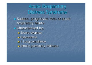
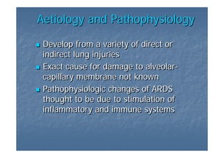
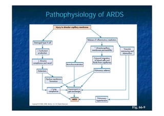
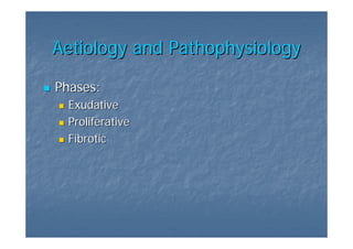
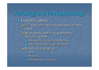
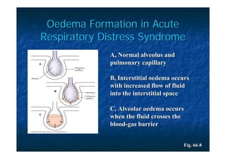
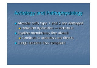
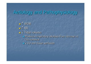
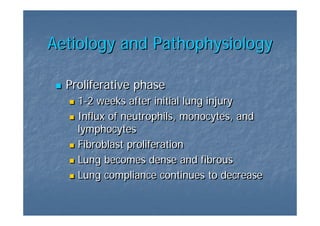
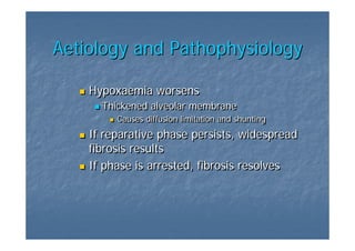
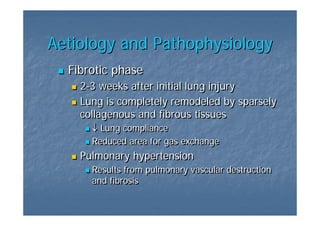
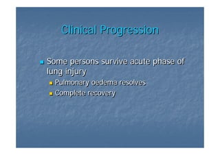
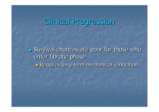
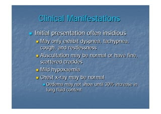
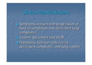
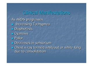
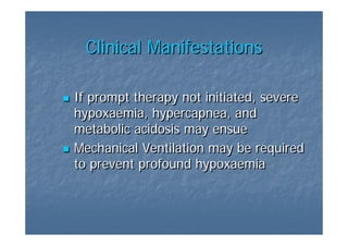
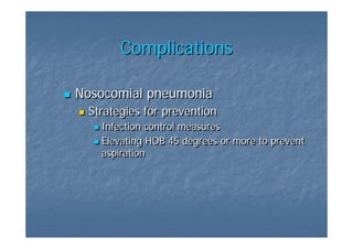
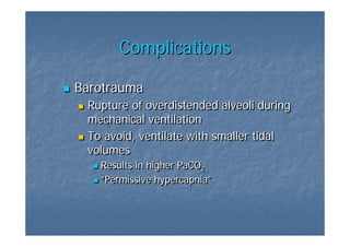
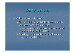
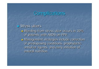
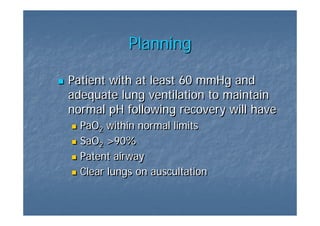
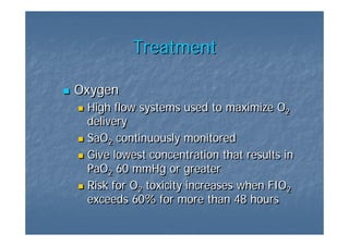
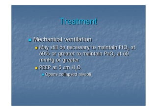
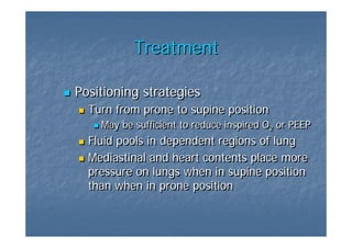
Acute Respiratory
 Distress Syndrome

         (ARDS)

Presented by Melinda C. Jordan
Acute Respiratory
       Distress Syndrome
Sudden, progressive form of acute
respiratory failure
Characterised by:
  Severe dyspnea
  Hypoxaemia
  ↓ Lung compliance
  Diffuse pulmonary infiltrates
Clinical Conditions Associated with
     the development of ARDS
Sepsis
Severe Trauma
Pulmonary contusion
Pneumonia
Near drowning
Massive transfusion
Fat embolism
Aetiology and Pathophysiology

 Develop from a variety of direct or
 indirect lung injuries
 Exact cause for damage to alveolar-
                            alveolar-
 capillary membrane not known
 Pathophysiologic changes of ARDS
 thought to be due to stimulation of
 inflammatory and immune systems
Pathophysiology of ARDS


                          Fig. 66-9
Aetiology and Pathophysiology

Phases:
 Exudative
 Proliferative
 Fibrotic
Aetiology and Pathophysiology
  Exudative phase
    1-7 days after direct lung injury or host
    1-7
    insult
    Neutrophils adhere to pulmonary
    microcirculation
      Damage to vascular endothelium
      Increased capillary permeability
    Results in leakage of
      H2O
      Protein
      Inflammatory chemical
Oedema Formation in Acute
Respiratory Distress Syndrome
             A, Normal alveolus and
             pulmonary capillary

             B, Interstitial oedema occurs
             with increased flow of fluid
             into the interstitial space

             C, Alveolar oedema occurs
             when the fluid crosses the
             blood-gas barrier


                                       Fig. 66-8
Aetiology and Pathophysiology
 Alveolar cells type 1 and 2 are damaged
   Surfactant dysfunction → atelectasis
 Hyaline membranes line alveoli
   Contribute to atelectasis and fibrosis
 Lungs become less compliant
Aetiology and Pathophysiology

   ↑ WOB
   ↑ RR
   ↓ Tidal volume
     Produces respiratory alkalosis from increase in
     CO2 removal
        2
     ↓ CO and tissue perfusion
Aetiology and Pathophysiology

  Proliferative phase
    1-2 weeks after initial lung injury
    1-2
    Influx of neutrophils, monocytes, and
    lymphocytes
    Fibroblast proliferation
    Lung becomes dense and fibrous
    Lung compliance continues to decrease
Aetiology and Pathophysiology

    Hypoxaemia worsens
      Thickened alveolar membrane
         Causes diffusion limitation and shunting
    If reparative phase persists, widespread
    fibrosis results
    If phase is arrested, fibrosis resolves
Aetiology and Pathophysiology
  Fibrotic phase
    2-3 weeks after initial lung injury
    2-3
    Lung is completely remodeled by sparsely
    collagenous and fibrous tissues
      ↓ Lung compliance
      Reduced area for gas exchange
    Pulmonary hypertension
      Results from pulmonary vascular destruction
      and fibrosis
Clinical Progression

Some persons survive acute phase of
lung injury
  Pulmonary oedema resolves
  Complete recovery
Clinical Progression


Survival chances are poor for those who
enter fibrotic phase
  Requires long-term mechanical ventilation
           long-term
Clinical Manifestations
Initial presentation often insidious
  May only exhibit dyspnea, tachypnea,
  cough, and restlessness
  Auscultation may be normal or have fine,
  scattered crackles
  Mild hypoxaemia
  Chest x-ray may be normal
         x-ray
    Oedema may not show until 30% increase in
    lung fluid content
Clinical Manifestations

Symptoms worsen with progression of
fluid accumulation and decreased lung
compliance
Evident discomfort and WOB
Pulmonary function tests reveal
decreased compliance and lung volume
Clinical Manifestations
As ARDS progresses
   Increasing Tachypnea
  Diaphoresis
  Cyanosis
  Pallor
  Decreases in sensorium
  Chest x-ray termed whiteout or white lung
         x-ray
  due to consolidation
Clinical Manifestations

If prompt therapy not initiated, severe
hypoxaemia, hypercapnea, and
hypoxaemia,
metabolic acidosis may ensue
Mechanical Ventilation may be required
to prevent profound hypoxaemia
Complications

Nosocomial pneumonia
 Strategies for prevention
   Infection control measures
   Elevating HOB 45 degrees or more to prevent
   aspiration
Complications

Barotrauma
 Rupture of overdistended alveoli during
 mechanical ventilation
 To avoid, ventilate with smaller tidal
 volumes
   Results in higher PaCO2
                         2
   “Permissive hypercapnia”
   “Permissive hypercapnia”
Complications
Volu-pressure trauma
Volu-pressure
  Occurs when large tidal volumes used to
  ventilate noncompliant lungs
    Alveolar fractures and movement of fluids and
    proteins into alveolar spaces
  Avoid by using smaller tidal volumes or
  pressure ventilation
Complications

Stress ulcers
  Bleeding from stress ulcer occurs in 30%
  of patients with ARDS on PPV
  Management strategies include correction
  of predisposing conditions, prophylactic
  antiulcer agents, and early initiation of
  enteral nutrition
Complications

Renal failure
  Occurs from decreased renal tissue
  oxygenation from hypotension, hypoxemia,
  or hypercapnia
  May also be caused by nephrotoxic drugs
  used for infections associated with ARDS
Nursing Assessment
History of lung disease, Smoking
Restlessness
Agitation
Pale, cool, clammy or warm, flushed
skin
Shallow breathing with increased
respiratory rate
Use of accessory muscles
Nursing Assessment

Tachycardia progressing to bradycardia
Extra heart sounds
Abnormal breath sounds
Hypertension progressing to
hypotension
Nursing Assessment

Somnolence, confusion, delirium
Changes in pH, PaCO2, PaO2, SaO2
Decreased tidal volume, FVC
Abnormal x-ray
         x-ray
Planning

Patient with at least 60 mmHg and
adequate lung ventilation to maintain
normal pH following recovery will have
  PaO2 within normal limits
      2
  SaO2 >90%
      2
  Patent airway
  Clear lungs on auscultation
Treatment

Oxygen
 High flow systems used to maximize O2   2
 delivery
 SaO2 continuously monitored
      2
 Give lowest concentration that results in
 PaO2 60 mmHg or greater
      2
 Risk for O2 toxicity increases when FIO2
           2                             2
 exceeds 60% for more than 48 hours
Treatment

Mechanical ventilation
  May still be necessary to maintain FIO2 at
                                        2
  60% or greater to maintain PaO2 at 60
                                  2
  mmHg or greater
  PEEP at 5 cm H2O2
    Opens collapsed alveoli
Treatment

Positioning strategies
  Turn from prone to supine position
    May be sufficient to reduce inspired O2 or PEEP
                                          2
  Fluid pools in dependent regions of lung
  Mediastinal and heart contents place more
  pressure on lungs when in supine position
  than when in prone position
Medical Supportive Therapy

Maintenance of cardiac output and
tissue perfusion
  Continuous hemodynamic monitoring
  Arterial catheter
Medical Supportive Therapy

Use of inotropic drugs may be
necessary
Hemoglobin usually kept at levels >9 or
10 with SaO2 >90%
              >90%
  Packed RBCs
Maintenance of fluid balance
Evaluation
No abnormal breath sounds
Effective cough and expectoration
Normal respiratory rate, rhythm, and
depth
Synchronous thoracoabdominal
movement
Appropriate use of accessory muscles
Evaluation

PaO2 and PaCO2 within normal ranges
Maintenance of weight or weight gain
Serum albumin and protein within
normal ranges

ARDS Overview

  • 1.
    Acute Respiratory DistressSyndrome (ARDS) Presented by Melinda C. Jordan
  • 2.
    Acute Respiratory Distress Syndrome Sudden, progressive form of acute respiratory failure Characterised by: Severe dyspnea Hypoxaemia ↓ Lung compliance Diffuse pulmonary infiltrates
  • 3.
    Clinical Conditions Associatedwith the development of ARDS Sepsis Severe Trauma Pulmonary contusion Pneumonia Near drowning Massive transfusion Fat embolism
  • 4.
    Aetiology and Pathophysiology Develop from a variety of direct or indirect lung injuries Exact cause for damage to alveolar- alveolar- capillary membrane not known Pathophysiologic changes of ARDS thought to be due to stimulation of inflammatory and immune systems
  • 5.
  • 6.
    Aetiology and Pathophysiology Phases: Exudative Proliferative Fibrotic
  • 7.
    Aetiology and Pathophysiology Exudative phase 1-7 days after direct lung injury or host 1-7 insult Neutrophils adhere to pulmonary microcirculation Damage to vascular endothelium Increased capillary permeability Results in leakage of H2O Protein Inflammatory chemical
  • 8.
    Oedema Formation inAcute Respiratory Distress Syndrome A, Normal alveolus and pulmonary capillary B, Interstitial oedema occurs with increased flow of fluid into the interstitial space C, Alveolar oedema occurs when the fluid crosses the blood-gas barrier Fig. 66-8
  • 9.
    Aetiology and Pathophysiology Alveolar cells type 1 and 2 are damaged Surfactant dysfunction → atelectasis Hyaline membranes line alveoli Contribute to atelectasis and fibrosis Lungs become less compliant
  • 10.
    Aetiology and Pathophysiology ↑ WOB ↑ RR ↓ Tidal volume Produces respiratory alkalosis from increase in CO2 removal 2 ↓ CO and tissue perfusion
  • 11.
    Aetiology and Pathophysiology Proliferative phase 1-2 weeks after initial lung injury 1-2 Influx of neutrophils, monocytes, and lymphocytes Fibroblast proliferation Lung becomes dense and fibrous Lung compliance continues to decrease
  • 12.
    Aetiology and Pathophysiology Hypoxaemia worsens Thickened alveolar membrane Causes diffusion limitation and shunting If reparative phase persists, widespread fibrosis results If phase is arrested, fibrosis resolves
  • 13.
    Aetiology and Pathophysiology Fibrotic phase 2-3 weeks after initial lung injury 2-3 Lung is completely remodeled by sparsely collagenous and fibrous tissues ↓ Lung compliance Reduced area for gas exchange Pulmonary hypertension Results from pulmonary vascular destruction and fibrosis
  • 14.
    Clinical Progression Some personssurvive acute phase of lung injury Pulmonary oedema resolves Complete recovery
  • 15.
    Clinical Progression Survival chancesare poor for those who enter fibrotic phase Requires long-term mechanical ventilation long-term
  • 16.
    Clinical Manifestations Initial presentationoften insidious May only exhibit dyspnea, tachypnea, cough, and restlessness Auscultation may be normal or have fine, scattered crackles Mild hypoxaemia Chest x-ray may be normal x-ray Oedema may not show until 30% increase in lung fluid content
  • 17.
    Clinical Manifestations Symptoms worsenwith progression of fluid accumulation and decreased lung compliance Evident discomfort and WOB Pulmonary function tests reveal decreased compliance and lung volume
  • 18.
    Clinical Manifestations As ARDSprogresses Increasing Tachypnea Diaphoresis Cyanosis Pallor Decreases in sensorium Chest x-ray termed whiteout or white lung x-ray due to consolidation
  • 19.
    Clinical Manifestations If prompttherapy not initiated, severe hypoxaemia, hypercapnea, and hypoxaemia, metabolic acidosis may ensue Mechanical Ventilation may be required to prevent profound hypoxaemia
  • 20.
    Complications Nosocomial pneumonia Strategiesfor prevention Infection control measures Elevating HOB 45 degrees or more to prevent aspiration
  • 21.
    Complications Barotrauma Rupture ofoverdistended alveoli during mechanical ventilation To avoid, ventilate with smaller tidal volumes Results in higher PaCO2 2 “Permissive hypercapnia” “Permissive hypercapnia”
  • 22.
    Complications Volu-pressure trauma Volu-pressure Occurs when large tidal volumes used to ventilate noncompliant lungs Alveolar fractures and movement of fluids and proteins into alveolar spaces Avoid by using smaller tidal volumes or pressure ventilation
  • 23.
    Complications Stress ulcers Bleeding from stress ulcer occurs in 30% of patients with ARDS on PPV Management strategies include correction of predisposing conditions, prophylactic antiulcer agents, and early initiation of enteral nutrition
  • 24.
    Complications Renal failure Occurs from decreased renal tissue oxygenation from hypotension, hypoxemia, or hypercapnia May also be caused by nephrotoxic drugs used for infections associated with ARDS
  • 25.
    Nursing Assessment History oflung disease, Smoking Restlessness Agitation Pale, cool, clammy or warm, flushed skin Shallow breathing with increased respiratory rate Use of accessory muscles
  • 26.
    Nursing Assessment Tachycardia progressingto bradycardia Extra heart sounds Abnormal breath sounds Hypertension progressing to hypotension
  • 27.
    Nursing Assessment Somnolence, confusion,delirium Changes in pH, PaCO2, PaO2, SaO2 Decreased tidal volume, FVC Abnormal x-ray x-ray
  • 28.
    Planning Patient with atleast 60 mmHg and adequate lung ventilation to maintain normal pH following recovery will have PaO2 within normal limits 2 SaO2 >90% 2 Patent airway Clear lungs on auscultation
  • 29.
    Treatment Oxygen High flowsystems used to maximize O2 2 delivery SaO2 continuously monitored 2 Give lowest concentration that results in PaO2 60 mmHg or greater 2 Risk for O2 toxicity increases when FIO2 2 2 exceeds 60% for more than 48 hours
  • 30.
    Treatment Mechanical ventilation May still be necessary to maintain FIO2 at 2 60% or greater to maintain PaO2 at 60 2 mmHg or greater PEEP at 5 cm H2O2 Opens collapsed alveoli
  • 31.
    Treatment Positioning strategies Turn from prone to supine position May be sufficient to reduce inspired O2 or PEEP 2 Fluid pools in dependent regions of lung Mediastinal and heart contents place more pressure on lungs when in supine position than when in prone position
  • 32.
    Medical Supportive Therapy Maintenanceof cardiac output and tissue perfusion Continuous hemodynamic monitoring Arterial catheter
  • 33.
    Medical Supportive Therapy Useof inotropic drugs may be necessary Hemoglobin usually kept at levels >9 or 10 with SaO2 >90% >90% Packed RBCs Maintenance of fluid balance
  • 34.
    Evaluation No abnormal breathsounds Effective cough and expectoration Normal respiratory rate, rhythm, and depth Synchronous thoracoabdominal movement Appropriate use of accessory muscles
  • 35.
    Evaluation PaO2 and PaCO2within normal ranges Maintenance of weight or weight gain Serum albumin and protein within normal ranges