This document defines acute respiratory distress syndrome (ARDS) and outlines its diagnostic criteria, etiologies, pathophysiology, stages of progression, clinical manifestations, management including mechanical ventilation strategies, complications, prognosis, and bibliography of references. ARDS is characterized by hypoxemic respiratory failure due to diffuse pulmonary edema from acute lung injury. The management focuses on supportive care including mechanical ventilation with low tidal volumes and PEEP to prevent further lung injury while the underlying condition is treated. The mortality rate for ARDS remains high but has decreased to around 40% with standardized treatment protocols.
Based on the information provided, this patient is experiencing hypoxic respiratory failure and is not adequately compensating despite high-flow oxygen therapy. Mechanical ventilation would be indicated to support oxygenation and ventilation until the underlying pneumonia improves with treatment.
1) Respiratory failure is a condition where the lungs cannot properly oxygenate the blood and remove carbon dioxide, classified as Type I (hypoxemic) or Type II (hypercapnic).
2) It can result from problems affecting gas exchange in the lungs, respiratory control centers in the brain, or the chest wall muscles.
3) Common causes of Type I respiratory failure include pneumonia, ARDS, and severe asthma, while Type II is often due to conditions that decrease breathing, such as COPD.
Acute Respiratory Distress Syndrome (ARDS) is a clinical syndrome characterized by hypoxemia, bilateral pulmonary infiltrates, and respiratory failure. The document defines ARDS and discusses its etiology, pathophysiology, clinical features, diagnosis, and evidence-based treatment recommendations. Key points include low tidal volume ventilation to minimize lung injury, conservative fluid management, use of PEEP to recruit alveoli while limiting pressures, and treating the underlying cause of ARDS. Outcomes remain poor with high mortality rates, though some patients fully recover lung function over time.
Anesthetic consideration in smokers,alcoholics and addictsAftab Hussain
Anaesthetic consideration in smokers alcoholic and drug addicts. As an anaesthesiologist we must be aware with the problems associated with their management and interaction with anaesthetics.
The document discusses acute respiratory failure, which can be either hypercapnic or hypoxemic. It provides details on the causes, pathophysiology, and assessment of different types of respiratory failure including acute respiratory distress syndrome. It also describes a case study of a patient with respiratory failure and discusses their management.
This document discusses acute respiratory distress syndrome (ARDS). It begins by defining ARDS and describing its signs and symptoms. It then discusses the history of ARDS definitions and criteria. It outlines the pathophysiology and three phases of ARDS. Treatment strategies covered include mechanical ventilation, monitoring, infection control, and specific therapies. Prognosis and risk factors are also summarized.
The document discusses respiratory failure and its management. It begins by defining respiratory failure and describing the types. It then lists common causes and presents results of diagnostic tests for a patient, including abnormal blood gases, imaging findings, and clinical signs. Treatment for this patient's respiratory failure included mechanical ventilation, bronchodilators, diuretics, and oxygen therapy. Complications of respiratory failure mentioned include cardiac or respiratory arrest.
Slideshow is from the University of Michigan Medical School's M2 Respiratory sequence
View additional course materials on Open.Michigan:
openmi.ch/med-M2Resp
Non-cardiogenic pulmonary edema, also known as acute respiratory distress syndrome (ARDS), is characterized by severe arterial hypoxemia, bilateral alveolar infiltrates on chest imaging, and a normal left atrial pressure. It results from increased vascular permeability in the lungs leading to fluid accumulation in the alveoli and impaired gas exchange. The underlying pathophysiology involves diffuse damage to the alveolar-capillary membrane by various insults such as sepsis, infection, or injury. This disrupts the normal barrier function and leads to flooding of the lungs with protein-rich fluid.
Acute respiratory failure occurs when the pulmonary system can no longer meet the body's metabolic demands. It can be hypoxaemic, with an oxygen level below 60 mmHg, or hypercapnic, with a carbon dioxide level over 50 mmHg. Respiratory failure results from issues in oxygen intake and carbon dioxide removal, due to problems in ventilation, perfusion matching, gas exchange, or other pathologies. It is monitored clinically and with blood tests, and treated by addressing the underlying cause, providing supportive oxygen therapy or ventilation support like CPAP or mechanical ventilation if needed.
This document discusses acute respiratory distress syndrome (ARDS) and respiratory failure. It provides details on:
- The diagnostic criteria for ARDS including hypoxemia, bilateral infiltrates on chest x-ray, and no left atrial hypertension.
- The clinical course of ARDS involving exudative, proliferative and fibrotic phases in the first few weeks.
- Treatment focuses on mechanical ventilation to prevent alveolar collapse while avoiding ventilator induced lung injury through strategies like PEEP and prone positioning.
- Respiratory failure is defined as inability to maintain adequate gas exchange and can be type 1 (hypoxemia without hypercapnia) or type 2 (hypoxemia with
1) ARDS is characterized by increased permeability of the alveolar capillary membrane leading to pulmonary edema and reduced lung compliance. It is caused by direct or indirect lung injury and results in hypoxemia.
2) The management of ARDS focuses on supportive care including mechanical ventilation with small tidal volumes, fluid management, and treatment of the underlying condition.
3) The pathology of ARDS involves three phases - exudative, proliferative and fibrotic - resulting in diffuse alveolar damage and impaired gas exchange. Prognosis depends on severity of the initial lung injury and development of complications.
This document discusses chronic respiratory failure and various treatment strategies. It covers topics such as acute respiratory failure, exacerbations of chronic obstructive airway disease, ventilation techniques, adjunctive treatments, lung recruitment strategies, hypercapnia, and lung replacement options including extracorporeal membrane oxygenation and novelung. The overall approach outlined is to maximize protective ventilation using adjuncts if needed, employ novelung for elevated carbon dioxide, and consider extracorporeal membrane oxygenation for continuing hypoxia. Reversibility of the patient's condition is emphasized over simply extending life.
Respiratory failure occurs when the lungs fail to effectively oxygenate the blood or remove carbon dioxide. It is classified as type 1 (hypoxic but normal CO2 levels) or type 2 (hypoxic and elevated CO2 levels). Type 1 is more common and caused by conditions like pneumonia that affect only part of the lungs. Type 2 involves more generalized lung damage. Acute respiratory failure develops rapidly while chronic failure progresses over days or longer. Treatment depends on the underlying cause but may include supplemental oxygen, mechanical ventilation, treating infection, or lung transplantation in severe cases.
Respiratory failure is a condition in which your blood doesn't have enough oxygen or has too much carbon dioxide. Sometimes you can have both problems. When you breathe, your lungs take in oxygen. The oxygen passes into your blood, which carries it to your organs
respiratory failure
typel respiratory failure
typell respiratory failure
clinical presentation
diagnosis
managament
oxygen
This document provides an overview of respiratory failure, including its causes, types, symptoms, diagnosis, and management. It begins by defining respiratory failure as the failure of the respiratory system in gas exchange functions of oxygenation and carbon dioxide elimination. Respiratory failure is then classified based on PaO2 and PaCO2 levels into hypoxemic (Type I) and hypercapnic (Type II) types. Common causes, clinical features, investigations, and general management principles are discussed for respiratory failure. Key aspects of managing hypoxemia and hypercapnia are also summarized.
Respiratory failure occurs when the lungs fail to effectively oxygenate the blood or remove carbon dioxide. It can be caused by conditions that decrease lung function or increase oxygen needs. Symptoms include shortness of breath, confusion, and bluish skin. Diagnosis involves assessing symptoms, risk factors, and tests like blood gases, imaging, and pulmonary function tests. Management focuses on treating the underlying cause, correcting gas exchange abnormalities through oxygen supplementation or ventilation, and preventing complications. Nursing care monitors the patient's condition and provides interventions to address issues like impaired gas exchange, low cardiac output, poor nutrition, and anxiety.
This document discusses respiratory distress and respiratory failure. Respiratory distress refers to increased work of breathing, while respiratory failure is the inability of the lungs to provide oxygen or remove carbon dioxide. Respiratory failure can be acute or chronic. It can occur due to problems with the respiratory pump (central nervous system issues, muscle weakness) or due to airway/lung dysfunction (conditions affecting gas exchange like asthma, pneumonia). Proper monitoring of patients with respiratory distress or failure includes clinical examination, blood gas analysis, and oximetry. Immediate treatment of acute respiratory failure focuses on oxygenation and ventilation. Chronic respiratory failure often has a more insidious onset and requires careful monitoring, especially during sleep or illness.
Acute respiratory distress syndrome (ARDS) is a condition that damages the lungs and prevents sufficient oxygen from entering the bloodstream. It is caused by injury to the lungs from direct or indirect insults. In ARDS, the alveolar-capillary membrane is compromised, allowing fluid to enter the lungs. Treatment involves intensive care, use of a ventilator to deliver oxygen under pressure, and addressing the underlying cause while preventing further lung injury. The goal is to support breathing and remove fluid from the damaged lungs.
This document discusses acute respiratory distress syndrome (ARDS) and respiratory failure. It provides details on:
- The diagnostic criteria for ARDS including hypoxemia, bilateral infiltrates on chest x-ray, and no left atrial hypertension.
- The clinical course of ARDS involving exudative, proliferative and fibrotic phases in the first few weeks.
- Treatment focuses on mechanical ventilation to prevent alveolar collapse and ventilator induced lung injury while maintaining low pressures.
- Respiratory failure is defined as inability to maintain adequate gas exchange and can be type 1 (hypoxemia without hypercapnia) or type 2 (hypoxemia with hypercapnia). Causes
This document describes two types of respiratory failure:
Type I is characterized by hypoxemia due to ventilation-perfusion mismatching or impaired gas exchange. Type II involves hypercapnia due to decreased minute ventilation and failure to remove carbon dioxide.
For Type I respiratory failure, supplemental oxygen may improve hypoxemia while non-invasive ventilation or intubation may be needed to support work of breathing. For Type II failure, non-invasive ventilation is first indicated to support breathing, while intubation and mechanical ventilation are needed if non-invasive methods fail.
This document discusses respiratory failure, including its causes, types, and management. Respiratory failure occurs when inadequate gas exchange prevents normal oxygen and carbon dioxide levels in the blood. It can result from conditions affecting breathing muscles/nerves or lung tissue damage. The two main types are hypoxemic respiratory failure, where oxygen levels are too low, and hypercapnic respiratory failure, where carbon dioxide levels are too high. Management involves oxygen therapy, positioning, clearing secretions, and potentially positive pressure ventilation.
This document discusses Acute Respiratory Distress Syndrome (ARDS), a clinical syndrome characterized by severe lung inflammation and injury leading to hypoxemia. It is most commonly caused by pneumonia, sepsis, aspiration, or trauma. The pathogenesis involves an initial exudative inflammatory phase, followed by a proliferative phase and possible fibrotic phase. Diagnostic tests include blood gases, chest X-rays, and CT scans. Treatment focuses on treating the underlying cause, administering oxygen, antibiotics, and corticosteroids. Nursing management centers around pulmonary toilet, monitoring fluid balance, improving breathing and nutrition, and mobilizing the patient.
Design of artificial respiratory modelShîvãm Gûptå
Design of Artificial Respiratory Model.. Know about the respiratory system.
The respiratory system consists of the upper respiratory tract (nasal passages), the airway conduction system (larynx, trachea, bronchi, bronchioles and terminal bronchioles), and the lower respiratory tract (alveolar ducts and alveoli). Not all segments of the respiratory system mature at the same pace. The olfactory epithelium matures earliest by PND 7. The lung, however, is not considered mature until PND 21, when alveolarization and microvascular maturation are complete. This chapter will discuss the embryological development (briefly), adult histomorphology, and postnatal histologic development of each major component of the respiratory system.
Respiratory failure occurs when the lungs fail to oxygenate the blood or eliminate carbon dioxide. It can be classified as type I (hypoxemic) or type II (hypercapnic). Common causes include pneumonia, COPD, pulmonary embolism, and cardiac failure. Diagnosis involves blood gas analysis, imaging, and identifying the underlying cause. Management focuses on treating the cause, supporting oxygenation and ventilation, and mechanical ventilation if needed. Type II respiratory failure requires careful oxygen therapy to prevent worsening acidosis.
This document discusses respiratory failure, defined as inadequate oxygenation, ventilation, or both to meet metabolic demands. It can be classified as type 1 (hypoxemic) or type 2 (hypercapnic) respiratory failure. Risk factors include age, smoking, lung disease, and neurological or muscular disorders. Pathophysiology involves ventilation-perfusion mismatching, right-to-left shunting, or hypoventilation. Causes include pneumonia, pulmonary embolism, neuromuscular disorders, and acute respiratory distress syndrome. The control of breathing and gas exchange physiology are also summarized.
This document discusses how social media has changed notions of what is private and public information. It notes that knowledge is now accessible with a click of a button, and fact and fiction are harder to distinguish online. The document advocates for businesses to implement social media policies to provide guidance on appropriate employee online behavior, as 54% of companies still ban social media despite evidence this approach is ineffective. It presents the goals of AssistNZ to empower communities and businesses through social media education and planning.
Strategic leadership is the ability to anticipate change, maintain flexibility, and empower others to create strategic change. Effective strategic leaders shape a firm's strategic intent, mission, and actions through visioning and influencing behaviors. They must effectively manage a firm's human capital and resources. While the job of a CEO is challenging, some strategic leaders have achieved great success, like the CEOs of Avon and Applied Materials discussed in the article, by adapting to changes, taking risks, and nurturing an effective culture. However, many CEOs have also failed due to arrogance, hubris, and lack of ethics. Effective strategic leadership is critical for a firm to achieve competitive advantage and above-average returns.
Attempting to Jump the Largest Agile HurdleMatt Badgley
Agile Culture can be a tough thing -- this presentation was my attempt to have a discussion on how organizations can address the challenges around adopting an agile mindset; which would hopefully lead to successful agile software development.
The Beijing Tiantan Puhua Hospital is the largest neuroscience specialty hospital in Beijing. It was originally a joint venture ward within the renowned Tiantan Hospital but opened as an independent hospital in 2005. It operates in conjunction with American, Chinese, and other international partners. The hospital utilizes international standards and facilities alongside experts from China's top hospitals to provide comprehensive neuroscience and other healthcare services. It is also a leader in neural stem cell research and treatment.
The document discusses the concept of servant leadership as exemplified by Jesus Christ. It defines servant leadership as serving others unselfishly while empowering them to accomplish God's purpose. The document provides examples of both positive and negative leadership from Scripture. It emphasizes that true leadership requires having a servant's attitude and focusing on others rather than pursuing one's own agenda. The document concludes with a checklist for evaluating whether one's leadership demonstrates the principles of servant leadership.
The document discusses agile documentation and how it can coexist with agile principles. It introduces the speaker and their background. Several tips are provided for creating documentation in an agile manner, such as focusing on the minimum needed, documenting decisions, and treating documentation like a backlog item. User stories and acceptance criteria are discussed as important agile requirements techniques. The document advocates for documentation that will actually be used and read.
Shooting For The Stars, A Discussion About Our Current State of AgileMatt Badgley
The document discusses the benefits of meditation for reducing stress and anxiety. Regular meditation practice can help calm the mind and body by lowering heart rate and blood pressure. Studies have shown that meditating for just 10-20 minutes per day can have significant positive impacts on both mental and physical health over time.
Learning to Fly - Finding the Keys to Engagement - Agile & Beyond 2016Matt Badgley
We are in the midst of a purpose drive revolution that is changing the way we do business, the way we work, and the way teams get stuff done. Now it is time, we make sure that everyone understands why.
Servant leadership is exercising godly leadership by serving and empowering others to accomplish God's purpose, as Jesus did by washing his disciples' feet. It involves placing others' needs above your own and having eternal values rather than seeking power, control or personal gain. True biblical leadership requires first being a servant and earning others' respect through love and care, rather than force of personality. The document provides examples of servant leadership from scripture and evaluates the reader's leadership based on biblical principles of serving, empowering others, and placing God's purpose above personal agendas.
The document discusses the benefits of exercise for mental health. Regular physical activity can help reduce anxiety and depression and improve mood and cognitive functioning. Exercise boosts blood flow and levels of neurotransmitters and endorphins which elevate and stabilize mood.
Dr. Jing Li, MD
Publications:
• Han X, et al. Stem cell therapy for malignant glioma: Current status and future
perspectives. Cancer Lett. 2014
• Han X, et al. Neural stem cell transplantation for the treatment of glioma. J
Neurooncol. 2013
• Han X, et al. Clinical management of cerebral aneurysms. Chin Med J. 2012
• Han X, et al. Intraoperative ultrasound guidance for brain tumor resection. J
Neurosurg. 2011
• Han X, et al. Glioma vaccine: current status and future prospects. J Clin Neurosci.
2010
• Han X, et al. Gene
Monomax is a professional conference and event organizer based in Russia that has over 18 years of experience managing over 900 international conferences and 100 association conventions. They provide full conference management services including financial management, registration, on-site coordination, accommodation, and producing flexible reports for organizing committees. Monomax has experience managing a wide range of conferences from 30 to 2500 participants on topics such as polar research, ice hockey, nanosystems, and cerebrovascular pathology.
Getting Blood From a Turnip: The Art of Facilitation Made Fun and ProductiveMatt Badgley
The document discusses the results of a study on the impact of climate change on global wheat production. Researchers found that rising temperatures will significantly reduce wheat yields across different regions of the world by the end of the century. Under a high emissions scenario, wheat production is projected to decrease between 6-27% globally depending on the region, posing substantial risks to global food security.
Shortkeynote at the CoCreation und Collaboration Workshopjovoto GmbH
Crowdstorming, or brainstorming at scale, allows companies to outsource creative tasks to a global talent pool. Jovoto is a platform that facilitates crowdstorming. It structures the entire process from briefing to evaluation. This ensures unlimited access to talent outside an organization. Crowdstorming results in a broad range of ideas due to its large pool of participants and fosters better motivation and results through voluntary participation. Jovoto manages the idea generation, collaboration, and filtering process to identify the best solutions for clients.
PATHOGENESIS AND MANAGEMENT OF ARDS-2.pptxdevanshi92
1) ARDS results from increased lung vascular permeability leading to accumulation of fluid and protein in the lungs. This causes diffuse pulmonary edema and hypoxemia that is resistant to oxygen therapy.
2) Treatment involves identifying and treating the underlying cause, using mechanical ventilation with low tidal volumes and limiting inspiratory pressures to prevent further lung injury, and maintaining adequate oxygen levels through techniques like PEEP.
3) The ARMA trial showed that a ventilation strategy using low tidal volumes and plateau pressures reduced mortality in ARDS patients, establishing this approach as the standard of care.
Acute Respiratory Distress Syndrome (ARDS) is a sudden, progressive form of respiratory failure characterized by severe dyspnea, hypoxemia, and decreased lung compliance. It develops from direct or indirect lung injuries and is thought to be caused by stimulation of the inflammatory and immune systems, resulting in leakage of fluid into the lungs. The clinical progression of ARDS involves exudative, proliferative, and fibrotic phases that can lead to respiratory failure if not promptly treated with oxygen supplementation, mechanical ventilation, and other supportive therapies.
Acute respiratory failure occurs when the respiratory system fails to maintain adequate gas exchange. There are two main types: hypoxemic respiratory failure, characterized by low oxygen levels, and acute ventilatory failure, characterized by high carbon dioxide levels. Hypoxemic failure is most common and can result from conditions that impair gas exchange like pneumonia or pulmonary edema. Ventilatory failure involves impaired breathing and can be caused by conditions that increase breathing workload like COPD. Diagnosis involves blood gas analysis and imaging. Treatment focuses on supporting oxygenation and ventilation through oxygen supplementation, ventilation support, and treating underlying causes.
Acute respiratory failure occurs when the respiratory system fails to maintain adequate gas exchange. There are two main types: hypoxemic respiratory failure, characterized by low oxygen levels (PaO2) with normal or low carbon dioxide (PaCO2) levels; and ventilatory (hypercapnic) respiratory failure, characterized by high PaCO2 levels. Hypoxemic failure is most common and can result from conditions that impair gas exchange like pneumonia or pulmonary edema. Ventilatory failure involves impaired ventilation and can be caused by conditions that obstruct airflow like COPD. Diagnosis involves blood gas analysis and imaging. Treatment focuses on supporting oxygenation and ventilation through oxygen supplementation, ventilation support, and treating the underlying cause.
1. ARDS is an acute respiratory failure condition characterized by diffuse pulmonary infiltrates from non-cardiac causes. It was formally defined in 1992 to include acute onset, bilateral lung infiltrates, and low pulmonary artery wedge pressure.
2. ARDS results from lung injury from direct or indirect causes, leading to inflammation and accumulation of fluid in the lungs. This makes breathing difficult and prevents the lungs from providing oxygen to the blood.
3. Management focuses on supportive care including mechanical ventilation with low tidal volumes, permissive hypercapnia, and optimizing oxygen levels with PEEP and supplemental oxygen. Other strategies include prone positioning, inhaled nitric oxide, and extracorporeal membrane oxygenation in severe cases.
This document provides information on Acute Respiratory Distress Syndrome (ARDS), including its diagnostic criteria, clinical presentation, associated conditions, investigations, management, and new treatments being researched. ARDS is defined by acute onset hypoxemia, bilateral lung opacities on CXR, and respiratory failure not fully explained by cardiac failure. Common causes include sepsis, trauma, aspiration, and transfusions. Management involves treating the underlying condition, supportive care including mechanical ventilation with low tidal volumes, and cardiovascular support. New areas of research include gene therapy, enhancing edema clearance, nitric oxide donors, targeting vascular permeability, and modulating inflammation.
This document discusses oxygen therapy and oxygen toxicity. It aims to provide guidelines for oxygen therapy including indications, goals, delivery methods and their advantages/disadvantages. Prolonged high concentration oxygen can cause pulmonary toxicity similar to ARDS. The optimal oxygen concentration is the lowest that maintains tissue oxygenation below 60% FIO2 to avoid toxicity. Measures like mechanical ventilation and antioxidants can help prevent toxicity from prolonged high concentration oxygen.
This document discusses various medical gases including oxygen, carbon dioxide, helium, and their properties, preparation, storage, clinical uses, and safety considerations. It provides details on oxygen therapy including indications, assessment, delivery methods, complications like toxicity, and monitoring. The oxygen cascade and hypoxia types are explained. Carbon dioxide properties and uses in anesthesia are covered. Helium properties and use in partial airway obstruction are outlined. Methods of gas analysis are also summarized.
1. Respiratory failure occurs when the respiratory system fails in its gas exchange function, resulting in low oxygen and high carbon dioxide levels in the blood.
2. It can be acute, coming on suddenly from conditions like pneumonia, or chronic from ongoing diseases like COPD.
3. Treatment depends on the type of failure - oxygen therapy for hypoxemic respiratory failure and ventilation support like non-invasive ventilation for hypercapnic respiratory failure. Physiotherapy focuses on clearing secretions, maintaining strength, and mobilization to facilitate weaning from ventilation.
The document discusses targeting low levels of PaO2 in patients with ARDS. It notes that while specific PaO2 levels cannot be defined, values between 60-75 mmHg are generally appropriate, and 50-60 mmHg may be tolerated in healthy young patients. Lower values between 40-50 mmHg should not be desirable. The key consideration is the "clinical price" of interventions to increase PaO2, such as increased FiO2 or mean airway pressures. As long as tissue oxygenation is maintained, permissive hypoxemia in ARDS appears to be well tolerated without evidence of tissue hypoxia.
The document provides information about acute respiratory distress syndrome (ARDS). It begins with a brief history of ARDS and provides the clinical definition. It describes the diagnostic criteria and etiology, including that most cases are caused by sepsis, pneumonia, or trauma. It then discusses the normal lung physiology and pathophysiology of ARDS, which involves three phases: exudative, proliferative, and fibrotic. The management section outlines the principles of therapy to provide adequate gas exchange while avoiding secondary injury, including mechanical ventilation protocols, fluid management, and other strategies. It concludes with a discussion of prognosis and recent advances in ARDS management such as protective ventilation strategies.
This document provides an overview of respiratory failure, including its definition, types, causes, patient presentation, investigations, management, and complications. There are four types of respiratory failure: type I involves hypoxemic failure due to issues with oxygenation; type II involves hypercapneic failure due to ventilation issues; type III occurs perioperatively due to lung collapse; and type IV is due to respiratory muscle hypoperfusion in shock. The management of respiratory failure involves treating the underlying cause, providing oxygen support, and potentially mechanical ventilation. Outcomes depend on the severity of acidosis and underlying illnesses.
1) Acute respiratory distress syndrome (ARDS) is a life-threatening lung condition caused by direct or indirect injury to the lungs whereby the alveolar capillary membrane becomes damaged and permeable, resulting in pulmonary edema.
2) ARDS is characterized by hypoxemia, reduced lung compliance, and diffuse pulmonary infiltrates seen on chest imaging.
3) Treatment involves supportive care in an intensive care unit including mechanical ventilation, supplemental oxygen, and positioning therapies like prone positioning to improve oxygenation.
This document discusses respiratory failure, including its classification, pathophysiology, clinical presentation, evaluation, complications, and management. Respiratory failure is classified as type 1 (hypoxemic) or type 2 (hypercapnic) based on blood gas abnormalities. Common causes include lung disease, disorders of the nervous system or respiratory muscles. Signs may include dyspnea, cyanosis, confusion. Evaluation includes blood gases, imaging, and tests to identify the underlying cause. Complications affect multiple organ systems. Management focuses on correcting hypoxemia and hypercapnia through supportive measures like oxygen supplementation or mechanical ventilation, as well as treating the underlying condition.
1. The document discusses acute respiratory distress syndrome (ARDS), describing its pathophysiology, causes, diagnosis, treatment and prognosis.
2. ARDS is characterized by hypoxemia, reduced lung compliance and diffuse pulmonary infiltrates leading to respiratory failure. Common causes include sepsis, pneumonia and trauma.
3. Treatment involves treating the underlying cause, supportive care including mechanical ventilation with low tidal volumes, and managing fluid levels and oxygenation. Prognosis depends on severity of illness, with reported mortality ranging from 41-65%.
Pediatric ARDS is a common cause of respiratory failure in children. It is defined by acute onset hypoxemia that cannot be explained by cardiac failure, with bilateral lung opacities on chest imaging. Management involves controlling the underlying cause, lung protective ventilation with low tidal volumes, permissive hypercapnia, prone positioning, and consideration of recruitment maneuvers, HFOV, surfactant, inhaled nitric oxide, or ECMO in severe cases. Noninvasive ventilation may be tried initially for mild disease but intubation is often required for more severe pediatric ARDS. The goals of management are to maintain adequate oxygenation and ventilation while minimizing ventilator induced lung injury.
This document discusses acute respiratory distress syndrome (ARDS). It defines ARDS according to the Berlin definition and describes its etiology, risk factors, pathogenesis, clinical features, and treatment approaches. Regarding treatment, the main focus is lung protective ventilation with low tidal volumes, optimizing PEEP levels, permissive hypercapnia, and conservative fluid management when possible. Other supportive strategies discussed include prone positioning, neuromuscular blockade, inhaled vasodilators, glucocorticoids, and evaluating patients daily for spontaneous breathing trials to guide weaning from mechanical ventilation.
Pediatrics notes about "Acute Respiratory Failure". These notes were published in 2018.
You can download them from
- Telegram: https://t.me/pediatric_notes_2018
- Mediafire: http://www.mediafire.com/folder/u5u60m184t9z7/Pediatric_Notes_2018
This document discusses hypoxia and oxygen therapy. It begins by providing historical background on the discovery of oxygen. It then describes the oxygen cascade and factors that can affect oxygen levels at different points. Various methods of oxygen delivery are presented, including low-flow and high-flow systems. Key goals and indications for oxygen therapy are outlined. Precautions for oxygen toxicity are discussed. The document concludes by providing guidance on selecting an appropriate oxygen delivery system based on the patient and clinical situation.
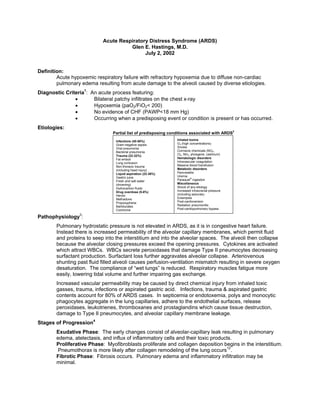
1. Acute Respiratory Distress Syndrome (ARDS)
Glen E. Hastings, M.D.
July 2, 2002
Definition:
Acute hypoxemic respiratory failure with refractory hypoxemia due to diffuse non-cardiac
pulmonary edema resulting from acute damage to the alveoli caused by diverse etiologies.
Diagnostic Criteria1: An acute process featuring:
• Bilateral patchy infiltrates on the chest x-ray
• Hypoxemia (paO2/FiO2< 200)
• No evidence of CHF (PAWP<18 mm Hg)
• Occurring when a predisposing event or condition is present or has occurred.
Etiologies:
2
Partial list of predisposing conditions associated with ARDS
Infections (40-50%) Inhaled toxins
Gram-negative sepsis O2 (high concentrations)
Viral pneumonia Smoke
Bacterial pneumonia Corrosive chemicals (NO2,
Trauma (22-32%) Cl2, NH3, phosgene, cadmium)
Fat emboli Hematologic disorders
Lung contusion Intravascular coagulation
Non-thoracic trauma Massive blood transfusion
(including head injury) Metabolic disorders
Liquid aspiration (22-36%) Pancreatitis
Gastric juice Uremia
Fresh and salt water Paraquat® ingestion
(drowning) Miscellaneous
Hydrocarbon fluids Shock of any etiology
Drug overdose (5-8%) Increased intracranial pressure
Heroin (including seizures)
Methadone Eclampsia
Propoxyphene Post-cardioversion
Barbiturates Radiation pneumonitis
Colchicine Post-cardiopulmonary bypass
Pathophysiology3:
Pulmonary hydrostatic pressure is not elevated in ARDS, as it is in congestive heart failure.
Instead there is increased permeability of the alveolar capillary membranes, which permit fluid
and proteins to seep into the interstitium and into the alveolar spaces. The alveoli then collapse
because the alveolar closing pressures exceed the opening pressures. Cytokines are activated
which attract WBCs. WBCs secrete peroxidases that damage Type II pneumocytes decreasing
surfactant production. Surfactant loss further aggravates alveolar collapse. Arteriovenous
shunting past fluid filled alveoli causes perfusion-ventilation mismatch resulting in severe oxygen
desaturation. The compliance of “wet lungs” is reduced. Respiratory muscles fatigue more
easily, lowering tidal volume and further impairing gas exchange.
Increased vascular permeability may be caused by direct chemical injury from inhaled toxic
gasses, trauma, infections or aspirated gastric acid. Infections, trauma & aspirated gastric
contents account for 80% of ARDS cases. In septicemia or endotoxemia, polys and monocytic
phagocytes aggregate in the lung capillaries, adhere to the endothelial surfaces, release
peroxidases, leukotrienes, thromboxanes and prostaglandins which cause tissue destruction,
damage to Type II pneumocytes, and alveolar capillary membrane leakage.
Stages of Progression4
Exudative Phase: The early changes consist of alveolar-capillary leak resulting in pulmonary
edema, atelectasis, and influx of inflammatory cells and their toxic products.
Proliferative Phase: Myofibroblasts proliferate and collagen deposition begins in the interstitium.
Pneumothorax is more likely after collagen remodeling of the lung occurs12.
Fibrotic Phase: Fibrosis occurs. Pulmonary edema and inflammatory infiltration may be
minimal.
2. ARDS- Page 2
Exudative Phase: Clinical Manifestations:
Early: Tachypnea followed by dyspnea; decreased pO2 & pCO2 because of ventilation-perfusion
mismatch and impairment of diffusion. X-ray and chest exam may be normal. Oxygen
by mask elevates the arterial pO2.
Later: Cyanosis, dyspnea, tachypnea, rales, tubular breath sounds, and diffuse alveolar x-ray
infiltrates. O2 by mask may no longer be sufficient because of A/V shunting past
collapsed alveoli. Mechanical ventilation with PEEP may be required. Rising CO2 with
deteriorating pO2 and deteriorating tidal volumes indicates a poor prognosis.
Management:
A. There is no specific treatment. Mechanical ventilation buys time for the lung to heal itself.
Supportive measures include:
• Maintenance of oxygenation (usually with mechanical ventilation with PEEP)
• Avoidance of barotrauma caused by excessive volume or plateau pressures
• Identification and treatment of the underlying cause
• Monitoring and maintaining hemodynamic stability and pO2 of the tissues.
• Prevention of nosocomial infections
• Maintenance of nutritional status & skin integrity.
• Maintain blood glucose between 70 & 100 mg/dL14.
B. Maintenance of Oxygenation:
• Goal: Maintenance of the pO2 at ≥60 mm Hg and ≥90% oxygen saturation
• Before intubation (Traditional Method):
- Start with 5-10 L/minute of 100% O2 by non-rebreather mask.
- Monitor arterial pO2
- Intubate if pO2 deteriorates despite maximum (+10 L/min) of 100% O2
- Bi-PAP may be used to bridge to ventilator management.
• After intubation (Low Volume, Low Plateau Pressure Method):
- The ventilator is set in the volume-assist-control mode.
- Start with tidal volume setting of 10cc/Kg of (calculated) lean body
weight, (Larger tidal volumes may produce alveolar over-distention and
injury), and lower respiratory rate (12-15/minute).
12,13
Calculations of Lean Body Weight
Men: 50 + 0.91(centimeters of height – 152.4)
Women: 45.5 + 0.91(centimeters of height – 152.4)
- Reduce tidal volume by 1cc/Kg/hour to about 6cc/Kg, as necessary to
maintain plateau pressure ≤ 30cmH2O & peak pressures ≤40.
- Increase the ventilatory rate up to 35/minute if required to keep the
arterial pH at 7.3 – 7.45.
- Set FiO2: initially at 100% and decrease as soon as possible to “safe
levels” below 60%.
- Initial PEEP might reasonably be 5 cmH2O and increased as necessary
to 24-25 cmH2O. PEEP values up to 32mmHg are permitted12,13.
- Plateau pressures may exceed 30 cmH2O if the pH is less than 7.15 or
tidal volume<4cc/Kg,
• The goals are to maintain pO2 at 55-80 cmH2O (O2 saturation at 88-95%), pH at 7.3-
7.45 and plateau pressures not to exceed 35 cmH2O12. Plateau pressure is
measured 0.5 second after the end of inspiration. It reflects the pressure
experienced at the alveolar level. Peak pressure, by contrast may be distorted by
mucous or other obstructions in the airway. They should not exceed 40cmH2O.
- Bicarbonate infusions are allowed to prevent acidosis.
- Consider sedation if patient fights the ventilator & activates pop off valve.
- Decrease FiO2 to lowest level that will keep the PaO2 ≥55 mm Hg.
3. ARDS- Page 3
Indication for PEEP6
• If the FiO2 can't be decreased to 60% or lower, add PEEP in 3 to 5 cmH2O
increments up to a maximum of about 32 cmH2O.
Determinants of optimal PEEP6
• Maximum PEEP is 32cmH2O12,13. Above this level, alveolar distention may increase
vascular resistance, increase shunting and thereby decrease PaO2. As a general
rule, set PEEP at level where the product of Cardiac Output x PaO2 is greatest.
• PaO2 response
• Degree of decrease of cardiac output related to PEEP.
Use of the Swan-Ganz catheter:
• The only reliable way to follow hemodynamic changes is with a Swan-Ganz catheter.
• Wedge pressures and intracardiac pressures must be corrected for PEEP. Measure
vascular pressures just before inflation. Subtract half the value of PEEP; the
remainder is equal to the wedge pressure.
• Keep the wedge pressure as low as possible while maintaining the cardiac output,
BP, and urine output.
• Mixed venous blood is the best indicator of the adequacy of O2 delivery (Arterial pO2
may be falsely elevated in sepsis because of systemic shunts)
Rationale for Using the Low Volume, Low Plateau Pressure Method7,12,13:
• Traditionally recommended tidal volumes of 10-15cc/Kg are substantially above the
6-7cc/Kg tidal volumes of normal people. Such high volumes produce lung damage
in rats & may well do so in humans. Using lower volumes necessitates higher
ventilatory rates in order to prevent acidosis and hypercapnia.
Anticipated Outcomes Using Low Tidal Volumes to Maintain Low Plateau Pressures:
• A recently published multi-center trial found an overall mortality of 31% among
patients managed with lower tidal volume ventilation, and lower average plateau
pressures (e.g. up to 35cmH2O) compared to a mortality rate of 39.8% among those
managed with traditional tidal volumes12.
D. Unconventional Therapies3:
• Positioning of the Patient as a Therapeutic Maneuver:
- Atelectasis produced by the weight of gravity is an important cause of vascular
shunting in ARDS so the strategy of positioning the patient so the less involved
lung is on the bottom in order to diminish arteriovenous shunting has been
reported. The technique is to alternate patients from supine to prone position.
The strategy works best when used early with severely hypoxemic patients.6
• Inverse-ratio ventilation and high frequency ventilation using breathing frequencies
up to 300/minute have shown no benefit over conventional therapies.
• Extra corporeal membrane oxygenation in which a catheter is inserted in the vena
cava and back into the aorta after oxygenation has shown no survival advantage.
• Exogenous surfactant improves survival in the neonatal respiratory distress
syndrome, but not in ARDS.
• Inhaled nitric oxide (NO) improves initial gas exchange but doesn’t affect mortality8.
• Monoclonal antibodies directed against the lipid A component of bacterial endotoxin,
TNF-α, and IL-1 have not been shown to improve survival in ARDS.
• Cyclooxygenase inhibitors and leukotriene inhibitors have improved survival in
animal models and in being investigated.
• Steroids do not improve survival or oxygenation7 in ARDS unless 2o to fat
embolism.4,7.
Complications:
• Left ventricular failure
• Pneumonia
• Disseminated Intravascular Coagulation (DIC Syndrome)
• Bronchial obstruction
4. ARDS- Page 4
• Pneumothorax or tension pneumothorax
• Pneumomediastinum
Prognosis:
Depends on etiology, ARDS caused by drug overdose has low mortality. Overall 18% of ARDS
patients die of respiratory failure. The first 3 days’ mortality is usually secondary to the underlying
condition. Seventy-three percent of deaths over the first 3 days occur because of sepsis or
multiorgan failure.9 Mortality approaches 100% with multiorgan failure. Overall mortality was
+50% for many years and has decreased to around 40% in most recent series. Of those who
survive, about 50% recover completely, 25% have mild lung impairment, 20% are moderately
impaired, and 5% are severely impaired.4
Bibliography:
1. Bernard, GR, Artigas A, Brigham KL, et al: The American-European Consensus Conference on ARDS.
Definitions, mechanisms, relevant outcomes, and clinical trial coordination. Am J Resp Crit Care Med
1994;149:818-824.
2. Flick MR: Pulmonary edema and acute lung injury, Chapter 56 in Murry JF &
Nadel JA (eds) Textbook of Respiratory Medicine, ed 2. Philadelphia, W B Saunders Co, 1994, p 1740.
3. Fulkerson WJ, MacIntyre N, Stamler J, Crapo JD: Pathogenesis and treatment of the
adult respiratory distress syndrome Arch Int Med 1996;156:29-38
4. Weidemann HP, Tai YT: Adult respiratory distress syndrome (ARDS): Current
management, future directions. Cleve Clin J Med 1997;64(7):365-372.
5. Schuster DP, Kollef MH: Acute Respiratory Distress Syndrome. Disease-a-Month 1996;42(5):265-328.
6. Blanch L, Mancebo J. Perez,M, Martinez M, et al: Short term effects of prone position in
critically ill patients with acute respiratory distress syndrome. Intensive Care Med 1997;23(10)1033-
1039.
7. Kollef MH, Schuster DP: The acute respiratory distress syndrome N Eng J Med 1995;
332(1):27-37.
8. Troncy E, Collet J-P, Shapiro S, Guimond J-G, Blair L, Charbonneau M, Blaise G:
Should we treat acute respiratory distress syndrome with inhaled nitric oxide? Lancet
1997;350:111-112.
9. Montgomery AB, Stager MA, Carrico CJ, Hudson LD: Causes of mortality in patients with acute respiratory
distress syndrome. Am Rev Resp Dis 1985;132:485-9.
10. Stewart TE, Meade MO, Cook DJ, Granton JT, et al: Evaluation of a ventilation strategy to prevent barotrauma
in patients at high risk for acute respiratory distress syndrome. N Engl J Med 1998;338:355-61
11. Amato MBP, Barbas CSV, Medeiros DM Magaldi RB, et al.: Effect of a protective-ventilation strategy on
mortality in the acute respiratory distress syndrome. N Engl J Med 1998;338:347-54.
12. The Acute Respiratory Distress Syndrome Network: Ventilation with lower tidal volumes as compared with
traditional tidal volumes for acute lung injury and the acute respiratory distress syndrome. N Engl J Med.
2000;342:1301-8.
13. Ware LB, Matthay MA: The acute respiratory distress syndrome. N Engl J Med. 2000;342:1334-49.
14. van den Berghe G, Wouters P, Weekers F, Verwaest C, et al. Intensive insulin therapy in the critically ill
patients. N Engl J Med. 2001;345(19):1359-67.