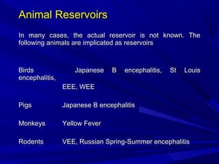
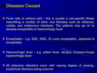
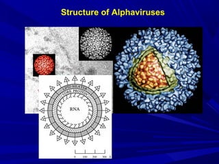
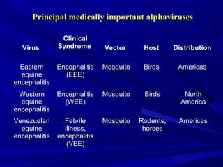
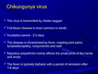
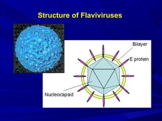
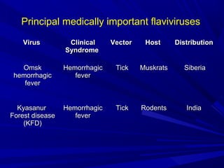
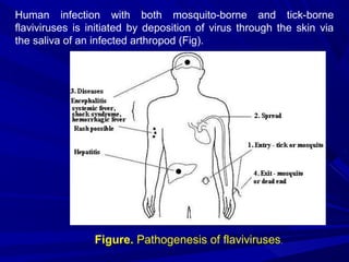
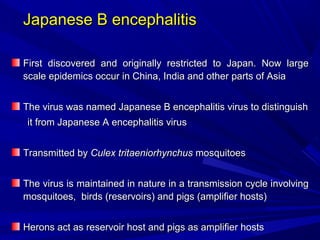
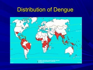
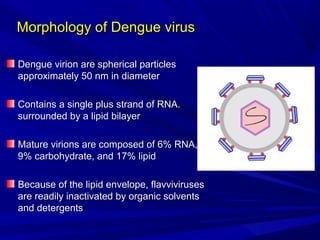
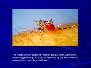
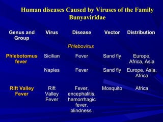
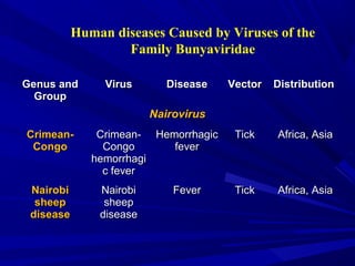
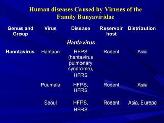
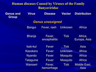
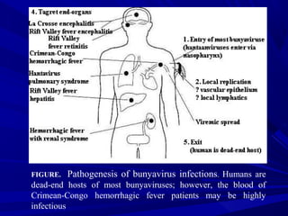
Arboviruses
Arboviruses
Arthropod-borne viruses (arboviruses) are viruses that can
be transmitted to man by arthropod vectors
The WHO definition
The WHO definition
Viruses that are maintained in nature principally, or to an
important extent, through biological transmission between
susceptible vertebrate host by hematophagous arthropods or
through transovarian and possibly venereal transmission in
arthropods”
arthropods”
 They can multiply in the tissues of the arthropod without evidence of
disease or damage
 The vector acquires a lifelong infection through the ingestion of blood
from a viremic vertebrate
 All arboviruses have an RNA genome, and most have a
lipid-containing envelope and consequently are inactivated by
ether or sodium deoxycholate
 Inclusion in this group is based on ecological and epidemiological
considerations and hence it contains viruses of diverse physical
and chemical properties
 Though taxonomically unacceptable, the name “arbovirus” is a
useful biological concept
 Togaviridae
Togaviridae Genus
Genus Alphavirus
Alphavirus
 Flaviviridae
Flaviviridae Genus
Genus Flavivirus
Flavivirus
 Bunyaviridae
Bunyaviridae Genus
Genus Bunyavirus
Bunyavirus
 Reoviridae
Reoviridae Genus
Genus Orbivirus
Orbivirus
 Rhabdoviridae
Rhabdoviridae Genus
Genus Vesiculovirus
Vesiculovirus
 Orthomyxoviridae
Orthomyxoviridae
Approximately 80 arboviruses known to cause human disease
Classification
Virus Reservoir Vector Disease
Chikungunya Monkeys Mosquito Chikungunya fever
Dengue Monkeys,
Man
Mosquito Dengue
haemorrhagic fever
Japanese B
encephalitis
Wild birds,
pigs
Mosquito Encephalitis
Kyasanur forest
disease
Forest birds,
animals
Tick Haemorrhagic fever
Sindbis - Mosquito Sindbis fever
Arboviruses prevalent in India
General properties
The arboviruses share some common biological properties
1. All members produce fatal encephalitis in suckling mice after
intracerebral inoculation
2. They possess haemagglutinin and agglutinate erythrocytes of goose
or day-old chicks
3. They can be grown in tissue cultures of primary cells like chick
embryo fibroblasts or continuous cell lines like vero, and in cultures
of appropriate insect tissues
4. They may also be isolated in the yolk sac or CAM of chick embryo
5. In general, arboviruses are readily inactivated at room temperature
and by bile salts, ether and other lipid solvents
Arthropod Vectors
Arthropod Vectors
Mosquitoes
Mosquitoes
Japanese encephalitis, dengue, yellow fever, Rift valley fever
St. Louis encephalitis, EEE, WEE, VEE etc
Ticks
Ticks
Crimean-Congo haemorrhagic fever,
Crimean-Congo haemorrhagic fever, Kyasanur forest disease
Kyasanur forest disease
and various tick-borne encephalitis etc.
Sandflies
Sandflies
Sicilian sandfly fever
Examples of Arthropod Vectors
Aedes aegyti
Ixodid Ticks
Phlebotomine Sandfly
Culex Mosquito
Animal Reservoirs
Animal Reservoirs
In many cases, the actual reservoir is not known. The
following animals are implicated as reservoirs
Birds
Birds Japanese B encephalitis, St Louis
Japanese B encephalitis, St Louis
encephalitis,
encephalitis,
EEE, WEE
EEE, WEE
Pigs
Pigs Japanese B encephalitis
Japanese B encephalitis
Monkeys
Monkeys Yellow Fever
Yellow Fever
Rodents
Rodents VEE, Russian Spring-Summer encephalitis
VEE, Russian Spring-Summer encephalitis
Pathogenesis
When an infected vector bites a suitable host, the virus
is injected into the capillary circulation
Virus comes in contact with susceptible target cells such as
endothelial cells of capillaries, monocytes, macrophages
and cells of RES
After replication in endothelial cells and RE cells, a
secondary viraemia usually results leading to infection of
target organs such as brain, skin, musculature and liver,
depending on the tissue tropism
The virus reaches the brain by infecting small blood vessels
of the brain or choroid plexus
Diseases Caused
Diseases Caused
Fever with or without rash
Fever with or without rash - this is usually a non-specific illness
- this is usually a non-specific illness
resembling a number of other viral illnesses such as influenza,
rubella, and enterovirus infections. The patients may go on to
develop encephalitis or haemorrhagic fever
Encephalitis
Encephalitis - e.g. EEE, WEE, St Louis encephalitis, Japanese B
- e.g. EEE, WEE, St Louis encephalitis, Japanese B
encephalitis
encephalitis
Haemorrhagic fever
Haemorrhagic fever - e.g. yellow fever, dengue, Crimean-Congo
- e.g. yellow fever, dengue, Crimean-Congo
haemorrhagic fever
haemorrhagic fever
All arbovirus infections occur with varying degree of severity,
subclinical infections being common
Structure of Alphaviruses
Principal medically important alphaviruses
Virus
Virus
Clinical
Clinical
Syndrome
Syndrome Vector
Vector Host
Host Distribution
Distribution
Eastern
Eastern
equine
equine
encephalitis
encephalitis
Encephalitis
Encephalitis
(EEE)
(EEE)
Mosquito
Mosquito Birds
Birds Americas
Americas
Western
Western
equine
equine
encephalitis
encephalitis
Encephalitis
Encephalitis
(WEE)
(WEE)
Mosquito
Mosquito Birds
Birds North
North
America
America
Venezuelan
Venezuelan
equine
equine
encephalitis
encephalitis
Febrile
Febrile
illness,
illness,
encephalitis
encephalitis
(VEE)
(VEE)
Mosquito
Mosquito Rodents,
Rodents,
horses
horses
Americas
Americas
Virus
Virus
Clinical
Clinical
Syndrome
Syndrome Vector
Vector Host
Host Distribution
Distribution
Chikungunya
Chikungunya
(CHIK)
(CHIK)
Febrile
Febrile
illness, rash,
illness, rash,
arthralgia
arthralgia
Mosquito
Mosquito humans
humans Africa,
Africa,
India,
India,
Southeast
Southeast
Asia
Asia
O’nyong-
O’nyong-
nyong (ONN)
nyong (ONN)
Febrile
Febrile
illness, rash,
illness, rash,
arthralgia
arthralgia
Mosquito
Mosquito Primates
Primates Africa
Africa
Sindbis (SIN)
Sindbis (SIN) Febrile
Febrile
illness, rash,
illness, rash,
arthralgia
arthralgia
Mosquito
Mosquito Birds
Birds Nothern
Nothern
Europe,
Europe,
Africa, Asia,
Africa, Asia,
Australia
Australia
Semliki
Semliki
Forest
Forest
Febrile
Febrile
illness, rare
illness, rare
encephalitis
encephalitis
Mosquito
Mosquito Birds
Birds Africa
Africa
Chikungunya virus
 The virus is transmitted by Aedes aegypti
 Full-blown disease is most common in adults
 Incubation period - 2-3 days
 The disease is chracterised by fever, crippling joint pains,
lymphadenopathy, conjunctivitis and rash
 Migratory polyarthritis mainly affects the small joints of the hands
and wrists
 The fever is typically biphasic with a period of remission after
1-6 days
 A maculopapular rash is common and most intense on the trunk
and limbs that may desquamate
 Haemorrhagic manifestations are seen in some patients
 Chickungunya is the native word for the disease in which the
patient lies ‘doubled up’ due to severe joint pains
 The virus first appeared in India in 1963 when it caused extensive
epidemics in calcutta, Madras and other areas
 There is no animal reservoir for the virus
 No vaccine is available
Structure of Flaviviruses
Virus
Virus Clinical
Clinical
Syndrome
Syndrome
Vector
Vector Host
Host Distribution
Distribution
Dengue
Dengue
(DEN)
(DEN)
Febrile
Febrile
illness, rash,
illness, rash,
hemorrhagic
hemorrhagic
fever, shock
fever, shock
syndrome
syndrome
Mosquito
Mosquito Humans
Humans Tropics,
Tropics,
worldwide
worldwide
Yellow fever
Yellow fever
(YF)
(YF)
Hemorrhagic
Hemorrhagic
fever,
fever,
hepatitis
hepatitis
Mosquito
Mosquito Primates,
Primates,
humans
humans
Africa, South
Africa, South
America
America
St. Louis
St. Louis
encephalitis
encephalitis
(SLE)
(SLE)
Encephalitis
Encephalitis Mosquito
Mosquito Birds
Birds Americas
Americas
Principal medically important flaviviruses
Virus
Virus Clinical
Clinical
Syndrome
Syndrome
Vector
Vector Host
Host Distribution
Distribution
Japanese
Japanese
encephalitis
encephalitis
(JE)
(JE)
Encephalitis
Encephalitis Mosquito
Mosquito Pigs, birds
Pigs, birds India, China,
India, China,
Japan,
Japan,
South-East
South-East
Asia
Asia
West Nile
West Nile Febrile
Febrile
illness
illness
Mosquito
Mosquito Birds
Birds Africa, Middle
Africa, Middle
East, Europe
East, Europe
Tick-borne
Tick-borne
encephalitis
encephalitis
(TBE)
(TBE)
Encephalitis
Encephalitis Tick
Tick Rodent
Rodent Europa, Asia
Europa, Asia
Principal medically important flaviviruses
Virus
Virus Clinical
Clinical
Syndrome
Syndrome
Vector
Vector Host
Host Distribution
Distribution
Omsk
Omsk
hemorrhagic
hemorrhagic
fever
fever
Hemorrhagic
Hemorrhagic
fever
fever
Tick
Tick Muskrats
Muskrats Siberia
Siberia
Kyasanur
Kyasanur
Forest disease
Forest disease
(KFD)
(KFD)
Hemorrhagic
Hemorrhagic
fever
fever
Tick
Tick Rodents
Rodents India
India
Principal medically important flaviviruses
Human infection with both mosquito-borne and tick-borne
flaviviruses is initiated by deposition of virus through the skin via
the saliva of an infected arthropod (Fig).
Figure. Pathogenesis of flaviviruses.
Japanese B encephalitis
First discovered and originally restricted to Japan. Now large
scale epidemics occur in China, India and other parts of Asia
The virus was named Japanese B encephalitis virus to distinguish
it from Japanese A encephalitis virus
Transmitted by
Transmitted by Culex tritaeniorhynchus
Culex tritaeniorhynchus mosquitoes
mosquitoes
The virus is maintained in nature in a transmission cycle involving
mosquitoes, birds (reservoirs) and pigs (amplifier hosts)
Herons act as reservoir host and pigs as amplifier hosts
Clinical features
 Most human infections are subclinical: the inapparent to clinical
cases is 500-1000:1
 Incubation period: 5-15 days
 The course of the disease in man may be divided into three stages
1.Prodromal stage
2.Acute encephalitic stage
3.Late stage and sequelae
Prodromal stage
The onset of illness is usually acute and symptoms include fever,
headache and vomiting
Acute encephalitic stage
 After 1-6 days, signs of encephalitis characterised by neck rigidity,
convulsions, altered sensorium and coma appear
Late stage and sequelae
 Convalescence may be prolonged and residual neurological deficits
may not be uncommon
 Case fatality rate varies between 20-40%, but it may reach 58% and
over in some epidemics
 Residual neurological damage may persist in about 50% of survivors
 The disease is usually diagnosed by serology
 No specific therapy is available
Prevention
Preventive measures include mosquito control and establishment
of piggeries away from residential areas
 A formalin inactivated mouse brain vaccine using the Nakayama
strain has been employed for human immunisation
 A live attenuated vaccine prepared in hamster kidney cell line
is also available
Yellow fever
Yellow Fever
Yellow Fever
 Yellow fever is a mosquito-borne acute febrile illness
Yellow fever is a mosquito-borne acute febrile illness
accompanied by hepatic necrosis
 It occurs mainly in tropical Africa and Latin America
It occurs mainly in tropical Africa and Latin America
 It does not exist in India
It does not exist in India
 The name has been derived from ‘yellow quarantine flag’ used
The name has been derived from ‘yellow quarantine flag’ used
by the ships during 17
by the ships during 17th
th
century to warn the presence, on board
of this infection
of this infection
 Yellow fever occurs in 2 major forms: urban and jungle (sylvatic)
Yellow fever occurs in 2 major forms: urban and jungle (sylvatic)
cycle
cycle
 In the urban cycle, man serves both as reservoir and as definitive
host, the virus being transmitted by Aedes aegypti mosquito
 In the forest or sylvatic cycle, wild monkeys act as reservoirs and
several species of forest mosquitos are vectors. Human cases occur
only when humans trespass into the forest or when monkeys raid
villages
Pathogenesis
After introduction into the skin by the mosquito-bite, the
virus multiplies locally and spreads to the local lymphnodes
where it multiplies
From the lymphnodes, it enters the circulating blood. The
virus starts appearing in blood 3-6 days after the bite of
infected mosquito and viraemia lasts for 4-5 days
From blood, the virus becomes localised in the liver,
spleen, kidney, bonemarrow and myocardium, where
it may persist for days
The lesions of yellow fever are due to the localization and
propagation of the virus in a particular organs
Clinical features
 After an incubation period of 3-6 days, patient develops fever with
chills, headache, myalgia and vomiting
 Most cases are mild in nature, especially in the endemic areas, in
whom the disease may present as undifferentiated fever without
jaundice
 The pulse is usually slow despite a high temperature
 In 15-20% of cases, the disease progresses to a more serious form
with jaundice, albuminuria, renal failure and haemorrhagic
manifestations and the patient may die of hepatic and renal failure
Laboratory diagnosis
Diagnosis is usually clinical; laboratory diagnosis is
made for confirmation
1.Detection of viral antigen
2.Isolation of virus
3.Postmortem diagnosis
4.Serology
Detection of viral antigen
Viral antigen or nucleic acid can be detected in tissue
specimen using ELISA, PCR, and immunohistochemistry
Isolation of virus
Virus can be isolated from blood in the first 4 days after
onset or from postmortem tissue by intracerebral inoculation
of mice or inoculating cell lines
Postmortem diagnosis
Can be made histologically
There is severe midzonal degeneration, necrosis and
acidophilic inclusion bodies seen in the liver
Serology
During first week of illness, IgM antibody can be
detected by ELISA
Prophylaxis
 There is no antiviral drug against yellow fever
The control of urban yellow fever can be achieved by eradicating
the vector mosquito
 Two vaccines have been developed for human use
1. The french neurotropic vaccine (Dakar) produced from infected
mouse brain
2. 17D vaccine developed by Theiler in 1937 by passaging the Asibi
strain serially in mouse embryo and whole chick embryo tissues
and then in chick embryo tissue from which the central nervous
tissue has been removed
Dengue Virus
Dengue
Dengue
The word dengue is derived from the
The word dengue is derived from the Swahili Ki denga pepo
Swahili Ki denga pepo
meaning a sudden seizure by a demon
Dengue fever is clinically similar to the illness caused by the
chikungunya and O’nyong-nyong viruses
Dengue virus is widely distributed in the Caribbean region,
south east asia
south east asia
In India first outbreak of dengue was recorded in 1812
In New Delhi, outbreaks of dengue fever reported in
1967,1970,1982, &1996
1967,1970,1982, &1996
Distribution of Dengue
Morphology of Dengue virus
Dengue virion are spherical particles
approximately 50 nm in diameter
Contains a single plus strand of RNA.
surrounded by a lipid bilayer
Mature virions are composed of 6% RNA,
9% carbohydrate, and 17% lipid
Because of the lipid envelope, flavviviruses
are readily inactivated by organic solvents
and detergents
and detergents
 Three viral proteins are associated with virions
 The E (envelope), M (membrane) and C (capsid) proteins
The E protein is the major surface protein of the viral particle and
mediates virus-cell membrane fusion. Antibodies that neutralize
virus infectivity usually recognize this protein and mutations in E
can affect virulence
 M protein is a small proteolytic fragment which is important for
maturation of the virus into an infectious form
 C protein is a component nucleocapsid
Etiology types
Etiology types
Four distinct antigenically related serotypes ( 1to 4) of dengue
virus of the family flaviviridae are etiologically responsible
Infection in human by one serotypes produces life long immunity
against re-infection by the same serotype
All 4 types of dengue viruses are present in India, more than one
type of dengue virus has been occasionally recovered from a
patient
patient
Subsequent infection with other serotypes may result in a severe
illness i. e., dengue haemorrhagic fever or dengue shock
syndrome
syndrome
Some genetic variants within each serotype appear to be more
virulent or have greater epidemic potential
The most common epidemic vector of dengue in the world is the
Aedes aegypti mosquito. It can be identified by the white bands or
scale patterns on its legs and thorax.
Aedes aegypti
• Dengue transmitted by infected female mosquito
• Primarily a daytime feeder
• Lives around human habitation
• Lays eggs and produces larvae preferentially in artificial containers
1.The virus is inoculated into
humans with the mosquito
saliva
2.The virus localizes and
replicates in various organs,
for example, local lymph
nodes, liver, spleen and the
thymus
3.The virus is then released
from these tissues into the
blood
4.Via the blood, the virus
spreads throughout the body
to infect other lymphatic
tissues and organs, which is
accompanied by symptoms
Pathogenesis
5.The mosquito ingests blood containing the virus
6.The virus replicates in the mosquito midgut, the ovaries, nerve
tissue and fat body. It then escapes into the body cavity, and later
infects the salivary glands
7.The virus replicates in the salivary glands and when the
mosquito bites another human, the cycle continues
Clinical features
The disease may occur in two forms
1. Classical dengue fever (break-bone fever)
2. Dengue in more serious forms with haemorrhagic manifestations
(DHF/DSS)
Classical dengue fever
 This usually affects older children and adults
 It has relatively benign course with fever, headache, retrobulbar
pain, conjunctival infection, pain in muscles and bones,
lymphadenopathy and maculopapular rash
 The fever is typically biphasic (saddle back)
 Incubation period is 5 – 8 days
 A maculopapular rash generally appears on 3rd
or 4th
day
 The febrile illness lasts for about 10 days after which recovery is
generally complete. It is rarely fatal
Other manifestations
 Dengue may also occur in more serious forms, with haemorrhagic
manifestations or with shock
 DHF/DSS remains mostly confined among children of 5 -10 years
age group in area where multiple dengue viruses cause disease
 It appers to be hyperimmune response
 On reinfection with a different serotype of dengue virus, antibody
formed against the first virus reacts with the second serotype virus
forming immune complexes (virus-antibody complex)
 In DHF/DSS, initial symptoms are like those of dengue fever but
associated with haemorrhagic rash, thrombocytopenia and shock
 The moratality rate is 5 -10 %
 The disease is more often found in epidemic form in Thailand, South
-East Asia and India where dengue serotypes are regularly present
 All four types of dengue virus are present in India
Clinical Case Definition for Dengue Fever
Classical Dengue fever or Break bone fever is an acute febrile viral
disease frequently presenting with headaches, bone or joint pain,
muscular pains,rash,and leucopenia
Clinical Case Definition for Dengue Hemorrhagic Fever
4 Necessary Criteria:
1. Fever, or recent history of acute fever
2. Hemorrhagic manifestations
3. Low platelet count (100,000/mm3 or less)
4. Objective evidence of “leaky capillaries:”
• elevated hematocrit (20% or more over baseline)
• low albumin
• pleural or other effusions
Clinical Case Definition for Dengue Shock Syndrome
 4 criteria for DHF
+
 Evidence of circulatory failure manifested indirectly by all of the
following
•Rapid and weak pulse
•Narrow pulse pressure (< 20 mm Hg) OR
hypotension for age
•Cold, clammy skin and altered mental status
•Frank shock is direct evidence of circulatory failure
Hemorrhagic Manifestations of Dengue
•Skin hemorrhages:
petechiae, purpura, ecchymoses
•Gingival bleeding
•Nasal bleeding
•Gastrointestinal bleeding:
Hematemesis, melena, hematochezia
•Hematuria
•Increased menstrual flow
Laboratory diagnosis
Specimens
1) For antibody detection – serum
2) For antigen detection – serum
3) For isolation of virus and PCR
a) Serum
b) Plasma
c) Whole blood (washed buffy coat)
d) Autopsy tissues
e) Mosquitoes collected in nature
Haematological diagnosis
 Thrombocytopenia (1,00,000 cells or less per mm3
)
 Haemoconcentration (> 20 % rise in haematocrit)
Microbiological diagnosis
Isolation of virus is difficult hence serology plays a major role in
diagnosis
1. Detection of antibody
 Demonstration of IgM antibody in serum provides early diagnosis
 IgM antibody appears 5 days after onset of symptoms and persists
for one to three months
 Detection of four fold rise in IgG titre in paired sera taken at an
interval of ten days or more is confirmatory
2. Detection of NS1 antigen
 Immunochromatographic test is available for detection of NS1 antigen
(nonstructural protein 1)
 It is a rapid test and detects antigen on the first day of fever
3. Isolation of virus
 Virus isolation can be done by inoculating clinical specimen into
mosquitoes, mosquitoes cell lines (C6/36 or AP-61 cells) or
suckling mice
4. PCR
 Viral RNA can be detected in clinical specimens by RT-PCR
Dengue fever Management
There is no specific antiviral treatment
The management is essentially supportive and symptomatic
The key to success is frequent monitoring and changing
strategies depending on clinical and laboratory evaluations
Bed rest is advisable during the acute febrile phase
Antipyretics or cold sponging should be used to keep the body
temperature < 40
temperature < 400
0
C
C
Analgesics and mild sedation may be required to control pain
Prophylaxis
 Control measures include elimination of mosquitoes
 No effective vaccine is available
 In order to avoid the DHF/DSS in immunised persons, a live
attenuated vaccine containing all four dengue serotypes is under
clinical trials
Tick-borne Flaviviruses
1.Tick-borne encephalitis viruses
a) Russian spring-summer encephalitis
b) Powassan virus
2. Tick-borne haemorrhagic fevers
a) Kyasanur Forest Disease (KFD)
b) Omsk haemorrhagic fever
Kyasanur Forest Disease (KFD)
 Febrile disease associated with hemorrhages that appeared in
Kyasanur Forest of Karnataka in 1957 as a fatal epizootic affecting
monkeys, along with a severe prostrating illness in some of the
villagers in the area
 Antigenically related to the RSSE virus
 Birds and small mammals are believed to be the reservoirs of the
virus
 Virus is transmitted by bite of tick (Haemaphysalis spinigera)
 Ticks may also act as the reservoir hosts as virus is transmitted
transovarially in them
 Monkeys act as amplifier hosts
Clinical features
 Incubation period varies from 3 – 7 days
 Patient develops fever of sudden onset with headache, vomiting,
conjunctivitis, myalgia and severe prostration
 Some patients also develop haemorrhages into the skin, mucosa,
alimentary canal, chest cavity and also in viscera
 Epistaxis may occur in some cases
 Case fatality is about 5 %
Control
 Control of ticks
 The population at risk should be vaccinated with killed KFD vaccine
 Personnel protection – protection of individuals by adequate clothing
and insect repellents
Bunyaviridae is a family of arthropod-borne or rodent-borne,
spherical, enveloped RNA viruses. Bunyaviruses are
responsible for a number of febrile diseases in humans and
other vertebrates. They have either a rodent host or an
arthropod vector and a vertebrate host
Genus and
Genus and
Group
Group
Virus
Virus Disease
Disease Vector
Vector Distributi
Distributi
on
on
Bunyavirus
Bunyavirus
Bunyamwera
Bunyamwera Bunyamwera
Bunyamwera Fever
Fever Mosquito
Mosquito Africa
Africa
Bwamba
Bwamba Bwamba
Bwamba Fever ,
Fever ,
Rash
Rash
Mosquito
Mosquito Africa
Africa
California
California California
California
encephalitis
encephalitis
Encep
Encep
ha-litis
ha-litis
Mosquito
Mosquito North
North
America
America
Simbu
Simbu Shuni
Shuni Fever
Fever Mosquito
Mosquito Africa,
Africa,
Asia
Asia
Human diseases Caused by Viruses
of the Family Bunyaviridae
Human diseases Caused by Viruses of the Family
Bunyaviridae
Genus and
Genus and
Group
Group
Virus
Virus Disease
Disease Vector
Vector Distribution
Distribution
Phlebovirus
Phlebovirus
Phlebotomus
Phlebotomus
fever
fever
Sicilian
Sicilian Fever
Fever Sand fly
Sand fly Europe,
Europe,
Africa, Asia
Africa, Asia
Naples
Naples Fever
Fever Sand fly
Sand fly Europe, Asia,
Europe, Asia,
Africa
Africa
Rift Valley
Rift Valley
Fever
Fever
Rift
Rift
Valley
Valley
Fever
Fever
Fever,
Fever,
encephalitis,
encephalitis,
hemorrhagic
hemorrhagic
fever,
fever,
blindness
blindness
Mosquito
Mosquito Africa
Africa
Human diseases Caused by Viruses of the
Family Bunyaviridae
Genus and
Genus and
Group
Group
Virus
Virus Disease
Disease Vector
Vector Distribution
Distribution
Nairovirus
Nairovirus
Crimean-
Crimean-
Congo
Congo
Crimean-
Crimean-
Congo
Congo
hemorrhagi
hemorrhagi
c fever
c fever
Hemorrhagic
Hemorrhagic
fever
fever
Tick
Tick Africa, Asia
Africa, Asia
Nairobi
Nairobi
sheep
sheep
disease
disease
Nairobi
Nairobi
sheep
sheep
disease
disease
Fever
Fever Tick
Tick Africa, Asia
Africa, Asia
Human diseases Caused by Viruses of the
Family Bunyaviridae
Genus and
Genus and
Group
Group
Virus
Virus Disease
Disease Reservoir
Reservoir
host
host
Distribution
Distribution
Hantavirus
Hantavirus
Hanntavirus
Hanntavirus Hantaan
Hantaan HFPS
HFPS
(hantavirus
(hantavirus
pulmonary
pulmonary
syndrome),
syndrome),
HFRS
HFRS
Rodent
Rodent Asia
Asia
Puumala
Puumala HFPS,
HFPS,
HFRS
HFRS
Rodent
Rodent Asia
Asia
Seoul
Seoul HFPS,
HFPS,
HFRS
HFRS
Rodent
Rodent Asia, Europe
Asia, Europe
Human diseases Caused by Viruses of the Family
Bunyaviridae
Genus and
Genus and
Group
Group
Virus
Virus Disease
Disease Vector
Vector Distribution
Distribution
Genus unassigned
Genus unassigned
Bangui
Bangui Fever, rash
Fever, rash Unknown
Unknown Africa
Africa
Bhanja
Bhanja Fever,
Fever,
encephalitis
encephalitis
Tick
Tick Africa,
Africa,
Europa, Asia
Europa, Asia
Issk-kul
Issk-kul Fever
Fever Tick
Tick Asia
Asia
Kasokero
Kasokero Fever
Fever Unknown
Unknown Africa
Africa
Nyando
Nyando Fever
Fever Mosquito
Mosquito Africa
Africa
Tataguine
Tataguine Fever
Fever Mosquito
Mosquito Africa
Africa
Wanowri
Wanowri Fever,
Fever,
hemorrhage
hemorrhage
Tick
Tick Middle East,
Middle East,
Asia
Asia
FIGURE. Pathogenesis of bunyavirus infections. Humans are
dead-end hosts of most bunyaviruses; however, the blood of
Crimean-Congo hemorrhagic fever patients may be highly
infectious
Signs of Crimean-Congo Hemorrhagic Fever
Laboratory diagnosis of arboviruses
Specimens
Blood, CSF, brain tissue may be used for isolation of virus
1. Virus isolation
a) Suckling mice
Specimens are inoculated intracerebrally into suckling mice
The animal develops fatal encephalitis
Most sensitive method for isolation of arboviruses
b) Tissue culture
 Vero, BHK-21 and mosquito cell lines are inoculated with specimens
 Growth of virus in cell cultures is identified by immunofluorescence,
haemagglutination inhibition, CFT, ELISA or neutralisation tests
2. Serology
 Usually used to make a diagnosis of arbovirus infections
3. Direct detection tests
 Methods for detection of antigen and nucleic acids are available
Prevention of arbovirus infections
Surveillance
Surveillance - of disease and vector populations
- of disease and vector populations
Control of vector
Control of vector - pesticides, elimination of breeding grounds
- pesticides, elimination of breeding grounds
Personal protection
Personal protection - screening of houses, bed nets, insect
- screening of houses, bed nets, insect
repellants.
repellants. When possible, wear protective clothing while outdoors
When possible, wear protective clothing while outdoors
Vaccination
Vaccination - available for a number of arboviral infections e.g.
- available for a number of arboviral infections e.g.
Yellow fever, Japanese encephalitis, Russian tick-borne
encephalitis
encephalitis
Treatment of arbovirus infections
No specific therapy
No specific therapy
Arboviral encephalitis treated by hospitalization, intravenous fluids,
respiratory support, prevention of secondary infections, and good
nursing care
nursing care

arboviruses.pdf

  • 1.
  • 2.
    Arthropod-borne viruses (arboviruses)are viruses that can Arthropod-borne viruses (arboviruses) are viruses that can be transmitted to man by arthropod vectors be transmitted to man by arthropod vectors The WHO definition The WHO definition Viruses that are maintained in nature principally, or to an Viruses that are maintained in nature principally, or to an important extent, through biological transmission between important extent, through biological transmission between susceptible vertebrate host by hematophagous arthropods or susceptible vertebrate host by hematophagous arthropods or through transovarian and possibly venereal transmission in through transovarian and possibly venereal transmission in arthropods” arthropods”
  • 3.
     They canmultiply in the tissues of the arthropod without evidence of disease or damage  The vector acquires a lifelong infection through the ingestion of blood from a viremic vertebrate  All arboviruses have an RNA genome, and most have a lipid-containing envelope and consequently are inactivated by ether or sodium deoxycholate  Inclusion in this group is based on ecological and epidemiological considerations and hence it contains viruses of diverse physical and chemical properties  Though taxonomically unacceptable, the name “arbovirus” is a useful biological concept
  • 4.
     Togaviridae Togaviridae Genus GenusAlphavirus Alphavirus  Flaviviridae Flaviviridae Genus Genus Flavivirus Flavivirus  Bunyaviridae Bunyaviridae Genus Genus Bunyavirus Bunyavirus  Reoviridae Reoviridae Genus Genus Orbivirus Orbivirus  Rhabdoviridae Rhabdoviridae Genus Genus Vesiculovirus Vesiculovirus  Orthomyxoviridae Orthomyxoviridae Approximately 80 arboviruses known to cause human disease Approximately 80 arboviruses known to cause human disease Classification
  • 5.
    Virus Reservoir VectorDisease Chikungunya Monkeys Mosquito Chikungunya fever Dengue Monkeys, Man Mosquito Dengue haemorrhagic fever Japanese B encephalitis Wild birds, pigs Mosquito Encephalitis Kyasanur forest disease Forest birds, animals Tick Haemorrhagic fever Sindbis - Mosquito Sindbis fever Arboviruses prevalent in India
  • 6.
    General properties The arbovirusesshare some common biological properties 1. All members produce fatal encephalitis in suckling mice after intracerebral inoculation 2. They possess haemagglutinin and agglutinate erythrocytes of goose or day-old chicks 3. They can be grown in tissue cultures of primary cells like chick embryo fibroblasts or continuous cell lines like vero, and in cultures of appropriate insect tissues 4. They may also be isolated in the yolk sac or CAM of chick embryo 5. In general, arboviruses are readily inactivated at room temperature and by bile salts, ether and other lipid solvents
  • 7.
    Arthropod Vectors Arthropod Vectors Mosquitoes Mosquitoes Japaneseencephalitis, dengue, yellow fever, Rift valley fever Japanese encephalitis, dengue, yellow fever, Rift valley fever St. Louis encephalitis, EEE, WEE, VEE etc St. Louis encephalitis, EEE, WEE, VEE etc Ticks Ticks Crimean-Congo haemorrhagic fever, Crimean-Congo haemorrhagic fever, Kyasanur forest disease Kyasanur forest disease and various tick-borne encephalitis etc. and various tick-borne encephalitis etc. Sandflies Sandflies Sicilian sandfly fever Sicilian sandfly fever
  • 8.
    Examples of ArthropodVectors Examples of Arthropod Vectors Aedes aegyti Ixodid Ticks Phlebotomine Sandfly Culex Mosquito
  • 9.
    Animal Reservoirs Animal Reservoirs Inmany cases, the actual reservoir is not known. The In many cases, the actual reservoir is not known. The following animals are implicated as reservoirs following animals are implicated as reservoirs Birds Birds Japanese B encephalitis, St Louis Japanese B encephalitis, St Louis encephalitis, encephalitis, EEE, WEE EEE, WEE Pigs Pigs Japanese B encephalitis Japanese B encephalitis Monkeys Monkeys Yellow Fever Yellow Fever Rodents Rodents VEE, Russian Spring-Summer encephalitis VEE, Russian Spring-Summer encephalitis
  • 10.
    Pathogenesis When an infectedvector bites a suitable host, the virus is injected into the capillary circulation Virus comes in contact with susceptible target cells such as endothelial cells of capillaries, monocytes, macrophages and cells of RES After replication in endothelial cells and RE cells, a secondary viraemia usually results leading to infection of target organs such as brain, skin, musculature and liver, depending on the tissue tropism The virus reaches the brain by infecting small blood vessels of the brain or choroid plexus
  • 11.
    Diseases Caused Diseases Caused Feverwith or without rash Fever with or without rash - this is usually a non-specific illness - this is usually a non-specific illness resembling a number of other viral illnesses such as influenza, resembling a number of other viral illnesses such as influenza, rubella, and enterovirus infections. The patients may go on to rubella, and enterovirus infections. The patients may go on to develop encephalitis or haemorrhagic fever develop encephalitis or haemorrhagic fever Encephalitis Encephalitis - e.g. EEE, WEE, St Louis encephalitis, Japanese B - e.g. EEE, WEE, St Louis encephalitis, Japanese B encephalitis encephalitis Haemorrhagic fever Haemorrhagic fever - e.g. yellow fever, dengue, Crimean-Congo - e.g. yellow fever, dengue, Crimean-Congo haemorrhagic fever haemorrhagic fever All arbovirus infections occur with varying degree of severity, All arbovirus infections occur with varying degree of severity, subclinical infections being common subclinical infections being common
  • 12.
  • 13.
    Principal medically importantalphaviruses Virus Virus Clinical Clinical Syndrome Syndrome Vector Vector Host Host Distribution Distribution Eastern Eastern equine equine encephalitis encephalitis Encephalitis Encephalitis (EEE) (EEE) Mosquito Mosquito Birds Birds Americas Americas Western Western equine equine encephalitis encephalitis Encephalitis Encephalitis (WEE) (WEE) Mosquito Mosquito Birds Birds North North America America Venezuelan Venezuelan equine equine encephalitis encephalitis Febrile Febrile illness, illness, encephalitis encephalitis (VEE) (VEE) Mosquito Mosquito Rodents, Rodents, horses horses Americas Americas
  • 14.
    Virus Virus Clinical Clinical Syndrome Syndrome Vector Vector Host HostDistribution Distribution Chikungunya Chikungunya (CHIK) (CHIK) Febrile Febrile illness, rash, illness, rash, arthralgia arthralgia Mosquito Mosquito humans humans Africa, Africa, India, India, Southeast Southeast Asia Asia O’nyong- O’nyong- nyong (ONN) nyong (ONN) Febrile Febrile illness, rash, illness, rash, arthralgia arthralgia Mosquito Mosquito Primates Primates Africa Africa Sindbis (SIN) Sindbis (SIN) Febrile Febrile illness, rash, illness, rash, arthralgia arthralgia Mosquito Mosquito Birds Birds Nothern Nothern Europe, Europe, Africa, Asia, Africa, Asia, Australia Australia Semliki Semliki Forest Forest Febrile Febrile illness, rare illness, rare encephalitis encephalitis Mosquito Mosquito Birds Birds Africa Africa
  • 15.
    Chikungunya virus  Thevirus is transmitted by Aedes aegypti  Full-blown disease is most common in adults  Incubation period - 2-3 days  The disease is chracterised by fever, crippling joint pains, lymphadenopathy, conjunctivitis and rash  Migratory polyarthritis mainly affects the small joints of the hands and wrists  The fever is typically biphasic with a period of remission after 1-6 days
  • 16.
     A maculopapularrash is common and most intense on the trunk and limbs that may desquamate  Haemorrhagic manifestations are seen in some patients  Chickungunya is the native word for the disease in which the patient lies ‘doubled up’ due to severe joint pains  The virus first appeared in India in 1963 when it caused extensive epidemics in calcutta, Madras and other areas  There is no animal reservoir for the virus  No vaccine is available
  • 17.
  • 18.
    Virus Virus Clinical Clinical Syndrome Syndrome Vector Vector Host HostDistribution Distribution Dengue Dengue (DEN) (DEN) Febrile Febrile illness, rash, illness, rash, hemorrhagic hemorrhagic fever, shock fever, shock syndrome syndrome Mosquito Mosquito Humans Humans Tropics, Tropics, worldwide worldwide Yellow fever Yellow fever (YF) (YF) Hemorrhagic Hemorrhagic fever, fever, hepatitis hepatitis Mosquito Mosquito Primates, Primates, humans humans Africa, South Africa, South America America St. Louis St. Louis encephalitis encephalitis (SLE) (SLE) Encephalitis Encephalitis Mosquito Mosquito Birds Birds Americas Americas Principal medically important flaviviruses
  • 19.
    Virus Virus Clinical Clinical Syndrome Syndrome Vector Vector Host HostDistribution Distribution Japanese Japanese encephalitis encephalitis (JE) (JE) Encephalitis Encephalitis Mosquito Mosquito Pigs, birds Pigs, birds India, China, India, China, Japan, Japan, South-East South-East Asia Asia West Nile West Nile Febrile Febrile illness illness Mosquito Mosquito Birds Birds Africa, Middle Africa, Middle East, Europe East, Europe Tick-borne Tick-borne encephalitis encephalitis (TBE) (TBE) Encephalitis Encephalitis Tick Tick Rodent Rodent Europa, Asia Europa, Asia Principal medically important flaviviruses
  • 20.
    Virus Virus Clinical Clinical Syndrome Syndrome Vector Vector Host HostDistribution Distribution Omsk Omsk hemorrhagic hemorrhagic fever fever Hemorrhagic Hemorrhagic fever fever Tick Tick Muskrats Muskrats Siberia Siberia Kyasanur Kyasanur Forest disease Forest disease (KFD) (KFD) Hemorrhagic Hemorrhagic fever fever Tick Tick Rodents Rodents India India Principal medically important flaviviruses
  • 21.
    Human infection withboth mosquito-borne and tick-borne flaviviruses is initiated by deposition of virus through the skin via the saliva of an infected arthropod (Fig). Figure. Pathogenesis of flaviviruses.
  • 22.
    Japanese B encephalitis JapaneseB encephalitis First discovered and originally restricted to Japan. Now large First discovered and originally restricted to Japan. Now large scale epidemics occur in China, India and other parts of Asia scale epidemics occur in China, India and other parts of Asia The virus was named Japanese B encephalitis virus to distinguish The virus was named Japanese B encephalitis virus to distinguish it from Japanese A encephalitis virus it from Japanese A encephalitis virus Transmitted by Transmitted by Culex tritaeniorhynchus Culex tritaeniorhynchus mosquitoes mosquitoes The virus is maintained in nature in a transmission cycle involving The virus is maintained in nature in a transmission cycle involving mosquitoes, birds (reservoirs) and pigs (amplifier hosts) mosquitoes, birds (reservoirs) and pigs (amplifier hosts) Herons act as reservoir host and pigs as amplifier hosts Herons act as reservoir host and pigs as amplifier hosts
  • 23.
    Clinical features  Mosthuman infections are subclinical: the inapparent to clinical cases is 500-1000:1  Incubation period: 5-15 days  The course of the disease in man may be divided into three stages 1.Prodromal stage 2.Acute encephalitic stage 3.Late stage and sequelae
  • 24.
    Prodromal stage The onsetof illness is usually acute and symptoms include fever, headache and vomiting Acute encephalitic stage  After 1-6 days, signs of encephalitis characterised by neck rigidity, convulsions, altered sensorium and coma appear Late stage and sequelae  Convalescence may be prolonged and residual neurological deficits may not be uncommon  Case fatality rate varies between 20-40%, but it may reach 58% and over in some epidemics  Residual neurological damage may persist in about 50% of survivors
  • 25.
     The diseaseis usually diagnosed by serology  No specific therapy is available Prevention Preventive measures include mosquito control and establishment of piggeries away from residential areas  A formalin inactivated mouse brain vaccine using the Nakayama strain has been employed for human immunisation  A live attenuated vaccine prepared in hamster kidney cell line is also available
  • 26.
  • 27.
    Yellow Fever Yellow Fever Yellow fever is a mosquito-borne acute febrile illness Yellow fever is a mosquito-borne acute febrile illness accompanied by hepatic necrosis accompanied by hepatic necrosis  It occurs mainly in tropical Africa and Latin America It occurs mainly in tropical Africa and Latin America  It does not exist in India It does not exist in India  The name has been derived from ‘yellow quarantine flag’ used The name has been derived from ‘yellow quarantine flag’ used by the ships during 17 by the ships during 17th th century to warn the presence, on board century to warn the presence, on board of this infection of this infection  Yellow fever occurs in 2 major forms: urban and jungle (sylvatic) Yellow fever occurs in 2 major forms: urban and jungle (sylvatic) cycle cycle
  • 28.
     In theurban cycle, man serves both as reservoir and as definitive host, the virus being transmitted by Aedes aegypti mosquito  In the forest or sylvatic cycle, wild monkeys act as reservoirs and several species of forest mosquitos are vectors. Human cases occur only when humans trespass into the forest or when monkeys raid villages
  • 29.
    Pathogenesis After introduction intothe skin by the mosquito-bite, the virus multiplies locally and spreads to the local lymphnodes where it multiplies From the lymphnodes, it enters the circulating blood. The virus starts appearing in blood 3-6 days after the bite of infected mosquito and viraemia lasts for 4-5 days From blood, the virus becomes localised in the liver, spleen, kidney, bonemarrow and myocardium, where it may persist for days The lesions of yellow fever are due to the localization and propagation of the virus in a particular organs
  • 30.
    Clinical features  Afteran incubation period of 3-6 days, patient develops fever with chills, headache, myalgia and vomiting  Most cases are mild in nature, especially in the endemic areas, in whom the disease may present as undifferentiated fever without jaundice  The pulse is usually slow despite a high temperature  In 15-20% of cases, the disease progresses to a more serious form with jaundice, albuminuria, renal failure and haemorrhagic manifestations and the patient may die of hepatic and renal failure
  • 31.
    Laboratory diagnosis Diagnosis isusually clinical; laboratory diagnosis is made for confirmation 1.Detection of viral antigen 2.Isolation of virus 3.Postmortem diagnosis 4.Serology
  • 32.
    Detection of viralantigen Viral antigen or nucleic acid can be detected in tissue specimen using ELISA, PCR, and immunohistochemistry Isolation of virus Virus can be isolated from blood in the first 4 days after onset or from postmortem tissue by intracerebral inoculation of mice or inoculating cell lines
  • 33.
    Postmortem diagnosis Can bemade histologically There is severe midzonal degeneration, necrosis and acidophilic inclusion bodies seen in the liver Serology During first week of illness, IgM antibody can be detected by ELISA
  • 34.
    Prophylaxis  There isno antiviral drug against yellow fever The control of urban yellow fever can be achieved by eradicating the vector mosquito  Two vaccines have been developed for human use 1. The french neurotropic vaccine (Dakar) produced from infected mouse brain 2. 17D vaccine developed by Theiler in 1937 by passaging the Asibi strain serially in mouse embryo and whole chick embryo tissues and then in chick embryo tissue from which the central nervous tissue has been removed
  • 35.
  • 36.
    Dengue Dengue The word dengueis derived from the The word dengue is derived from the Swahili Ki denga pepo Swahili Ki denga pepo meaning a sudden seizure by a demon meaning a sudden seizure by a demon Dengue fever is clinically similar to the illness caused by the Dengue fever is clinically similar to the illness caused by the chikungunya and O’nyong-nyong viruses chikungunya and O’nyong-nyong viruses Dengue virus is widely distributed in the Caribbean region, Dengue virus is widely distributed in the Caribbean region, south east asia south east asia In India first outbreak of dengue was recorded in 1812 In India first outbreak of dengue was recorded in 1812 In New Delhi, outbreaks of dengue fever reported in In New Delhi, outbreaks of dengue fever reported in 1967,1970,1982, &1996 1967,1970,1982, &1996
  • 37.
  • 38.
    Morphology of Denguevirus Morphology of Dengue virus Dengue virion are spherical particles Dengue virion are spherical particles approximately 50 nm in diameter approximately 50 nm in diameter Contains a single plus strand of RNA. Contains a single plus strand of RNA. surrounded by a lipid bilayer surrounded by a lipid bilayer Mature virions are composed of 6% RNA, Mature virions are composed of 6% RNA, 9% carbohydrate, and 17% lipid 9% carbohydrate, and 17% lipid Because of the lipid envelope, flavviviruses Because of the lipid envelope, flavviviruses are readily inactivated by organic solvents are readily inactivated by organic solvents and detergents and detergents
  • 39.
     Three viralproteins are associated with virions  The E (envelope), M (membrane) and C (capsid) proteins
  • 40.
    The E proteinis the major surface protein of the viral particle and mediates virus-cell membrane fusion. Antibodies that neutralize virus infectivity usually recognize this protein and mutations in E can affect virulence  M protein is a small proteolytic fragment which is important for maturation of the virus into an infectious form  C protein is a component nucleocapsid
  • 41.
    Etiology types Etiology types Fourdistinct antigenically related serotypes ( 1to 4) of dengue Four distinct antigenically related serotypes ( 1to 4) of dengue virus of the family flaviviridae are etiologically responsible virus of the family flaviviridae are etiologically responsible Infection in human by one serotypes produces life long immunity Infection in human by one serotypes produces life long immunity against re-infection by the same serotype against re-infection by the same serotype All 4 types of dengue viruses are present in India, more than one All 4 types of dengue viruses are present in India, more than one type of dengue virus has been occasionally recovered from a type of dengue virus has been occasionally recovered from a patient patient Subsequent infection with other serotypes may result in a severe Subsequent infection with other serotypes may result in a severe illness i. e., dengue haemorrhagic fever or dengue shock illness i. e., dengue haemorrhagic fever or dengue shock syndrome syndrome Some genetic variants within each serotype appear to be more Some genetic variants within each serotype appear to be more virulent or have greater epidemic potential virulent or have greater epidemic potential
  • 42.
    The most commonepidemic vector of dengue in the world is the Aedes aegypti mosquito. It can be identified by the white bands or scale patterns on its legs and thorax.
  • 43.
    Aedes aegypti • Denguetransmitted by infected female mosquito • Primarily a daytime feeder • Lives around human habitation • Lays eggs and produces larvae preferentially in artificial containers
  • 44.
    1.The virus isinoculated into humans with the mosquito saliva 2.The virus localizes and replicates in various organs, for example, local lymph nodes, liver, spleen and the thymus 3.The virus is then released from these tissues into the blood 4.Via the blood, the virus spreads throughout the body to infect other lymphatic tissues and organs, which is accompanied by symptoms Pathogenesis
  • 45.
    5.The mosquito ingestsblood containing the virus 6.The virus replicates in the mosquito midgut, the ovaries, nerve tissue and fat body. It then escapes into the body cavity, and later infects the salivary glands 7.The virus replicates in the salivary glands and when the mosquito bites another human, the cycle continues
  • 46.
    Clinical features The diseasemay occur in two forms 1. Classical dengue fever (break-bone fever) 2. Dengue in more serious forms with haemorrhagic manifestations (DHF/DSS)
  • 47.
    Classical dengue fever This usually affects older children and adults  It has relatively benign course with fever, headache, retrobulbar pain, conjunctival infection, pain in muscles and bones, lymphadenopathy and maculopapular rash  The fever is typically biphasic (saddle back)  Incubation period is 5 – 8 days  A maculopapular rash generally appears on 3rd or 4th day  The febrile illness lasts for about 10 days after which recovery is generally complete. It is rarely fatal
  • 48.
    Other manifestations  Denguemay also occur in more serious forms, with haemorrhagic manifestations or with shock  DHF/DSS remains mostly confined among children of 5 -10 years age group in area where multiple dengue viruses cause disease  It appers to be hyperimmune response  On reinfection with a different serotype of dengue virus, antibody formed against the first virus reacts with the second serotype virus forming immune complexes (virus-antibody complex)
  • 49.
     In DHF/DSS,initial symptoms are like those of dengue fever but associated with haemorrhagic rash, thrombocytopenia and shock  The moratality rate is 5 -10 %  The disease is more often found in epidemic form in Thailand, South -East Asia and India where dengue serotypes are regularly present  All four types of dengue virus are present in India
  • 50.
    Clinical Case Definitionfor Dengue Fever Classical Dengue fever or Break bone fever is an acute febrile viral disease frequently presenting with headaches, bone or joint pain, muscular pains,rash,and leucopenia Clinical Case Definition for Dengue Hemorrhagic Fever 4 Necessary Criteria: 1. Fever, or recent history of acute fever 2. Hemorrhagic manifestations 3. Low platelet count (100,000/mm3 or less) 4. Objective evidence of “leaky capillaries:” • elevated hematocrit (20% or more over baseline) • low albumin • pleural or other effusions
  • 51.
    Clinical Case Definitionfor Dengue Shock Syndrome  4 criteria for DHF +  Evidence of circulatory failure manifested indirectly by all of the following •Rapid and weak pulse •Narrow pulse pressure (< 20 mm Hg) OR hypotension for age •Cold, clammy skin and altered mental status •Frank shock is direct evidence of circulatory failure
  • 52.
    Hemorrhagic Manifestations ofDengue •Skin hemorrhages: petechiae, purpura, ecchymoses •Gingival bleeding •Nasal bleeding •Gastrointestinal bleeding: Hematemesis, melena, hematochezia •Hematuria •Increased menstrual flow
  • 53.
    Laboratory diagnosis Specimens 1) Forantibody detection – serum 2) For antigen detection – serum 3) For isolation of virus and PCR a) Serum b) Plasma c) Whole blood (washed buffy coat) d) Autopsy tissues e) Mosquitoes collected in nature
  • 54.
    Haematological diagnosis  Thrombocytopenia(1,00,000 cells or less per mm3 )  Haemoconcentration (> 20 % rise in haematocrit) Microbiological diagnosis Isolation of virus is difficult hence serology plays a major role in diagnosis 1. Detection of antibody  Demonstration of IgM antibody in serum provides early diagnosis  IgM antibody appears 5 days after onset of symptoms and persists for one to three months  Detection of four fold rise in IgG titre in paired sera taken at an interval of ten days or more is confirmatory
  • 55.
    2. Detection ofNS1 antigen  Immunochromatographic test is available for detection of NS1 antigen (nonstructural protein 1)  It is a rapid test and detects antigen on the first day of fever 3. Isolation of virus  Virus isolation can be done by inoculating clinical specimen into mosquitoes, mosquitoes cell lines (C6/36 or AP-61 cells) or suckling mice 4. PCR  Viral RNA can be detected in clinical specimens by RT-PCR
  • 56.
    Dengue fever Management Denguefever Management There is no specific antiviral treatment There is no specific antiviral treatment The management is essentially supportive and symptomatic The management is essentially supportive and symptomatic The key to success is frequent monitoring and changing The key to success is frequent monitoring and changing strategies depending on clinical and laboratory evaluations strategies depending on clinical and laboratory evaluations Bed rest is advisable during the acute febrile phase Bed rest is advisable during the acute febrile phase Antipyretics or cold sponging should be used to keep the body Antipyretics or cold sponging should be used to keep the body temperature < 40 temperature < 400 0 C C Analgesics and mild sedation may be required to control pain Analgesics and mild sedation may be required to control pain
  • 57.
    Prophylaxis  Control measuresinclude elimination of mosquitoes  No effective vaccine is available  In order to avoid the DHF/DSS in immunised persons, a live attenuated vaccine containing all four dengue serotypes is under clinical trials
  • 58.
    Tick-borne Flaviviruses 1.Tick-borne encephalitisviruses a) Russian spring-summer encephalitis b) Powassan virus 2. Tick-borne haemorrhagic fevers a) Kyasanur Forest Disease (KFD) b) Omsk haemorrhagic fever
  • 59.
    Kyasanur Forest Disease(KFD)  Febrile disease associated with hemorrhages that appeared in Kyasanur Forest of Karnataka in 1957 as a fatal epizootic affecting monkeys, along with a severe prostrating illness in some of the villagers in the area  Antigenically related to the RSSE virus  Birds and small mammals are believed to be the reservoirs of the virus  Virus is transmitted by bite of tick (Haemaphysalis spinigera)  Ticks may also act as the reservoir hosts as virus is transmitted transovarially in them  Monkeys act as amplifier hosts
  • 60.
    Clinical features  Incubationperiod varies from 3 – 7 days  Patient develops fever of sudden onset with headache, vomiting, conjunctivitis, myalgia and severe prostration  Some patients also develop haemorrhages into the skin, mucosa, alimentary canal, chest cavity and also in viscera  Epistaxis may occur in some cases  Case fatality is about 5 %
  • 61.
    Control  Control ofticks  The population at risk should be vaccinated with killed KFD vaccine  Personnel protection – protection of individuals by adequate clothing and insect repellents
  • 62.
    Bunyaviridae is afamily of arthropod-borne or rodent-borne, spherical, enveloped RNA viruses. Bunyaviruses are responsible for a number of febrile diseases in humans and other vertebrates. They have either a rodent host or an arthropod vector and a vertebrate host
  • 63.
    Genus and Genus and Group Group Virus VirusDisease Disease Vector Vector Distributi Distributi on on Bunyavirus Bunyavirus Bunyamwera Bunyamwera Bunyamwera Bunyamwera Fever Fever Mosquito Mosquito Africa Africa Bwamba Bwamba Bwamba Bwamba Fever , Fever , Rash Rash Mosquito Mosquito Africa Africa California California California California encephalitis encephalitis Encep Encep ha-litis ha-litis Mosquito Mosquito North North America America Simbu Simbu Shuni Shuni Fever Fever Mosquito Mosquito Africa, Africa, Asia Asia Human diseases Caused by Viruses of the Family Bunyaviridae
  • 64.
    Human diseases Causedby Viruses of the Family Bunyaviridae Genus and Genus and Group Group Virus Virus Disease Disease Vector Vector Distribution Distribution Phlebovirus Phlebovirus Phlebotomus Phlebotomus fever fever Sicilian Sicilian Fever Fever Sand fly Sand fly Europe, Europe, Africa, Asia Africa, Asia Naples Naples Fever Fever Sand fly Sand fly Europe, Asia, Europe, Asia, Africa Africa Rift Valley Rift Valley Fever Fever Rift Rift Valley Valley Fever Fever Fever, Fever, encephalitis, encephalitis, hemorrhagic hemorrhagic fever, fever, blindness blindness Mosquito Mosquito Africa Africa
  • 65.
    Human diseases Causedby Viruses of the Family Bunyaviridae Genus and Genus and Group Group Virus Virus Disease Disease Vector Vector Distribution Distribution Nairovirus Nairovirus Crimean- Crimean- Congo Congo Crimean- Crimean- Congo Congo hemorrhagi hemorrhagi c fever c fever Hemorrhagic Hemorrhagic fever fever Tick Tick Africa, Asia Africa, Asia Nairobi Nairobi sheep sheep disease disease Nairobi Nairobi sheep sheep disease disease Fever Fever Tick Tick Africa, Asia Africa, Asia
  • 66.
    Human diseases Causedby Viruses of the Family Bunyaviridae Genus and Genus and Group Group Virus Virus Disease Disease Reservoir Reservoir host host Distribution Distribution Hantavirus Hantavirus Hanntavirus Hanntavirus Hantaan Hantaan HFPS HFPS (hantavirus (hantavirus pulmonary pulmonary syndrome), syndrome), HFRS HFRS Rodent Rodent Asia Asia Puumala Puumala HFPS, HFPS, HFRS HFRS Rodent Rodent Asia Asia Seoul Seoul HFPS, HFPS, HFRS HFRS Rodent Rodent Asia, Europe Asia, Europe
  • 67.
    Human diseases Causedby Viruses of the Family Bunyaviridae Genus and Genus and Group Group Virus Virus Disease Disease Vector Vector Distribution Distribution Genus unassigned Genus unassigned Bangui Bangui Fever, rash Fever, rash Unknown Unknown Africa Africa Bhanja Bhanja Fever, Fever, encephalitis encephalitis Tick Tick Africa, Africa, Europa, Asia Europa, Asia Issk-kul Issk-kul Fever Fever Tick Tick Asia Asia Kasokero Kasokero Fever Fever Unknown Unknown Africa Africa Nyando Nyando Fever Fever Mosquito Mosquito Africa Africa Tataguine Tataguine Fever Fever Mosquito Mosquito Africa Africa Wanowri Wanowri Fever, Fever, hemorrhage hemorrhage Tick Tick Middle East, Middle East, Asia Asia
  • 68.
    FIGURE. Pathogenesis ofbunyavirus infections. Humans are dead-end hosts of most bunyaviruses; however, the blood of Crimean-Congo hemorrhagic fever patients may be highly infectious
  • 69.
    Signs of Crimean-CongoHemorrhagic Fever
  • 70.
    Laboratory diagnosis ofarboviruses Specimens Blood, CSF, brain tissue may be used for isolation of virus 1. Virus isolation a) Suckling mice Specimens are inoculated intracerebrally into suckling mice The animal develops fatal encephalitis Most sensitive method for isolation of arboviruses
  • 71.
    b) Tissue culture Vero, BHK-21 and mosquito cell lines are inoculated with specimens  Growth of virus in cell cultures is identified by immunofluorescence, haemagglutination inhibition, CFT, ELISA or neutralisation tests 2. Serology  Usually used to make a diagnosis of arbovirus infections 3. Direct detection tests  Methods for detection of antigen and nucleic acids are available
  • 72.
    Prevention of arbovirusinfections Prevention of arbovirus infections Surveillance Surveillance - of disease and vector populations - of disease and vector populations Control of vector Control of vector - pesticides, elimination of breeding grounds - pesticides, elimination of breeding grounds Personal protection Personal protection - screening of houses, bed nets, insect - screening of houses, bed nets, insect repellants. repellants. When possible, wear protective clothing while outdoors When possible, wear protective clothing while outdoors Vaccination Vaccination - available for a number of arboviral infections e.g. - available for a number of arboviral infections e.g. Yellow fever, Japanese encephalitis, Russian tick-borne Yellow fever, Japanese encephalitis, Russian tick-borne encephalitis encephalitis
  • 73.
    Treatment of arbovirusinfections Treatment of arbovirus infections No specific therapy No specific therapy Arboviral encephalitis treated by hospitalization, intravenous fluids, Arboviral encephalitis treated by hospitalization, intravenous fluids, respiratory support, prevention of secondary infections, and good respiratory support, prevention of secondary infections, and good nursing care nursing care