SlideShare a Scribd company logo
Approach to the
Cardiovascular
Examination
Carly Welch
Final Year Medical Student
University of Birmingham
Contents
 Basic approach to the CVS patient
 General observations
 Hand signs
 Pulse
 Blood Pressure
 Eyes & face
 Neck & JVP
 Legs
 Examination of the praecordium
 Observation
 Palpation
 Auscultation
 Extra things to do
 Case scenarios
 Summary
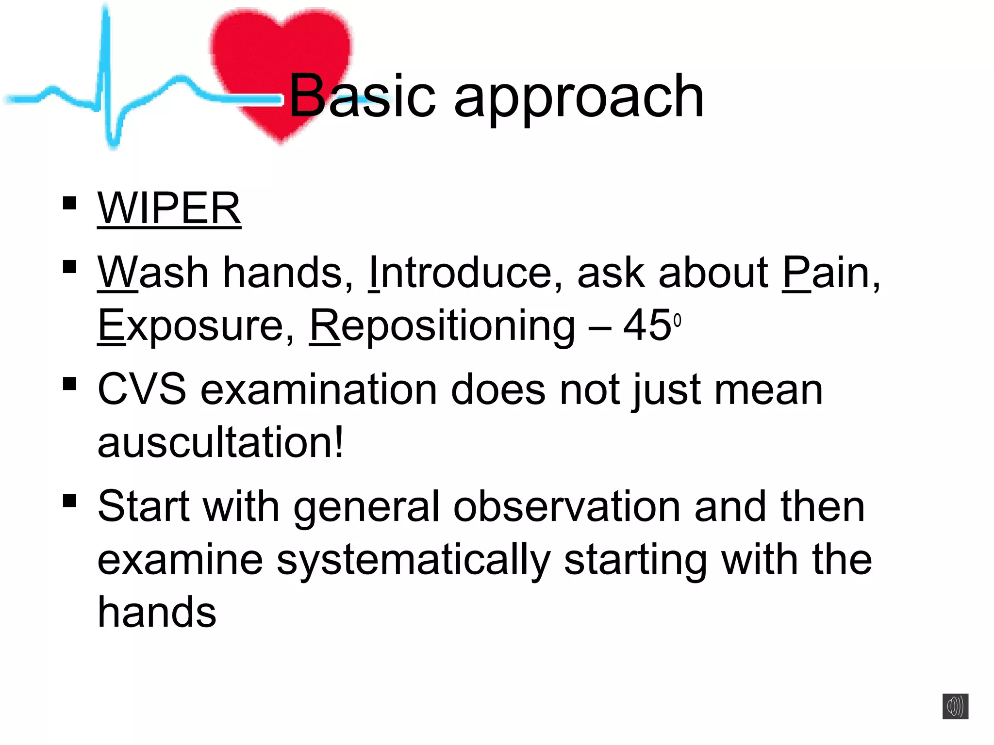
Basic approach
 WIPER
 Wash hands, Introduce, ask about Pain,
Exposure, Repositioning – 45o
 CVS examination does not just mean
auscultation!
 Start with general observation and then
examine systematically starting with the
hands
General observation
 Comfortable? Alert? Generally well or
unwell?
 Do they look breathless or cyanosed?
 Can you see any obvious scars or
pacemakers?
 Any clues around the bed e.g. GTN
sprays?
Hand signs
Clubbing
Koilonychia Palmar erythema
Splinter haemorrhages
Osler nodes Janeway lesions
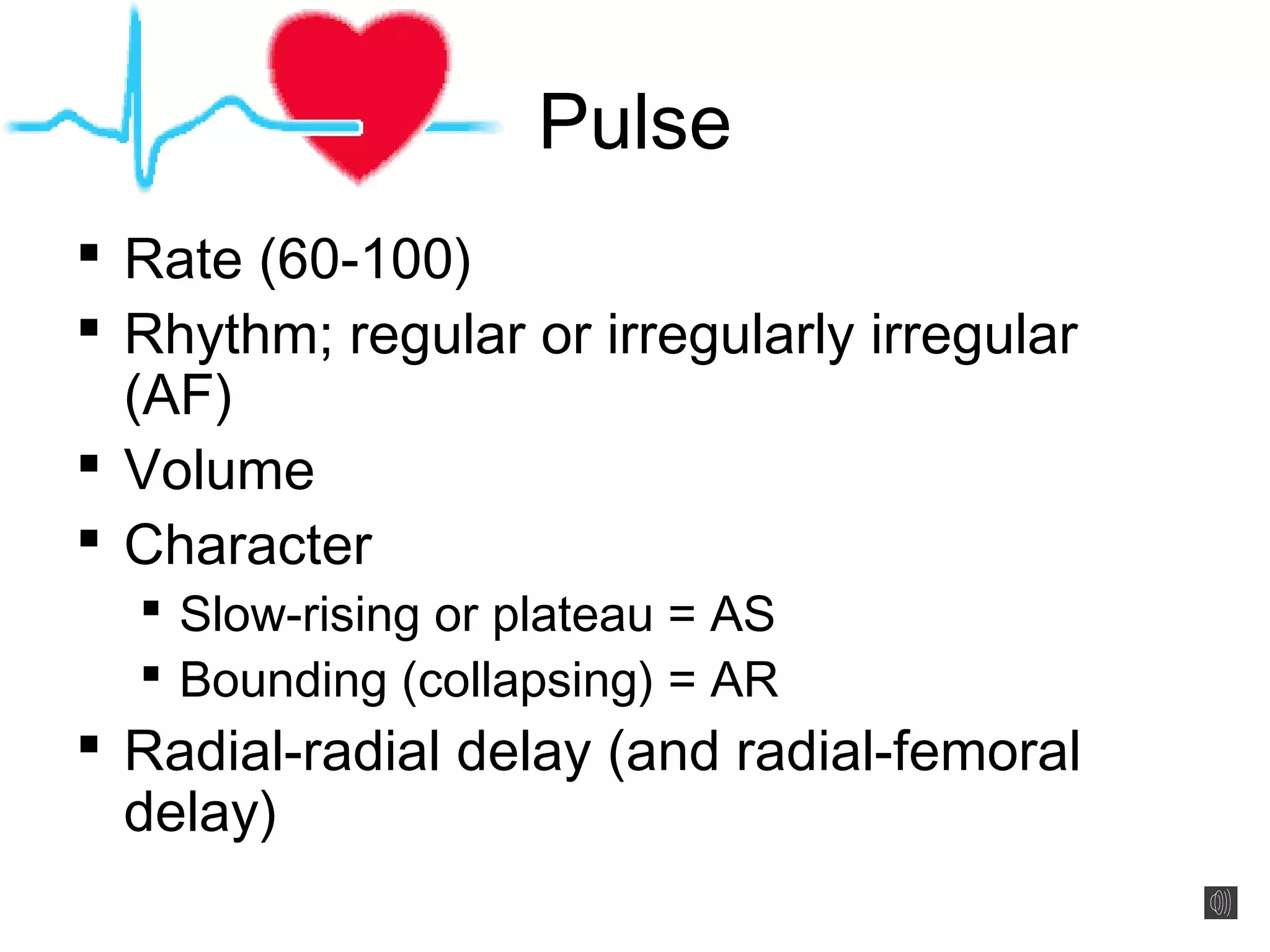
Pulse
 Rate (60-100)
 Rhythm; regular or irregularly irregular
(AF)
 Volume
 Character
 Slow-rising or plateau = AS
 Bounding (collapsing) = AR
 Radial-radial delay (and radial-femoral
delay)
The “Waterhammer” Pulse
 AKA Collapsing pulse or
Corrigan’s pulse
 Does not necessarily mean AR
 Many different techniques
described
 Basic principle is to elevate the
arm above the level of the heart
and to palpate the arm with the
palm of the hand
 Pulse feels forceful
Blood pressure
 Very useful in the acute situation!
 In stable patients:
 Are they hypertensive?
 Is there a wide or narrow pulse pressure?
Eyes & face
Face Eyes Mouth
General pallor
Malar flush
Corneal arcus
Xanthelasma
Conjunctival pallor
Central cyanosis
Poor dentition
Neck & JVP
 Corrigan’s sign – visible carotid pulsations
 de Musset’s sign – head nodding in time with carotid
pulsation
Jugular Venous Pressure
 Various waveforms – forget!
 Is it >4cm above sternal
angle or <4cm?
 Hepatojugular/
abdominojugular reflux –
warn patient first!
Legs
 Some prefer to
leave this until the
end
 Check for
peripheral oedema
– pitting or non-
pitting?
 Check for vein
harvesting scars
Observation of the praecordium
 Any scars?
 Central sternotomy or valve replacement
 Any pacemakers?
 Any obvious chest deformities?
 Also take this time to listen carefully for
the audible “click” of a mechanical valve
that may be heard without a stethoscope
Palpation
 Feel for any pacemakers or defibrillator devices
 Feel for apex beat (don’t be disheartened if you find this tricky!)
 Place flat of hand where you expect the apex to be (mid-
clavicular line 5th
intercostal space) and find point of maximum
pulsation
 Then count down from sternal angle to find the position of the
apex beat
 NB: remember dextracardia!
 Determine the character of the apex
 Tapping? Heaving? Thrusting? Jerky?
 Then feel for any parasternal heaves using the flat of your hand
 Finally feel for any thrills with ulnar border of your hand – these are
palpable murmurs; feel in all areas where you will listen with your
stethoscope (see next slide)
Auscultation
 Four areas to listen:
 Aortic = 2nd
intercostal space right sternal
edge
 Pulmonary = 2nd
intercostal space left
sternal edge
 Tricuspid = 4th
intercostal space left sternal
edge
 Mitral = apex beat
 Take your time and don’t panic!
 Don’t move on to the next area until you
are happy with what you have heard!
 Feel carotid pulse simultaneously; systolic
murmurs coincide with pulse
 Listen with diaphragm in all areas and bell
in mitral area
 Listen at the carotids with the bell for a
carotid bruit
System for heart sounds
1. Can I hear heart sounds?
2. Can I hear S1 and S2 equally?
3. Can I hear any other sounds?
 E.g. gallop rhythm: S3 “tenessee” or S4 “kentucky”
4. Can I hear any murmurs?
 Are they systolic or diastolic?
 Are they continuous or can I clearly hear S1 and S2
separately?
 If they are not continuous, are they early, middle or late in
timing?
 Where is the murmur loudest and can it be heard anywhere
else?
 Aortic murmurs radiate to the neck; mitral murmurs radiate
to the axilla
5. Can I put this information together?
Manoeuvres to accentuate
murmurs
 Ask the patient to roll onto their left side
 This will accentuate the murmur of MS so listen with the bell
 Ask the patient to lean forward and listen at the lower left
sternal edge
 This will accentuate aortic murmurs
 At the same time, take this opportunity to listen at the bases of
the lungs for any basal crackles suggestive of heart failure or
any sacral oedema
 Asking the patient to hold their breath in expiration will
accentuate aortic and mitral murmurs and in inspiration
will accentuate pulmonary and tricuspid murmurs
 This will be uncomfortable for the patient and make sure you tell
them they can breathe normally afterwards!
Extra things to do
 Take a full history!
 Blood pressure
 Even if you said it earlier as the examiner is likely to
forget!
 Feel for radial-femoral delay (if not done)
 Examine the respiratory system
 Measure oxygen sats
 Perform a 12-lead ECG
 Perform a chest X-ray
Case scenarios
 Likely cases for OSCEs include:
 Aortic stenosis/sclerosis
 Mitral regurgitation (or PSM of other cause)
 Aortic regurgitation (rarely!)
 A patient who has had a previous Coronary
Artery Bypass Graft (CABG)
 A patient who has had a previous valve
replacement
Aortic stenosis/sclerosis
 Signs to look for on examination:
 Slow-rising/ plateau pulse
 Narrow pulse pressure
 Ejection systolic murmur with radiation to the carotids
 Differential diagnosis:
 Aortic stenosis
 Aortic sclerosis
 (Pulmonary stenosis)
 Some common questions:
 How can you tell the difference between aortic
stenosis and aortic sclerosis?
 At what point might you consider valve replacement?
Pansystolic murmur
 Signs to look for on examination:
 Nature of apex beat
 Pansystolic murmur – note where it is loudest and any
radiation
 Pulsatile liver edge – TR!
 Differential diagnosis:
 Mitral regurgitation
 Tricuspid regurgitation
 Ventricular septal defect
 Some common questions:
 What do you think might have caused this murmur?
Aortic regurgitation
 Signs to look for on examination:
 Lots of eponyms; here are 3:
 Quincke’s sign = nail bed pulsations
 de Musset’s sign = head nodding
 Corrigan’s pulse = carotid pulsation
 Also: collapsing/ waterhammer pulse
 Wide pulse pressure
 Early diastolic murmur
 Differential diagnosis:
 (Pulmonary regurgitation)
 Some common questions:
 What do you think might have caused this murmur?
Summary
 The CVS examination demands a
systematic approach
 Many clues to clinical diagnosis can be
picked up before auscultation
 Try to stay calm and don’t rush
 If you are unable to palpate the apex beat
or hear heart sounds admit this and use it
as a learning opportunity
 Good luck and enjoy cardiology!!

More Related Content

What's hot

Gi exam by Dr.M.Mujeebullah
Gi exam by Dr.M.MujeebullahGi exam by Dr.M.Mujeebullah
Gi exam by Dr.M.Mujeebullah
Dr.Mujeebullah Mahboob
 
Clinical Examination of RS
Clinical Examination of RSClinical Examination of RS
Clinical Examination of RS
Prajwal Rk
 
Examination of gastrointestinal system by HX
Examination of gastrointestinal system by HXExamination of gastrointestinal system by HX
Examination of gastrointestinal system by HXDr. Rubz
 
Local chest examination
Local chest examinationLocal chest examination
Local chest examinationimangalal
 
Abdominal pain- all quadrants- case based learning
Abdominal pain-  all quadrants- case based learningAbdominal pain-  all quadrants- case based learning
Abdominal pain- all quadrants- case based learning
Selvaraj Balasubramani
 
Acute Abdomen Ppt
Acute Abdomen PptAcute Abdomen Ppt
Acute Abdomen Ppt
Kritz M Krishnan
 
Physical examination of cvs
Physical examination of cvsPhysical examination of cvs
Physical examination of cvsBitew Mekonnen
 
Palpitations
PalpitationsPalpitations
Palpitations
tabitha polamuri
 
Mitral stenosis
Mitral stenosisMitral stenosis
Mitral stenosis
Kavindya Fernando
 
Examination of the respiratory system
Examination of the respiratory systemExamination of the respiratory system
Examination of the respiratory system
Yapa
 
Cardiovascular history taking
Cardiovascular history takingCardiovascular history taking
Cardiovascular history taking
Ramachandra Barik
 
Exam Cvs 09.
Exam Cvs 09.Exam Cvs 09.
Exam Cvs 09.
Shaikhani.
 
Jugular Venous Pressure (JVP) Jugular Venous Pulse
Jugular Venous Pressure (JVP) Jugular Venous PulseJugular Venous Pressure (JVP) Jugular Venous Pulse
Jugular Venous Pressure (JVP) Jugular Venous Pulse
Arun Vasireddy
 
General physical Examination
General physical Examination General physical Examination
General physical Examination
Virendra Hindustani
 
History taking and physical examination for lower gastro intestinal bleed
History taking and physical examination for lower gastro intestinal bleedHistory taking and physical examination for lower gastro intestinal bleed
History taking and physical examination for lower gastro intestinal bleedAbino David
 
Heart sounds and murmur
Heart sounds and murmurHeart sounds and murmur
Heart sounds and murmur
Vitrag Shah
 
Heart murmurs
Heart murmursHeart murmurs
CNS examination
CNS examinationCNS examination
CNS examination
Walaa Manaa
 

What's hot (20)

Abdominal Exam
Abdominal ExamAbdominal Exam
Abdominal Exam
 
Gi exam by Dr.M.Mujeebullah
Gi exam by Dr.M.MujeebullahGi exam by Dr.M.Mujeebullah
Gi exam by Dr.M.Mujeebullah
 
Clinical Examination of RS
Clinical Examination of RSClinical Examination of RS
Clinical Examination of RS
 
Examination of gastrointestinal system by HX
Examination of gastrointestinal system by HXExamination of gastrointestinal system by HX
Examination of gastrointestinal system by HX
 
Local chest examination
Local chest examinationLocal chest examination
Local chest examination
 
Abdominal pain- all quadrants- case based learning
Abdominal pain-  all quadrants- case based learningAbdominal pain-  all quadrants- case based learning
Abdominal pain- all quadrants- case based learning
 
Acute Abdomen Ppt
Acute Abdomen PptAcute Abdomen Ppt
Acute Abdomen Ppt
 
Physical examination of cvs
Physical examination of cvsPhysical examination of cvs
Physical examination of cvs
 
Palpitations
PalpitationsPalpitations
Palpitations
 
Mitral stenosis
Mitral stenosisMitral stenosis
Mitral stenosis
 
Examination of the respiratory system
Examination of the respiratory systemExamination of the respiratory system
Examination of the respiratory system
 
Cardiovascular history taking
Cardiovascular history takingCardiovascular history taking
Cardiovascular history taking
 
Exam Cvs 09.
Exam Cvs 09.Exam Cvs 09.
Exam Cvs 09.
 
Jugular Venous Pressure (JVP) Jugular Venous Pulse
Jugular Venous Pressure (JVP) Jugular Venous PulseJugular Venous Pressure (JVP) Jugular Venous Pulse
Jugular Venous Pressure (JVP) Jugular Venous Pulse
 
General physical Examination
General physical Examination General physical Examination
General physical Examination
 
History taking and physical examination for lower gastro intestinal bleed
History taking and physical examination for lower gastro intestinal bleedHistory taking and physical examination for lower gastro intestinal bleed
History taking and physical examination for lower gastro intestinal bleed
 
Heart sounds and murmur
Heart sounds and murmurHeart sounds and murmur
Heart sounds and murmur
 
Acute coronary syndromes
Acute coronary syndromesAcute coronary syndromes
Acute coronary syndromes
 
Heart murmurs
Heart murmursHeart murmurs
Heart murmurs
 
CNS examination
CNS examinationCNS examination
CNS examination
 

Similar to Approach to the cardiovascular examination

Cardiac assessment ppt
Cardiac assessment pptCardiac assessment ppt
Cardiac assessment ppt
sudhir khuntia
 
chapter 3 CVS examination.pptx
chapter 3 CVS examination.pptxchapter 3 CVS examination.pptx
chapter 3 CVS examination.pptx
AbdiIsaq1
 
Cardiac Examination.ppt
Cardiac Examination.pptCardiac Examination.ppt
Cardiac Examination.ppt
ElizabethMvukuta
 
Auscultation.pptx
Auscultation.pptxAuscultation.pptx
Auscultation.pptx
Amrutha0013
 
Cardiolovascular Exam.pdf
Cardiolovascular Exam.pdfCardiolovascular Exam.pdf
Cardiolovascular Exam.pdf
SOLAHA
 
Cardiolovascular Exam.pdf
Cardiolovascular Exam.pdfCardiolovascular Exam.pdf
Cardiolovascular Exam.pdf
Sani191640
 
Health-Assessment-Chapter-10-Assessment-of-Cardiovascular-System.ppt
Health-Assessment-Chapter-10-Assessment-of-Cardiovascular-System.pptHealth-Assessment-Chapter-10-Assessment-of-Cardiovascular-System.ppt
Health-Assessment-Chapter-10-Assessment-of-Cardiovascular-System.ppt
deepshikakakoty
 
Cardiac auscultation
Cardiac auscultationCardiac auscultation
Cardiac auscultationhzllws
 
Presentation (2).pptx
Presentation (2).pptxPresentation (2).pptx
Presentation (2).pptx
GSaritha9
 
detailed first and second year OSCE stations
detailed first and second year OSCE stationsdetailed first and second year OSCE stations
detailed first and second year OSCE stationsmeducationdotnet
 
Ms mr
Ms mrMs mr
Cardiovascular examination
Cardiovascular examinationCardiovascular examination
Cardiovascular examination
Pritom Das
 
MULTI VALVULAR HEART DISEASE clinical presentation
MULTI VALVULAR HEART DISEASE clinical presentation MULTI VALVULAR HEART DISEASE clinical presentation
MULTI VALVULAR HEART DISEASE clinical presentation
Kurian Joseph
 
Amos assessment 1
Amos assessment 1Amos assessment 1
Amos assessment 1
MELIKIPROTICHAMOS
 
CARDIOVASCULAR EXAMINATION.pptx
CARDIOVASCULAR EXAMINATION.pptxCARDIOVASCULAR EXAMINATION.pptx
CARDIOVASCULAR EXAMINATION.pptx
Dr. Adanwali Hassan
 
percussion and Auscultation of cardiovascular system with heart sounds and mu...
percussion and Auscultation of cardiovascular system with heart sounds and mu...percussion and Auscultation of cardiovascular system with heart sounds and mu...
percussion and Auscultation of cardiovascular system with heart sounds and mu...
alok thakur
 

Similar to Approach to the cardiovascular examination (20)

2. cv assessment
2. cv assessment2. cv assessment
2. cv assessment
 
Cardiac assessment ppt
Cardiac assessment pptCardiac assessment ppt
Cardiac assessment ppt
 
chapter 3 CVS examination.pptx
chapter 3 CVS examination.pptxchapter 3 CVS examination.pptx
chapter 3 CVS examination.pptx
 
Cardiac Examination.ppt
Cardiac Examination.pptCardiac Examination.ppt
Cardiac Examination.ppt
 
Cardiovascular.330.Ss.09
Cardiovascular.330.Ss.09Cardiovascular.330.Ss.09
Cardiovascular.330.Ss.09
 
CVS
CVSCVS
CVS
 
Auscultation.pptx
Auscultation.pptxAuscultation.pptx
Auscultation.pptx
 
Cardiolovascular Exam.pdf
Cardiolovascular Exam.pdfCardiolovascular Exam.pdf
Cardiolovascular Exam.pdf
 
Cardiolovascular Exam.pdf
Cardiolovascular Exam.pdfCardiolovascular Exam.pdf
Cardiolovascular Exam.pdf
 
Health-Assessment-Chapter-10-Assessment-of-Cardiovascular-System.ppt
Health-Assessment-Chapter-10-Assessment-of-Cardiovascular-System.pptHealth-Assessment-Chapter-10-Assessment-of-Cardiovascular-System.ppt
Health-Assessment-Chapter-10-Assessment-of-Cardiovascular-System.ppt
 
Cardiac auscultation
Cardiac auscultationCardiac auscultation
Cardiac auscultation
 
Presentation (2).pptx
Presentation (2).pptxPresentation (2).pptx
Presentation (2).pptx
 
detailed first and second year OSCE stations
detailed first and second year OSCE stationsdetailed first and second year OSCE stations
detailed first and second year OSCE stations
 
Pe cardiovoascular exam
Pe cardiovoascular examPe cardiovoascular exam
Pe cardiovoascular exam
 
Ms mr
Ms mrMs mr
Ms mr
 
Cardiovascular examination
Cardiovascular examinationCardiovascular examination
Cardiovascular examination
 
MULTI VALVULAR HEART DISEASE clinical presentation
MULTI VALVULAR HEART DISEASE clinical presentation MULTI VALVULAR HEART DISEASE clinical presentation
MULTI VALVULAR HEART DISEASE clinical presentation
 
Amos assessment 1
Amos assessment 1Amos assessment 1
Amos assessment 1
 
CARDIOVASCULAR EXAMINATION.pptx
CARDIOVASCULAR EXAMINATION.pptxCARDIOVASCULAR EXAMINATION.pptx
CARDIOVASCULAR EXAMINATION.pptx
 
percussion and Auscultation of cardiovascular system with heart sounds and mu...
percussion and Auscultation of cardiovascular system with heart sounds and mu...percussion and Auscultation of cardiovascular system with heart sounds and mu...
percussion and Auscultation of cardiovascular system with heart sounds and mu...
 

More from meducationdotnet

Water and sanitation and their impact on health
Water and sanitation and their impact on healthWater and sanitation and their impact on health
Water and sanitation and their impact on healthmeducationdotnet
 
Health Care Worker Migration
Health Care Worker MigrationHealth Care Worker Migration
Health Care Worker Migrationmeducationdotnet
 
International Institutions
International InstitutionsInternational Institutions
International Institutionsmeducationdotnet
 
Haemochromotosis brief overview
Haemochromotosis brief overviewHaemochromotosis brief overview
Haemochromotosis brief overviewmeducationdotnet
 
Overview of Antidepressants
Overview of AntidepressantsOverview of Antidepressants
Overview of Antidepressantsmeducationdotnet
 
Review of orthopaedic services: Prepared for the Auditor General for Scotland...
Review of orthopaedic services: Prepared for the Auditor General for Scotland...Review of orthopaedic services: Prepared for the Auditor General for Scotland...
Review of orthopaedic services: Prepared for the Auditor General for Scotland...meducationdotnet
 
Sugammadex - a revolution in anaesthesia?
Sugammadex - a revolution in anaesthesia?Sugammadex - a revolution in anaesthesia?
Sugammadex - a revolution in anaesthesia?meducationdotnet
 

More from meducationdotnet (20)

No Title
No TitleNo Title
No Title
 
Spondylarthropathy
SpondylarthropathySpondylarthropathy
Spondylarthropathy
 
Diagnosing Lung cancer
Diagnosing Lung cancerDiagnosing Lung cancer
Diagnosing Lung cancer
 
Eczema Herpeticum
Eczema HerpeticumEczema Herpeticum
Eczema Herpeticum
 
The Vagus Nerve
The Vagus NerveThe Vagus Nerve
The Vagus Nerve
 
Water and sanitation and their impact on health
Water and sanitation and their impact on healthWater and sanitation and their impact on health
Water and sanitation and their impact on health
 
The ethics of electives
The ethics of electivesThe ethics of electives
The ethics of electives
 
Intro to Global Health
Intro to Global HealthIntro to Global Health
Intro to Global Health
 
WTO and Health
WTO and HealthWTO and Health
WTO and Health
 
Globalisation and Health
Globalisation and HealthGlobalisation and Health
Globalisation and Health
 
Health Care Worker Migration
Health Care Worker MigrationHealth Care Worker Migration
Health Care Worker Migration
 
International Institutions
International InstitutionsInternational Institutions
International Institutions
 
Haemochromotosis brief overview
Haemochromotosis brief overviewHaemochromotosis brief overview
Haemochromotosis brief overview
 
Ascities overview
Ascities overviewAscities overview
Ascities overview
 
Overview of the Liver
Overview of the LiverOverview of the Liver
Overview of the Liver
 
Overview of Antidepressants
Overview of AntidepressantsOverview of Antidepressants
Overview of Antidepressants
 
Gout Presentation
Gout PresentationGout Presentation
Gout Presentation
 
Review of orthopaedic services: Prepared for the Auditor General for Scotland...
Review of orthopaedic services: Prepared for the Auditor General for Scotland...Review of orthopaedic services: Prepared for the Auditor General for Scotland...
Review of orthopaedic services: Prepared for the Auditor General for Scotland...
 
Sugammadex - a revolution in anaesthesia?
Sugammadex - a revolution in anaesthesia?Sugammadex - a revolution in anaesthesia?
Sugammadex - a revolution in anaesthesia?
 
Ophthamology Revision
Ophthamology RevisionOphthamology Revision
Ophthamology Revision
 

Approach to the cardiovascular examination

  • 1. Approach to the Cardiovascular Examination Carly Welch Final Year Medical Student University of Birmingham
  • 2. Contents  Basic approach to the CVS patient  General observations  Hand signs  Pulse  Blood Pressure  Eyes & face  Neck & JVP  Legs  Examination of the praecordium  Observation  Palpation  Auscultation  Extra things to do  Case scenarios  Summary
  • 3. Basic approach  WIPER  Wash hands, Introduce, ask about Pain, Exposure, Repositioning – 45o  CVS examination does not just mean auscultation!  Start with general observation and then examine systematically starting with the hands
  • 4. General observation  Comfortable? Alert? Generally well or unwell?  Do they look breathless or cyanosed?  Can you see any obvious scars or pacemakers?  Any clues around the bed e.g. GTN sprays?
  • 5. Hand signs Clubbing Koilonychia Palmar erythema Splinter haemorrhages Osler nodes Janeway lesions
  • 6. Pulse  Rate (60-100)  Rhythm; regular or irregularly irregular (AF)  Volume  Character  Slow-rising or plateau = AS  Bounding (collapsing) = AR  Radial-radial delay (and radial-femoral delay)
  • 7. The “Waterhammer” Pulse  AKA Collapsing pulse or Corrigan’s pulse  Does not necessarily mean AR  Many different techniques described  Basic principle is to elevate the arm above the level of the heart and to palpate the arm with the palm of the hand  Pulse feels forceful
  • 8. Blood pressure  Very useful in the acute situation!  In stable patients:  Are they hypertensive?  Is there a wide or narrow pulse pressure?
  • 9. Eyes & face Face Eyes Mouth General pallor Malar flush Corneal arcus Xanthelasma Conjunctival pallor Central cyanosis Poor dentition
  • 10. Neck & JVP  Corrigan’s sign – visible carotid pulsations  de Musset’s sign – head nodding in time with carotid pulsation Jugular Venous Pressure  Various waveforms – forget!  Is it >4cm above sternal angle or <4cm?  Hepatojugular/ abdominojugular reflux – warn patient first!
  • 11. Legs  Some prefer to leave this until the end  Check for peripheral oedema – pitting or non- pitting?  Check for vein harvesting scars
  • 12. Observation of the praecordium  Any scars?  Central sternotomy or valve replacement  Any pacemakers?  Any obvious chest deformities?  Also take this time to listen carefully for the audible “click” of a mechanical valve that may be heard without a stethoscope
  • 13. Palpation  Feel for any pacemakers or defibrillator devices  Feel for apex beat (don’t be disheartened if you find this tricky!)  Place flat of hand where you expect the apex to be (mid- clavicular line 5th intercostal space) and find point of maximum pulsation  Then count down from sternal angle to find the position of the apex beat  NB: remember dextracardia!  Determine the character of the apex  Tapping? Heaving? Thrusting? Jerky?  Then feel for any parasternal heaves using the flat of your hand  Finally feel for any thrills with ulnar border of your hand – these are palpable murmurs; feel in all areas where you will listen with your stethoscope (see next slide)
  • 14. Auscultation  Four areas to listen:  Aortic = 2nd intercostal space right sternal edge  Pulmonary = 2nd intercostal space left sternal edge  Tricuspid = 4th intercostal space left sternal edge  Mitral = apex beat  Take your time and don’t panic!  Don’t move on to the next area until you are happy with what you have heard!  Feel carotid pulse simultaneously; systolic murmurs coincide with pulse  Listen with diaphragm in all areas and bell in mitral area  Listen at the carotids with the bell for a carotid bruit
  • 15. System for heart sounds 1. Can I hear heart sounds? 2. Can I hear S1 and S2 equally? 3. Can I hear any other sounds?  E.g. gallop rhythm: S3 “tenessee” or S4 “kentucky” 4. Can I hear any murmurs?  Are they systolic or diastolic?  Are they continuous or can I clearly hear S1 and S2 separately?  If they are not continuous, are they early, middle or late in timing?  Where is the murmur loudest and can it be heard anywhere else?  Aortic murmurs radiate to the neck; mitral murmurs radiate to the axilla 5. Can I put this information together?
  • 16. Manoeuvres to accentuate murmurs  Ask the patient to roll onto their left side  This will accentuate the murmur of MS so listen with the bell  Ask the patient to lean forward and listen at the lower left sternal edge  This will accentuate aortic murmurs  At the same time, take this opportunity to listen at the bases of the lungs for any basal crackles suggestive of heart failure or any sacral oedema  Asking the patient to hold their breath in expiration will accentuate aortic and mitral murmurs and in inspiration will accentuate pulmonary and tricuspid murmurs  This will be uncomfortable for the patient and make sure you tell them they can breathe normally afterwards!
  • 17. Extra things to do  Take a full history!  Blood pressure  Even if you said it earlier as the examiner is likely to forget!  Feel for radial-femoral delay (if not done)  Examine the respiratory system  Measure oxygen sats  Perform a 12-lead ECG  Perform a chest X-ray
  • 18. Case scenarios  Likely cases for OSCEs include:  Aortic stenosis/sclerosis  Mitral regurgitation (or PSM of other cause)  Aortic regurgitation (rarely!)  A patient who has had a previous Coronary Artery Bypass Graft (CABG)  A patient who has had a previous valve replacement
  • 19. Aortic stenosis/sclerosis  Signs to look for on examination:  Slow-rising/ plateau pulse  Narrow pulse pressure  Ejection systolic murmur with radiation to the carotids  Differential diagnosis:  Aortic stenosis  Aortic sclerosis  (Pulmonary stenosis)  Some common questions:  How can you tell the difference between aortic stenosis and aortic sclerosis?  At what point might you consider valve replacement?
  • 20. Pansystolic murmur  Signs to look for on examination:  Nature of apex beat  Pansystolic murmur – note where it is loudest and any radiation  Pulsatile liver edge – TR!  Differential diagnosis:  Mitral regurgitation  Tricuspid regurgitation  Ventricular septal defect  Some common questions:  What do you think might have caused this murmur?
  • 21. Aortic regurgitation  Signs to look for on examination:  Lots of eponyms; here are 3:  Quincke’s sign = nail bed pulsations  de Musset’s sign = head nodding  Corrigan’s pulse = carotid pulsation  Also: collapsing/ waterhammer pulse  Wide pulse pressure  Early diastolic murmur  Differential diagnosis:  (Pulmonary regurgitation)  Some common questions:  What do you think might have caused this murmur?
  • 22. Summary  The CVS examination demands a systematic approach  Many clues to clinical diagnosis can be picked up before auscultation  Try to stay calm and don’t rush  If you are unable to palpate the apex beat or hear heart sounds admit this and use it as a learning opportunity  Good luck and enjoy cardiology!!

Editor's Notes

  1. This presentation describes the basic cardiovascular examination. It is suitable for students in their early clinical years but may also be appropriate for students in their final year as revision for their OSCEs or students returning to clinical medicine after an intercalated degree.
  2. I will begin by explaining the basic approach to the cardiovascular system and will then work through some of the common and classical signs that you might find on peripheral examination. I will then move on to focus in more detail on the examination of the praecordium. Finally, I will talk through some important clinical scenarios that you are likely to encounter on the wards and in your examinations.
  3. This slide demonstrates the basic approach to the patient that is relevant not just to the cardiovascular examination, but to the examination of any patient. You will hear many acronyms mentioned to help you to remember how to start an examination – personally I prefer “WIPER”. This stands for “Wash hands”, “Introduce” yourself and gain consent from the patient, ask the patient if they are in any “Pain” – this is important for any examination and not just the abdominal examination, “Expose” the patient appropriately – this will depend on what the patient feels comfortable with but generally it is recommended that for women they may lower their gown or top but keep their bra on and finally “Reposition” – you will often find that in an OSCE situation patients are already positioned at 45o, but to show that you have considered this you can either simply comment on this or attempt to move the bed out of position and back into position again. I think one of the most important things that I’d like you to take away from this presentation is to remember that the cardiovascular examination does not just mean listening to heart sounds; you can gain an impressive amount of information about the patient before you have even placed your stethoscope on their chest. As with any examination, the best way to look slick and not to miss any important findings is to have a systematic approach starting with general examination and then focussing more specifically on areas, starting with the hands.
  4. Begin by observing the patient from the end of the bed. Try to get a general feel for this patient’s condition. Ask yourself some basic questions; for instance, are they alert or do they look drowsy or confused? Are they sitting comfortably or do they seem restless or distressed? As you progress throughout your training, you will develop a basic instinct about whether a patient looks well or unwell – try to answer this question in your head whenever you see a new patient. Without counting their respiratory rate, do you think they seem to be obviously breathless? Do they look pale or cyanosed? Then have a look to see if there is anything obvious that jumps out at you – any scars, pacemakers or lines in-situ? A good tip if you notice a central sternotomy scar is to look at the legs for any vein harvesting scars; if these are present it is very likely that they have had a previous Coronary Artery Bypass Graft (CABG). Finally, have a look around the bed for any extra clues that might be relevant, for instance a GTN spray would indicate Ischaemic Heart Disease.
  5. Now move on to examine the patients hands in more detail. There are many different hand signs you will hear discussed. This slide shows a selection of these. Look at both hands together, begin by examining the nails, then the fingers, then the hands, then ask the patient to turn their hands over and examine their palms. Clubbing is a clinical sign that seems to be relevant to all systems examinations, but that in practice is most relevant to the respiratory examination. It is thought to be caused by hypoxia, so its causes relate to that of hypoxia. In terms of the cardiovascular examination, the most important differential would be congenital cyanotic heart disease; unless you are working on a paediatric ward, it is unlikely that this is something that you will come across. Other causes include a left atrial myxoma, and, if you really want to impress your consultant, you can say that coarctation of the aorta results in clubbing in the toes but not in the hands. Although a lot of emphasis is put on hand signs, it is in fact true that most signs that are found in the hands can also be found in the feet. However, I feel people might question a somewhat unorthodox technique of starting in the feet! Koilonychia is a sign of anaemia – in particular iron deficiency. Palmar erythema is a relatively non-specific finding but may be caused by pregnancy or thyrotoxicosis (both causes of high output cardiac failure). Splinter haemorrhages again are very non-specific; they are most commonly caused by trauma but may also arise in infective endocarditis. Osler’s nodes and Janeway lesions are both seen in infective endocarditis. Osler’s nodes are painful, raised and found on the pulps of the fingers. Whereas, Janeway lesions tend to be painful, flat and found on the palms. Other signs that you might find in the hands include peripheral cyanosis and pallor of palmar creases with anaemia.
  6. Examination of the pulse is vitally important in the cardiovascular examination. There are 4 main points regarding the pulse that you should aim to comment on. The first of these is the rate – time this over 15sec and give as beats/min. A rate &amp;lt;60bpm can be considered as bradycardic and a rate &amp;gt;100bpm can be considered tachycardic. Causes of bradycardia include fit athletes when resting (if &amp;gt;40bpm), heart block of varying degrees and treatment with beta-blockers. Causes of tachycardia include a patient who is acutely unwell, certain arrhythmias and anxiety if mildly increased. Next, try to assess the rhythm, in practice the most likely arrhythmia that you will pick up is Atrial Fibrillation; if you suspect that the rhythm is not regular you should request an ECG. The third area that you may be able to comment on is the volume. This can be difficult and is best assessed at the carotid pulse. If you think that the pulse is weak or thready, it may be a sign that this patient is acutely unwell. The character of the pulse is also difficult to assess but may provide clues to the diagnosis of the patient; again, this is best assessed at the carotid pulse. In aortic stenosis the pulse is “slow-rising” and the pressure remains flat or “plateau”. In aortic regurgitation the pulse in bounding or “collapsing”. There are many other pulse characters that you may read about in textbooks, but these will be difficult to pick up in practice. When feeling the radial pulse in the cardiovascular examination, I think that it is always good practice to feel the pulse concurrently on both sides. The main reason for this is that the presence of a distal embolus on one side might lead to the absence of a pulse that would otherwise not have been detected if the pulse was only felt on one side. Radial-radial delay might be caused by coarctation of the aorta, however, you would be much more likely to detect this through radial-femoral delay.
  7. Examination for Watson’s “waterhammer” pulse is specific to the examination of the cardiovascular system and will likely score you extra marks if you perform it in an OSCE. The term “waterhammer” can be slightly confusing, as it refers to an old children’s toy that most people are unfamiliar with; the term collapsing is generally performed. There are a few different techniques for examining for the collapsing pulse described but they all involve the basic principle of elevating the patient’s arm above the level of their heart and palpating their arm with the palm of the hand. A collapsing pulse will be felt to hit the back of the hand. Remember to ask the patient if they have any pain in their shoulder before elevating their arm. Something that I wasn’t initially aware of is the significance of the collapsing pulse. It may occur with aortic regurgitation, but is often present in other causes of a hyperdynamic or high output state such as pregnancy, thyrotoxicosis, anaemia or Paget’s disease.
  8. Taking the blood pressure is very useful practically as a hypotensive patient is likely to be very seriously ill and requires immediate attention. However, in stable patients that you are likely to come across in OSCEs there are two main points to think about. Firstly, is this patient hypertensive? Long-term this may be associated with hypertensive or ischaemic heart disease. Secondly, what is the pulse pressure? The pulse pressure tends to be narrow in aortic stenosis and wide in aortic regurgitation.
  9. Now move up to inspect the patient’s face. It can seem slightly strange to simply look up and down at a patient’s face so what I like to do is to take a moment to explain to the patient that you would like to look in and around their eyes and mouth and at the same time gain a general idea of the appearance of their face. You will of course check for conjunctival pallor, however, Caucasian patients who are grossly pale may be clear from general observation. Malar flush is classically associated with mitral stenosis. I, myself, have seen this once so far, but it is important to bear in mind. With the patient’s permission lower their right eyelid with your right thumb. Some people might teach you to lower both eyelids, but this is actually unnecessary as any pallor that is present should be present bilaterally. Look around the iris for any corneal arcus and around the eyelids for any xanthelasma, which both indicate high cholesterol levels, although bear in mind that corneal arcus becomes more common with increasing age. Check around the mouth for any central cyanosis – show the examiner that you are looking for this by asking them to raise their tongue to the roof of their mouth. Check to see what their dentition is like as poor dentition has previously been associated with infective endocarditis.
  10. As mentioned previously, the carotid pulse is the ideal point to assess pulse volume and character. Observe for Corrigan’s pulse or de Musset’s sign, which are both signs of aortic regurgitation. The ability to reliably assess the JVP is something that, unfortunately, will only improve with experience. There are various abnormalities in its waveform that you will hear about for different conditions – in practice these are difficult to interpret by even the most experienced practitioner. When examining for the JVP ensure that the patient is tilted at a 45o angle. Ask them to turn away from you slightly (but not so much that it tenses their sternocleidomastoid). Firstly, ask yourself if you can see a JVP. If you can not, this may mean that it is grossly elevated so look around the jawline and above the ear. Alternatively, it may mean that the JVP is below the level of the clavicle. One way of ascertaining this is to apply pressure to the liver or epigastric region, which should cause the JVP to rise momentarily for visualisation. This is possibly the most uncomfortable part of the cardiovascular exam so make sure you warn the patient first! Finally, inability to see the JVP may be due to poor lighting or lack of experience. If you are unsure, ask a more senior colleague for advice. If you can see a JVP assess whether it is raised or lowered. A normal JVP should be 4cm above the sternal notch at the 45o angle. A raised JVP implies volume overload or heart failure. A lowered JVP implies that the patient is underfilled.
  11. I’ve included the examination of the legs at this stage as I want to cover it before going into more detail about the praecordium. In practice, I prefer to examine the legs at the end as this is the order I have become familiar with. What is important is that you have your own system that you can reliably follow through – this means that you can either examine the legs now, or wait until the end. To assess for pitting oedema in the legs apply firm pressure for 5seconds and release. Pitting oedema will leave an area of indentation that takes time to return to normal. You can also take this opportunity to check for any vein harvesting scars, most commonly of the long saphenous vein, that may indicate a previous CABG.
  12. Start by checking that the patient has adequate exposure. If you used a sheet to cover them originally, kindly inform them that you need to lower it. Observe the general appearance of the chest. Central sternotomy scars will be less difficult to miss, but look more specifically for any smaller scars, particularly around the apical region, which may indicate a previous valve replacement. Look in the left upper chest for any pacemakers or defibrillator devices. Whilst you are performing your observations, also take this opportunity to listen carefully for the audible “click” of a mechanical valve.
  13. Take a moment to feel for any pacemakers or defibrillators that might not be obvious from simple observation. Then, move on to feel the apex beat. Place the palm of your hand over the area of the 5th intercostal space, mid-clavicular line and locate the point of maximum intensity. Then count down to locate this position from the sternal angle, which is at the level of the 2nd intercostal space. This is something that can be difficult on some patients and it’s important not to become disheartened if you are unable to feel the apex beat. Reasons for why you might not be able to palpate the apex beat include dextracardia, so make sure you feel on the other side, obesity and the apex being obscured by a rib, so you can try turning the patient onto their left side for palpation. If you are able to feel the apex, note whether it is displaced or undisplaced and its character. Then feel for any parasternal heaves using the flat of the hand. Finally, feel for any thrills, which are simply palpable murmurs in the areas where you will place your stethoscope, as detailed on the next slide. I use the ulnar border of my right hand for this.
  14. This is the part that most people panic about the most. I realise it’s easier said than done, but my best advice is honestly not to panic, to take your time and to think about what you are hearing! There are four areas where you should listen initially, as shown on the diagram above. The aortic area is in the 2nd intercostal space at the right sternal edge. The pulmonary area is in the 2nd intercostal space at the left sternal edge. The tricuspid area is in the 4th intercostal space at the left sternal edge and the mitral area is the point of the apex beat, which is normally in the 5th intercostal space in the midclavicular line. Listen with the diaphragm in all areas and the bell in the mitral area, as the murmur of mitral stenosis will be heard better. If you are asked during teaching, these are not the locations of the valves but simply the points where the mumurs can be heard best. Palpate the carotid pulse simultaneously to listening to help determine the timing of any murmurs; systolic murmurs should coincide with the pulse. I prefer to listen in the order aortic, pulmonary, tricuspid, mitral, but others may prefer to listen in the reverse of this order starting with mitral and ending with aortic. It is also useful to listen at the carotids with the bell for the presence of a carotid bruit. If you do this, ask the patient to momentarily hold their breath to ensure you are not hearing transmitted sounds from the trachea.
  15. This slide details my own system for listening to heart sounds that I think through in my head in each area. I ask myself 5 main questions. The first question I ask myself is can I hear heart sounds? Sometimes, even this can be difficult due to transmitted sounds from the lungs or gastrointestinal tract, background noise, obesity or genuinely quiet heart sounds. Once I am happy that I am hearing heart sounds, I then ask myself if I can hear S1 and S2 equally or if I can hear one louder than the other or if I can’t hear one at all. Once I am happy with this I then listen for any other sounds such as an S3 or S4. I find the most useful way to decide which of these I am hearing is to time the added sound, S1 and S2 with the words “tenessee” and “kentucky”. With an S3, the added sound will correspond with the “see” in tenessee, whereas with an S4, the added sound will correspond with the “ken” in kentucky. Only now that I have this information do I actively start listening for murmurs. If I do hear a murmur, I ask myself several questions about it. Firstly, is it systolic or diastolic, which can be gaged by palpating the carotid pulse simultaneously. Secondly, is the murmur continuous or can I hear S1 and S2 separately? If it is not continuous then I determine its timing in terms of early, middle or late in systole or diastole. Finally, after listening in all areas, I decide where the murmur is loudest and if there is any radiation. The 5th question that I ask myself is quite advanced for those of you in your early clinical years, but is good to start thinking about, even if you are unable to answer it. Can I put all this information together to fit with a disease process that I am aware of?
  16. This slide details some of the manoeuvres that you can perform the help accentuate heart murmurs. Start performing these routinely as part of your cardiovascular examination as they will score you extra points in your OSCEs. You can accentuate the murmur of mitral stenosis by asking the patient to roll onto their left side – remember to listen with the bell when doing this. You can accentuate aortic murmurs by asking the patient to lean forward and listening at the lower left sternal edge. You can use this opportunity to listen posteriorly at the bases of the lungs for any basal crepitations which would suggest heart failure and to check for any sacral oedema. Asking the patient to hold their breath in expiration will accentuate aortic and mitral murmurs and asking them to hold their breath in inspiration will accentuate pulmonary and tricuspid murmurs. This will be uncomfortable for the patient so don’t ask them to hold their breath for too long and remember to tell them they can breathe normally afterwards!
  17. Here are some things that you should say you would like to perform at the end of your examination. Taking a full history will provide you with more information than any other examination or investigation you can perform. Taking the blood pressure is key to the cardiovascular system and even if you mentioned this earlier I would mention it again at the end as the examiner is likely to forget! If you did not feel for radial-femoral delay then you can say that you would like to do it at the end. Examining the respiratory system and measuring oxygen saturations will provide you with extra information. If you are asked for some first line investigations that you would like to perform, I would suggest that you request a 12-lead ECG and a chest X-ray, which will provide useful information for most cardiac conditions.
  18. This slide demonstrates some of the common scenarios that you will see in an OSCE scenario. This is of course not an exclusive list but remember that patients you see in OSCEs will be stable patients with chronic conditions.
  19. This is a very common OSCE scenario and also something that you are likely to come across during your time on the wards. Students in their OSCE examinations are often asked how it is possible to tell the difference between aortic stenosis and aortic sclerosis. Aortic stenosis is occlusion of the aortic valve that leads to reduced function. Aortic sclerosis can be thought of as a partial occlusion without impairment of function. Both cause an ejection systolic murmur. Some people might tell you that you can tell the difference between the two by listening for radiation to the carotids; however, aortic sclerosis can radiate to the carotids. There are several pointers that indicate aortic stenosis. The presence of a slow-rising pulse and a narrow pulse pressure indicate this. Additionally, careful listening may elicit a loud S2 on auscultation. The question of when you might consider a valve replacement is of course not something you will be deciding as a junior doctor, but what the examiner is trying to highlight is the importance of checking to see if the stenosis is symptomatic. Patients with aortic stenosis can remain asymptomatic for many years, but once symptoms develop, death often occurs within 5years if left untreated. Ask about angina and syncope.
  20. This is another very common OSCE scenario. There are three main differential diagnosis for the pansystolic murmur; these are mitral regurgitation, tricuspid regurgitation or a ventricular septal defect. To help you to differentiate between these diagnoses, try to determine where the murmur is at its loudest. Mitral regurgitation should be heard loudest at the apex and may radiate to the axilla. Tricuspid regurgitation and ventricular septal defects would both be heard loudest at the left sternal edge, but ventricular septal defects are more likely to be heard all throughout the praecordium. Additonally, with tricuspid regurgiation, a pulsatile liver edge may be present. It is useful to be aware of some of the causes of each these differential diagnoses. Causes of mitral regurgitation include mitral valve prolapse, ischaemic heart disease, rheumatic heart disease and Marfan’s syndrome. Causes of tricuspid regurgitation include rheumatic heart disease, endocarditis, various medications and certain connective tissue disorders. Ventricular septal defects can be both congenital and acquired. A loud congenital ventricular septal defect is likely to be small and, therefore, may be asymptomatic. Acquired ventricular septal defects may arise after septal infarctions.
  21. Aortic regurgitation would be an unusual station to see in an OSCE examination as it is less common and produces a diastolic murmur, which can be more difficult to pick up. The murmur is an early diastolic murmur and is often described as “the absence of silence in early diastole”. The reason why I have chosen to highlight this scenario is that there are a lot of peripheral signs that might be picked up. There are many eponymous signs that you will hear about; I’ve listed 3 of these above. Quincke’s sign indicates nail bed pulsations. De Musset’s sign is head bobbing in time with the pulse. Corrigan’s pulse is a description of the collapsing pulse of the carotid artery. Watson’s waterhammer pulse may be present and there is likely to be a wide pulse pressure. Around 40% of aortic regurgitation cases are caused by idiopathic aortic root dilatation. Around 15% are caused by a congenital bicuspid aortic valve. Other causes include connective tissue diseases and rheumatic fever.
  22. I hope that you have found this presentation useful and that it has made you more confident to examine patients on the wards or in your OSCEs. One of the most important things is to stick to a systematic approach. A good tip I have is to practice examination on a doll or cuddly toy, especially in preparation for your OSCEs. Try to stay calm and not to rush, especially when listening to heart sounds. Finally, if you don’t pick up the clinical signs, don’t be ashamed to admit this! I would appreciate your feedback on this presentation – whether positive or negative! Let me know if you think there was too much or too little information or even if I got it just right! Good luck with your time on the wards or in any exams you have coming up and thank you for listening!