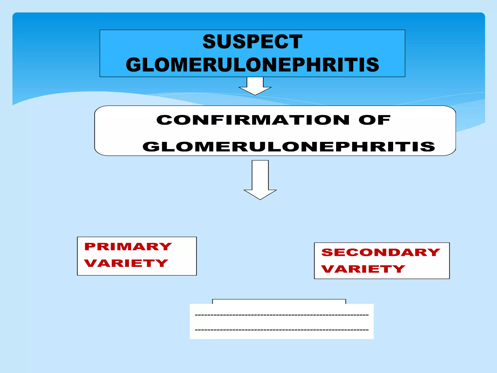
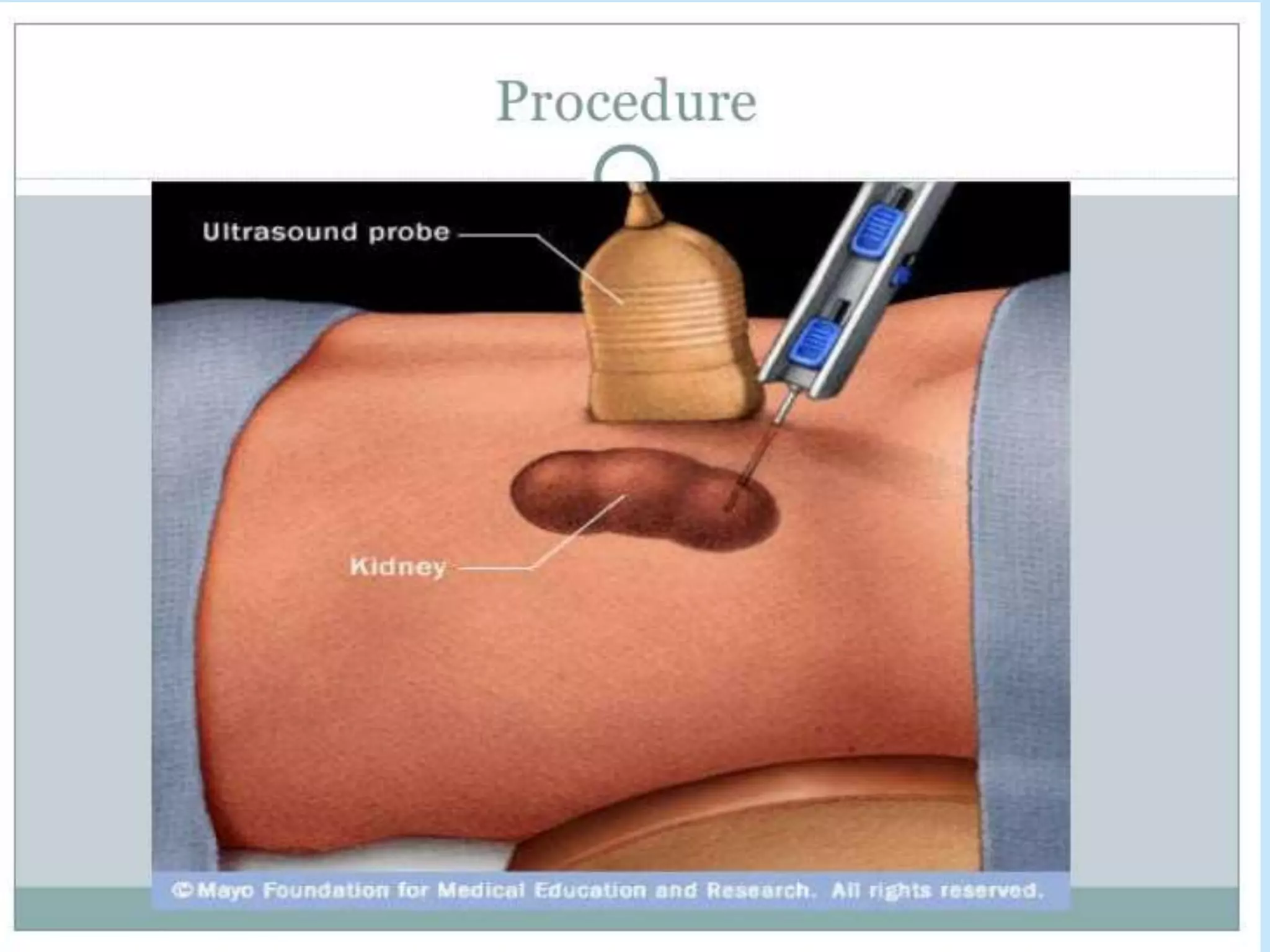
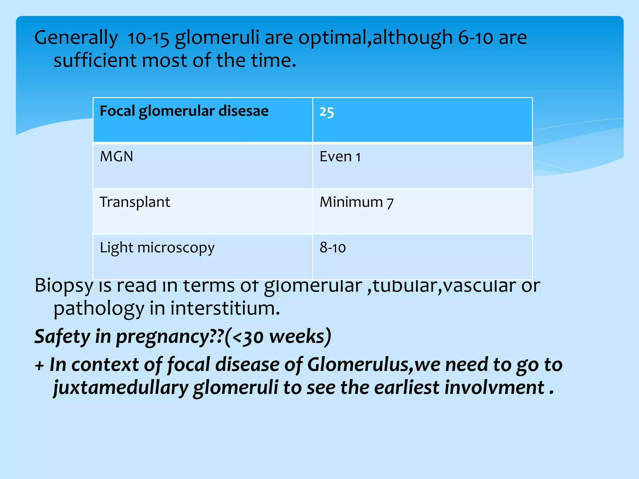
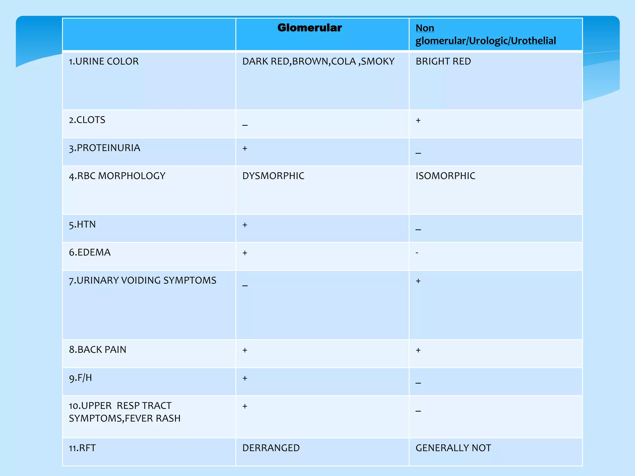
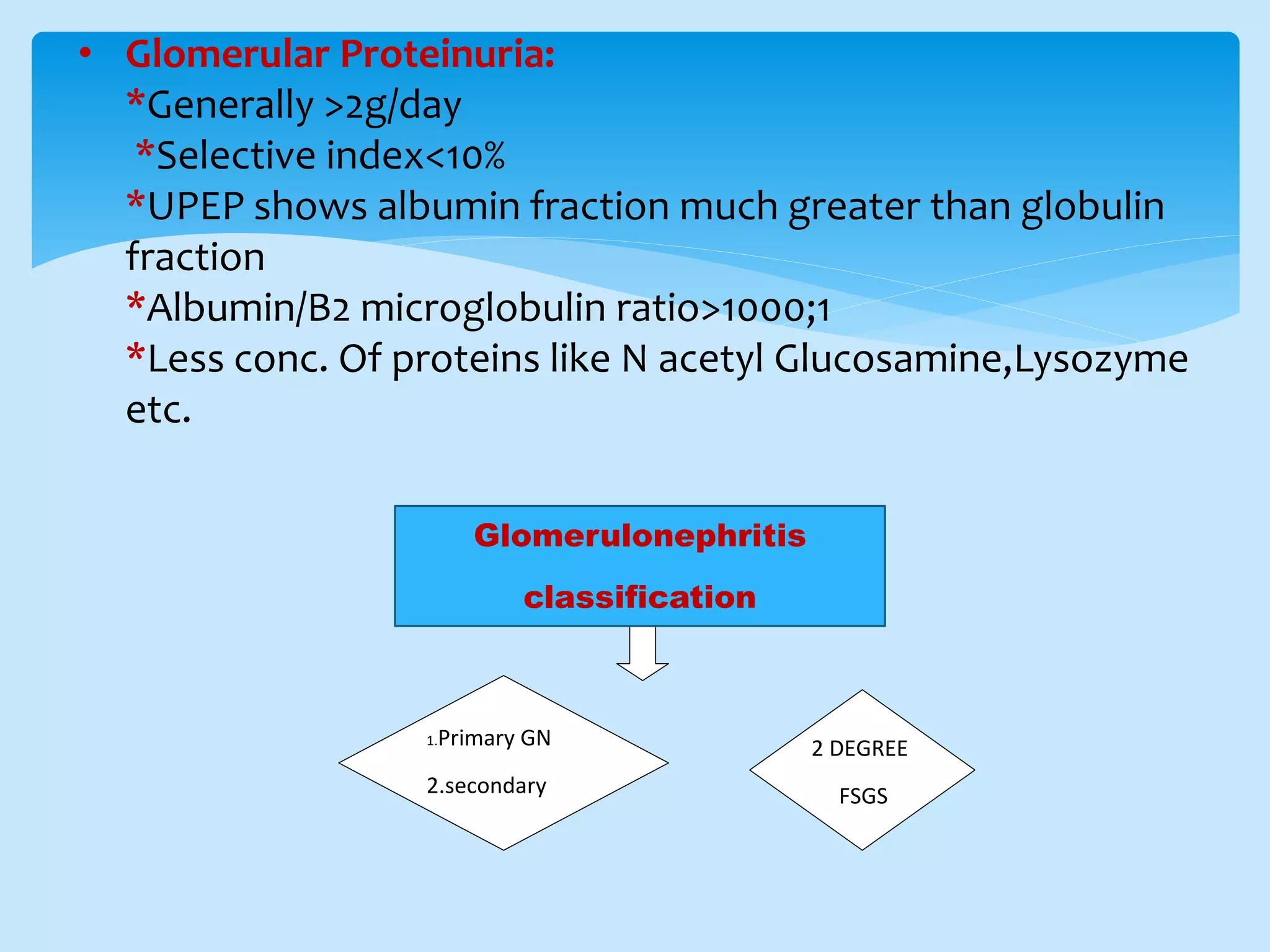
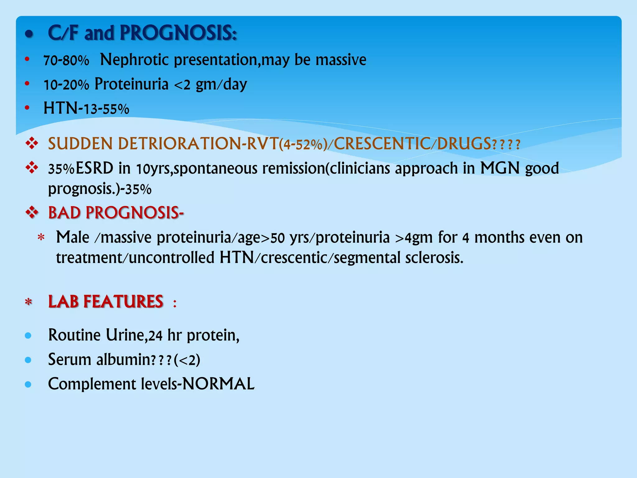
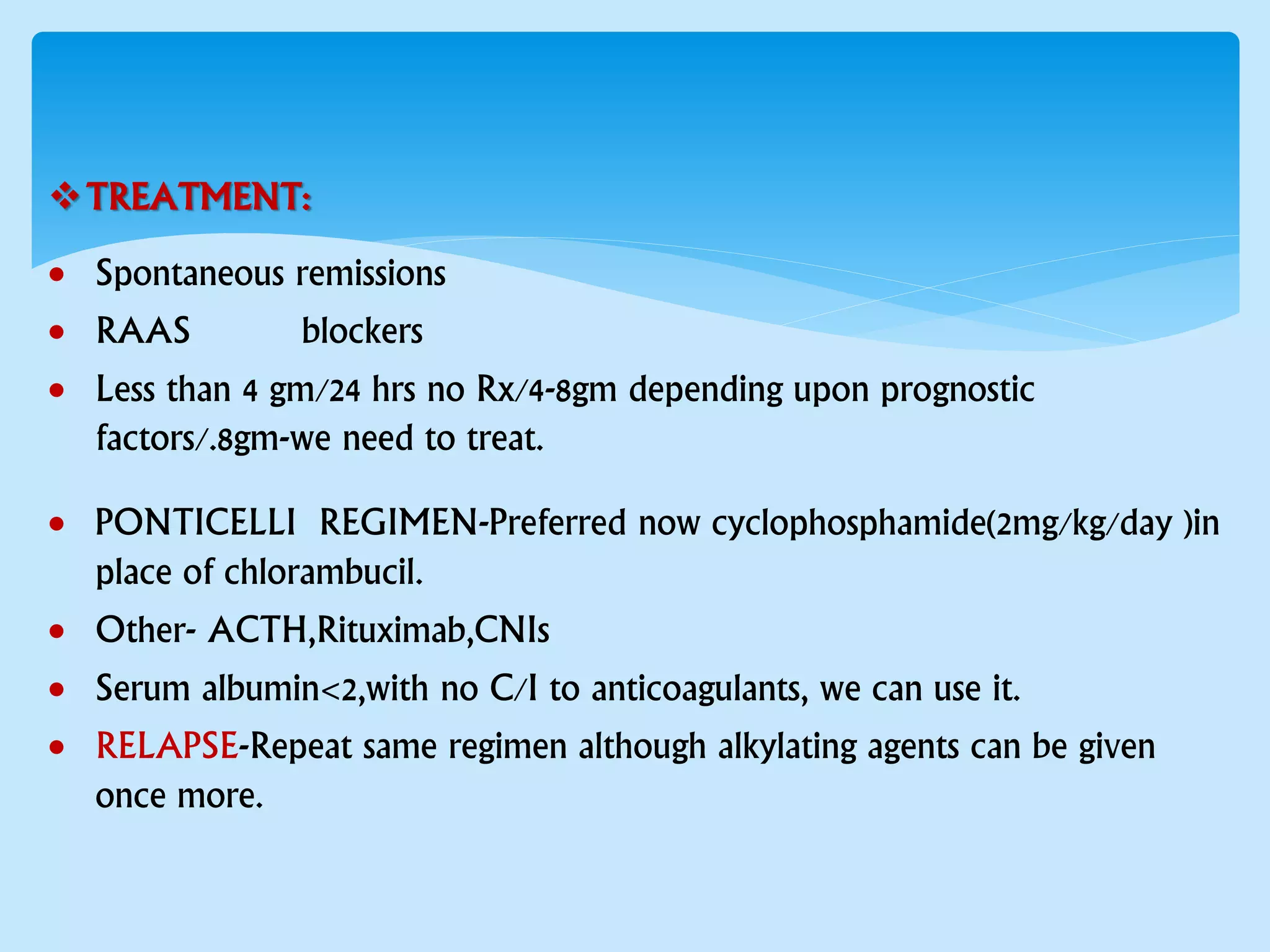
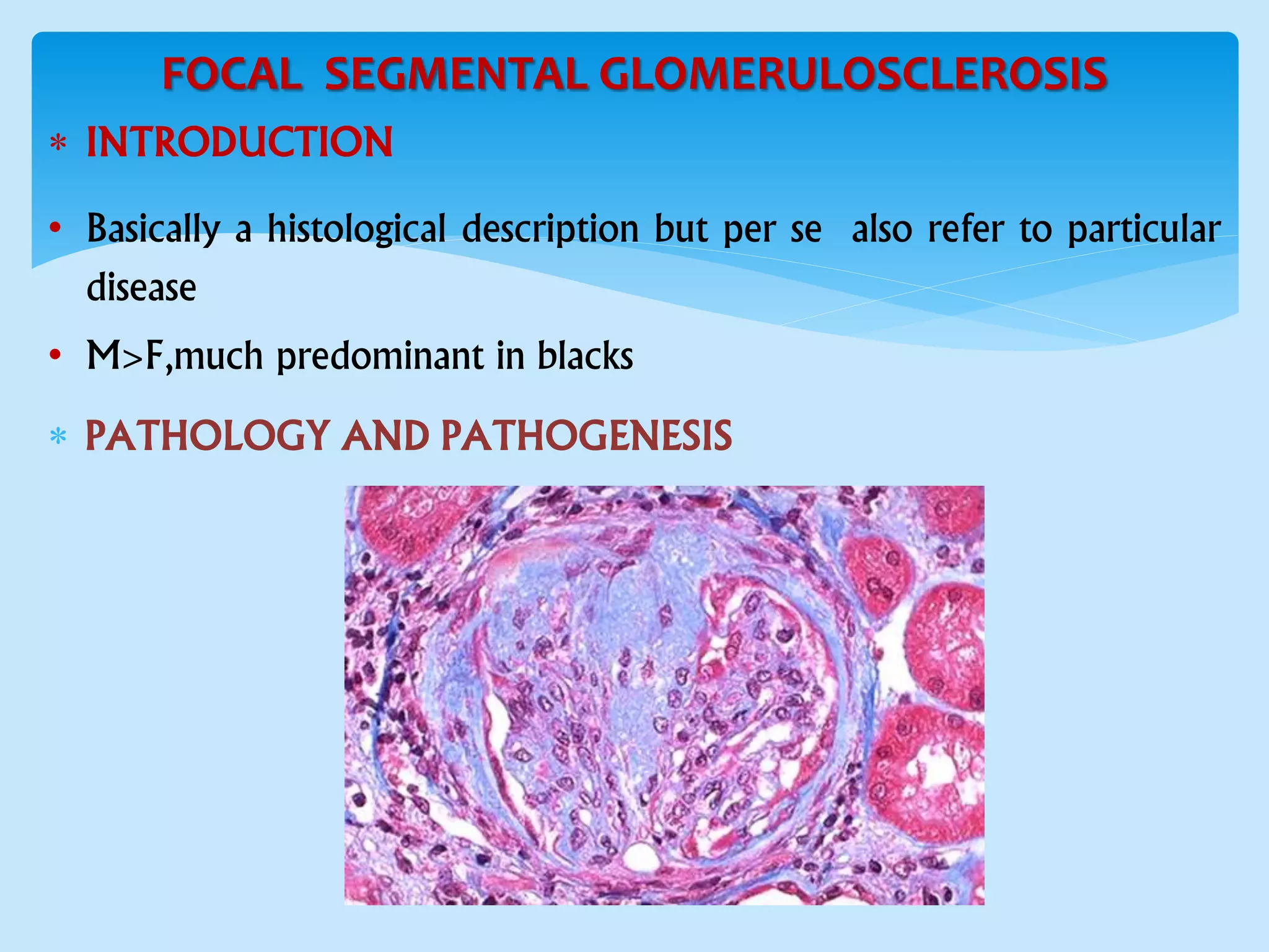
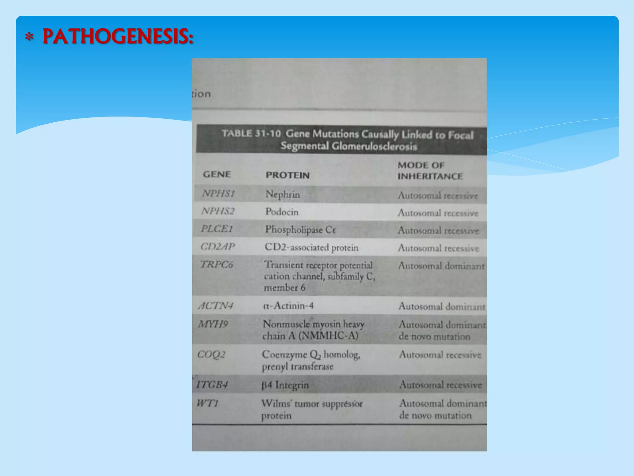
APPROACH TO PRIMARY
GLOMERULONEPHRITIS
DR GAURAV KUMAR(JR2)
DR PROF J.K.L.DAS UNIT
Basicdefinitions:
 Glomerulopathy: A set of disease affecting the glomeruli of
nephron.
 Glomerulitis:It accounts for inflammatory conditions of
glomerulus.
 Glomerulonephritis: Nephritis marked by inflammation of
glomerular capillaries.
primary: In which the kidney is the only or predominant organ
involved
Secondary: Accompanying systemic diseases.
CONFIRMATION OF
GLOMERULONEPHRITIS
PRIMARY
VARIETY
SECONDARY
VARIETY
2 DEGREE
FSGS
SUSPECT
GLOMERULONEPHRITIS
--------------------------------------------------------
--------------------------------------------------------
D/D of glomerulonephritis to be kept when a patient
presents with the following symptoms as a whole or rarely
when alone:
 Cola colored urine(1ml BLOOD in 1 l of
urine)(haemoglobinuria /myoglobinuria difference???by
Ammonium sulphate solubility test)
 Change in urine output to range from anuria to polyuria
 A newly onset of hypertension in patients or normotensive
patients
From bipedal edema to generalized anasarca
It can present as Acute nephritic
syndrome/Nephrotic/NephriticNephrotic/isolated
hematuria or proteinuria.
 Confirmation of diagnosis:
Glomerular hematuria:
 >3 RBC/hpf or >5RBC/microlitre
 >5% RBC are Acanthocytes,providing a specificity of 95% and
sensitivity although 50%.
 Accompanying RBC cast
 Accompanying albumin need to be 40% of total protein to support it
Keeping in view we must rule out :
 Glomerulo vascular disorders
 Atheroembolic disease
 Diabetes glomerulosclerosis
 Basement membrane disorder
 Warfarin nephropathy
History,physical
examination,U/A,urine
culture
Microscopic
hematuria(dipstick +ve
and rbc in urine)
non
glomerular
Suggestive of UTI No UTI
Glomerular
Quantitate
protein and
GFR,BIOPSY
BIOPSY BASICS
 Iverson and Brun performed first renal biopsy in 1951.
Indications of renal biopsy:
a.In context of glomerulonephritis
b.Systemic diseases with kidney involvement
c.Acute kidney disease
d.Renal transplant patients
e.Undiagnosed etiology
Performed with a kidney biopsy gun (preferably 16 G) under USG
guidance while the patient is in prone position
Generally 10-15 glomeruli are optimal,although 6-10 are
sufficient most of the time.
Biopsy is read in terms of glomerular ,tubular,vascular or
pathology in interstitium.
Safety in pregnancy??(<30 weeks)
+ In context of focal disease of Glomerulus,we need to go to
juxtamedullary glomeruli to see the earliest involvment .
Focal glomerular disesae 25
MGN Even 1
Transplant Minimum 7
Light microscopy 8-10
Glomerular Non
glomerular/Urologic/Urothelial
1.URINE COLOR DARK RED,BROWN,COLA ,SMOKY BRIGHT RED
2.CLOTS _ +
3.PROTEINURIA + _
4.RBC MORPHOLOGY DYSMORPHIC ISOMORPHIC
5.HTN + _
6.EDEMA + -
7.URINARY VOIDING SYMPTOMS _ +
8.BACK PAIN + +
9.F/H + _
10.UPPER RESP TRACT
SYMPTOMS,FEVER RASH
+ _
11.RFT DERRANGED GENERALLY NOT
• Glomerular Proteinuria:
*Generally >2g/day
*Selective index<10%
*UPEP shows albumin fraction much greater than globulin
fraction
*Albumin/B2 microglobulin ratio>1000;1
*Less conc. Of proteins like N acetyl Glucosamine,Lysozyme
etc.
1.Primary GN
2.secondary
GGN
2 DEGREE
FSGS
Glomerulonephritis
classification
Secondary causes of GN can be ruled out by:
 CBC,Hb1ac
 24 hour urine protein
 Serum albumin
 LDH
 C3,c4 level
 SPEP,free light chain Assay
 Viral markers,tropical infections profile
 ANA,ANCA,RA factor,cryoglobulins,APLA ,ds-DNAetc
 Drugs
EPIDEMIOLOGY
 First described by Munk in 1913 as lipoid nephrosis.
 Accounts for 70-90% of Nephrotic syndrome below 10yrs
 10-15% of adults and more propensity in age >60 yrs too,with more
complications.
MINIMAL CHANGE DISEASE
• In children M:F-2:1 or 3;1,Adults?
3. PATHOLOGY AND PATHOGENESIS:
 Effacement of foot process of podocytes(specific????) with
cytoskeleton clumping near BM.
EXTENT OF EFFACEMENT ~ DURATION OF ACTIVE NEPHROTIC SYNDROME THAN
WITH MAGNITUDE OF PROTEINURIA.
• Glomerular permiabilty factor-hemopexin/IL-13 modulated by T cells.
• Increased Ig E.
• Charge selectivity more imp.
 IMMUNOFLUORESCENCE-
• Low level mesangial staining for Ig M(Irregular Focal staining for Ig M and
C3?)
 CLINICAL FEATURES
 Classic presentation is nephrotic syndrome
 HTN(30 %children,50%in adults)
• Microscopic hematuria(20% children,33% adults)
• Atopy and allergy symptoms in 30-40%
• Decreased renal functions (5% in children,30% in adults)
LAB FEATURES
• Urinalysis
• Complement levels
• ESR
• Coagulation profile
• Lipid profile
• Selectivity index<10%?
• Serum Ig E levels
• TREATMENT
• Prednisolone-60mg /m2 in children,1mg/kg body wt (<80mg),treatment
continued for 6 weeks after complete remission(0.2gm/24 hrs) by DIPSTICK
test at home for consecutive 3 days.
• Post remission either alternate day therapy (to 40 mg /m2) or gradual
tapering (???? needed )Acute adrenal suppression may lead to relapse
• Steroid dependence(2 weeks within or during treatment aftr remission is
acheived)/frequent relapse(2 or more within 6 months)
 Cyclophosphamide-2mg/kg for 8-12 weeks /alternate is
cyclosporine/tacrolimus/MMF.
 Steroid resistant(up to 12 WEEKS in children and 16 weeks in adults)
• In children need to go for biopsy
• In adults or in children if its MCD,a regimen of Calcineurin inhibitors
followed by MMF .
Ig A NEPHROPATHY
 EPIDEMIOLOGY:
• Most common GN worldwide
• Described by Berger and Hinglais in late 1960s
• Male:female-2:1 to 6:1
• 2 nd to 3rd decade
• 30-40% to ESRD.
 GENETICS:
 Genetic and Environmental factors both
 D allele mutation of ACE gene in Asians.
 6th chromosome igan1 gene
 Galactosyl transferase gene ,sialyl transferase gene
 Selectin cluster gene
 PATHOLOGY AND PATHOGENESIS:
Immuno. Microscopy-
• 100% stains for Ig A/50 % for Ig G/Less consipicious C3
• If C1q +Ig A+Ig G+C3---LUPUS???
• More lambda chains (kappa chains in other)/light chain deposition
disease??-kappa predominant/renal amyloidosis have lambda
predominant
 Electron Microscopy-
 Mesangial deposits with mesangial matrix expansion and
hypercellularity.
 Light Microscopy-
 37% have focal proliferative glomerulonephritis,28% have DPGN
 Wide spread damage can present lupus like.
 Pathogenesis: Abnormal sialyation of N-ACETYL
GALACTOSAMINE stops conversion ie its replacement by
galactose ,this mutation in Ig A leads to functioning as Antigen
which binds to Ig G in serum and this circulating complex deposits in
glomeruli through mesangial receptors(transferrin) Ig A1 can also
bind to mesangium but it cant lead to proliferation
 ENVIROMENTAL CROSS REACTING ANTIGEN???
 CLINICAL FEATURES:(synpharyngitic presentation)
• 40-50% -Macroscopic hematuria often associated dysuria
• 10% cases-Acute renal insufficiency
• 30%-Asymptomatic microscopic hematuria
• 30% new onset proteinuria and HTN
• May be nephrotic syndrome like presentation
 Prognostic factors:
• Sustained hypertension
• Persistent proteinuria
• Impaired RFT
• Nephrotic like - all signifies Bad prognosis.
• Episodic macroscopic hematuria have good prognosis than
microscopic????(EPIOSDIC AND SELF RESOLVING)
 TORONTO FORMULA???
MAP and proteinuria
 Four independent parameters:
1. Mesangial hypercellularity-significant association with ESRD
2. Endocapillary hypercellularity-More benefit of immunosuppressive
3. Tubular atrophy
4. Segmental glomerulosclerosis
In IgA nephropathy there been a improved prognosis with moderate alcohol
consumption and mild jaundice.
 Laboratory finding:
• Nephritic pres.
• Complement levels,IgA/C3 ratio,C3 fragments
• Serum Ig A levels(Increased in 50%,importance???)
 Treatment Recommendations;
• For patients with proteinuria >0.5 gm/day..we need to treat
• Max tolerated ACE/ARBs to target protein excretion<0.5
• Steroids to come into practice when the prognostic factors are bad,but
GFR>60 and proteinuria >0.5after 3-6 months trial of ACEs.
• Still those with progressive renal insufficiency ,prednisolone with
cyclophosphamide followed by azathioprine should be used.
• Omega 3 fatty acids in view of their less side effect profile ,has made a
substantial benefit when used with ACE/ARBs.(12g /day)
• Recurrence after transplantation is 75-80% but graft loss is quite uncommon
except in those with bad prognostic factors.
MEMBRANOUS GLOMERULOPATHY
*In adult one of the commonest cause of Nephrotic syndrome.
* M:F-2:1,A:C-26:1
* Rule out secondaries-drugs,Ca lung and prostate MC,CTDs ,hep.
PATHOLOGY AND PATHOGENESIS
• EM-Ehrenerich and Churg staging
• Stage 1 from subepithelial deposition to stage 4 of irregular
electron dense deposition within irregular thickend BM.
 ?MESANGIAL DEPOSITS???
• IM-Ig G most prominent followed by Ig M and Ig A,C3 also present in 95%..but not that intense.
• LM-Diffuse capillary wall thickening with absence of significant glomerular hypercellularity.
 PATHOGENESIS- In situ immuncomplex depositon MC mechanism
1. Phospholipase A2 receptor of podocytes-MC 70% cases
2. Neutral endopeptidase of podocytes
3. Cationic bovine serum albumin
 MC antibody involved is Ig G4
 Circulating complex in SECONDARY DISEASE?
Role of complements???
• In complex deposited there is also paucity of complements,serum levels
are normal???
• Nephritogenic serums contain antibodies to react with Crry/DAF etc,less
production of factor H.
 C/F and PROGNOSIS:
• 70-80% Nephrotic presentation,may be massive
• 10-20% Proteinuria <2 gm/day
• HTN-13-55%
 SUDDEN DETRIORATION-RVT(4-52%)/CRESCENTIC/DRUGS????
 35%ESRD in 10yrs,spontaneous remission(clinicians approach in MGN good
prognosis.)-35%
 BAD PROGNOSIS-
 Male /massive proteinuria/age>50 yrs/proteinuria >4gm for 4 months even on
treatment/uncontrolled HTN/crescentic/segmental sclerosis.
 LAB FEATURES :
 Routine Urine,24 hr protein,
 Serum albumin???(<2)
 Complement levels-NORMAL
TREATMENT:
 Spontaneous remissions
 RAAS blockers
 Less than 4 gm/24 hrs no Rx/4-8gm depending upon prognostic
factors/.8gm-we need to treat.
 PONTICELLI REGIMEN-Preferred now cyclophosphamide(2mg/kg/day )in
place of chlorambucil.
 Other- ACTH,Rituximab,CNIs
 Serum albumin<2,with no C/I to anticoagulants, we can use it.
 RELAPSE-Repeat same regimen although alkylating agents can be given
once more.
FOCAL SEGMENTAL GLOMERULOSCLEROSIS
 INTRODUCTION
• Basically a histological description but per se also refer to particular
disease
• M>F,much predominant in blacks
 PATHOLOGY AND PATHOGENESIS
 LM - Focal and segmental glomerulosclerosis
• If we don’t get it,even focal tubulointerstitial injury with glomerular hypercellularity can
be used as surrogate marker.
• Microcystic change in tubular epithelial cells
• Endothelial tubuloreticular inclusions(others HIV,SLE,Interferon alpha,pamidronate)
 HISTOLOGICAL CLASSIFICATION
1.Classical variety/NOS variant-Proto type,MC
2.Cellular variant-Endocapillary hypercellualrity with capillary inclusion.
3.Tip variant-Alongside of proximal tubule, outer tip of glomerulus. Steroid responsive with
very good prognosis.
4.Collapsing variant-Torrential nephritic syndrome and rapid declining renal function.
5.Perihilar variant-Perihilar hyalinosis and sclerosis in >50%,often seen with hyperfiltration
injury .
 IM-
• IM staining is only shown by sclerotic glomeruli
• EM-V imp to rule out other causes of glomerular scarring.
 PATHOGENESIS:
• As shown above the genetic mutations
• Black race much vulnerable due to MYH9 SNPs ,APOL1 variants(protective against
T.Brucei) on chromosome 22.
• Glomerular permeability factor??
• Parvovirus and SV40 virus.
• Glomerular damages and inciting events
 C/F and PROGNOSTIC FACTORS:
• 80% Cases Nephrotic syndrome presentation
• Hematuria 50%/HTN-1/3rd cases
• Perihilar and collapsing variety early age , perihilar a/w hyperfiltration and
collapsing with torrential proteinuria
• Tip lesions –older peoples,although severe proteinuria like MCD,but most cases are
self resolving,or good remission with steroids.
• Poor prognosis are-
 Black male/severe proteinuria(>10gm/day)/Interstitial fibrosis on histology/no
responsive to rx/collapsing variant
 TREATMENT:
• Prednisolone 1mg/kg (max 80 mg) upto 16 weeks or till remission in
full dosage.If there is remission, we need to taper it to nil in 6
months.
• If we don’t get remission,prednisolone at 0.15 mg/kg to be continued
with cyclosporine(3-5 mg/kg) in two divided dosages, for 6 months.If
we get positive response continuation upto 12 months.
• If even not,we can try PEX(very good results in post
Tx),Tacrolimus,MMF.
POST STREPTOCCCAL
GLOMERULONEPHRITIS
 EPIDEMIOLOGY:
• Peak incidence 2-6 yrs,15% cases accounts between less than 2 and more than
40 yrs.M>F
• HLA –DRw4 are susceptible ones.
• Group A Lancefield (on Carbohydrate antigen) beta hemolytic streptococci
MC type 12 and 49,other Griffith M(on protein) type strains
1,2,3,4,18,52,55,57,58,59,60,61 are highly nephritogenic.Type 2,49,55,57 and 60
are associated with post impetigo,M type 49 can lead to nephritis after
both.Type C(Recent epidemic with strept. Zooepidemicus) and G has also
been implicated in few cases.
• Risk is about up to 15% with nephritogenic strains.(SEEGAL &EARL)
• Impetigo a/w epidemic PSGN ,Pharyngitic with sporadic mainly.
 C/F:
• Post Pharyngitic phase-7-21days
• Post impetigo-14-21 days
• Synpharyngitic <7 days.
• Classic nephritic presentation,HTN in around 75%,edema is presenting symptom in
2/3rd,with present in almost 90% cases.Flank pain is associated in about 50% cases
• Clinical manifestation usually resolves in 1-2 weeks,with complete resolution of
microscopic hematuria and proteinuria can take up to 8 weeks.
• Together PSGN and Rheumatic fever very rare but can be possible.
 LAB FINDING:
• R/E Urine- 5% of children and 20% adults have nephrotic range proteinuria.
• Streptozyme test (Anti streptococcal antibody assays.),CULTURE(10-70%),ASO
TITRE(30%),Anti DNAse-70%,Anti hyaluronidase -40%
• ASO titres (2/3rd of pharyngitic and 1/3rd of impetigo)and those not producing
,Anti –DNAse we should go for.
• In first week 90% of patients have depressed CH50 and C3 and normal C4.
 IM:
• Garland pattern suggestive of large closely opposed granular deposits on
capillary walls have nephrotic range proteinuria.
• Starry sky have scatterd granular deposits have less severe disease.
• Mesangial pattern with predominant c3 staining,corresponds to resolving
phase.
• Staining with Ig G as well as c3 indicates active on going disease,only with c3
indicates a sign of on going reemission.
 PATHOGENESIS;
• Implanted antigens leading to immune complex and complement
activation(alternate >classical )are most proposed ones.
• These imp antigens include Streptococcal pyogenic exotoxin B, Nephritis
associated plasmin receptor.
 TREATMENT:
• Supportive-Diuretics ,antibiotics,anti HTN,treatment of co morbidities
• Those presenting with crescentic GN and ARF too have very good recovery.
• Permanent renal failure occur in about 1% of children. Complete resolution
generally occur within 3-6 weeks of onset of nephritis.
• No evidence to date support that early treatment of
streptococcal disease either pharyngitis or cellulitis alter the
risk of PSGN
• Prognosis in elders is worse with high incidence of azotemia(up to
60%),nephrotic range proteinuria and ESRD.
RPGN:
NOMENCLATURE AND CATEGORAIZATION:
• Rapid loss of renal function(Upto 50% decrease in GFR) with features
of glomerulonephritis accompanied by oliguria/anuria.(<3months)
• Histologically ,crescents must form in >50% of all glomeruli.
• Inciting event leads to focal rupture of capillary walls.
• MC cause –Immune complex mediated GN,and same in children.
• In adults-Paucimmune variety.
• Rarest –Anti GBM.
• Immuncomplex variety and Anti GBM variety we can 1/3rd to ¼ th
cases +ve ANCA too.
Pictorial representation:
 Immune complex mediated:
• Evidence of special inciting GN ,systemic immunecomplex disease leading to it.
• Idiopathic crescentic immune complex.
 LM
• Along side of crescents in intact glomeruli we get varying combinations of capillary wall
thickening, endocapillary hypercellularity unlike in other two we get necrotizing
glomeruli,with total normal glomeruli in between,associated with sclerosis widespread.
• Crescents much less evident than in Anti GBM and pauciimmune ,so a/w good prognosis.
 IM:
• Mesangial IgA deposits?
• C3 dominant deposists??
• Fine granular Ig G4 deposits??
• Coarse granular capillary wall thickening??
• ANA +immunecomplex?
• ANA + anti GBM??

 TREATMENT:
 Influenced by the underlying etiology
 Idiopathic variety we go for-Immunosupressive therapy with pulse
methylprednisolone 3-5 days, followed by 1 mg/kg prednisolone
tapered over 2nd and 3 rd month to alternate day regimen until
completely discontinued.Rituximab has also been beneficial in many
trials.
 Anti GBM disesase:
 Two peaks-young onset in male (2 nd -3rd decade)/second peak in female in 6th-7th
decade.
 HLA-DR2
 PATHOGENESIS:
• Ig G1(MC) antibody formed against NC/Collagenase resistant alpha 3
domain of type 4 collagen.(alpha 4-TMD,alpha 5-Alport syndrome, alpha 1-
paraneoplastic syndromes)
• Anti GBM antibodies(>95%) breech only the monomer subunits of
hexameric quaternary structures of Nc1 which are otherwise cryptic in
normal individuals, exposed by some environmental factors, genetic
susceptibility.
• ANCA association in about 30% cases.
 MICROSCOPY:
• IM-Linear Ig G staining of GBM(diabetic glomerulosclerosis ,hypertensive
vascular disease,Fibrillary glomerulopathies???)
• LM-97% have some crescents,85% have more than 50% crescents
• Widespread necrotizing glomeruli with many crescents, sclerosis,
normal glomeruli in between.
 C/F and prognosis:
• Picture of GN and associated with pul. hemorrhages that may be subtle or
lifethreatning,same BM material may be found in pulmonary capillaries-GOOD
PASTURE SYNDROME.
• Haemorrhages more common in smokers.
 TREATMENT:
• Plasmapheresis(8-10timeneeded)+steroids+cyclophosphamide(3months
Rx)(i.v>oral??-less cumulative toxic dosage,less severe leucopenia)
• Patients who need dialysis becomes readily dialysis dependent.
• > 7 mg/dl creatinine with widespread glomerular and interstitial scarring,don’t get
benefit with immunosuppressive.
• If ANCA+ in same patients(cyclophosphamide is to be given )
• Once remission is achieved very rare relapse.
• Transplantation results are too good(Alport syndrome with alpha 3 antibody)-
Should wait for at least 6 months till serum antibodies are undetectable.
Paucimmune variety
 C-ANCA
 Antibody to proteinase 3(PR3)
 Found in primary granules of
neutrophils and lysosomes of
monocytes.
 This variety is associated with less
relapse. Although associated lung
and upper respiratory tract
involvement also signifies less
relapse.
 Characteristically
GRANULOMATOSIS WITH
POLYANGITIS have 80-90%
positivity with it.
 Association with patient exposed
to silica dust, alpha anti trypsin
deficiency.
 P-ANCA
 Antibody to MPO
 Found in primary granules of
neutrophils and lysosomes of
monocytes
 More relapse
 Notable examples are MICROSCOPIC
POLYANGITIS and CHURG
STRAUSS SYNDROME.
 Another pathogenic antibody being detected is lamp2 antibody.
 Around 1/3 rd cases presents as isolated clinical manifestation of
kidney disease,although we can get pathological findings in many of
cases.
Rx:
 Since mortality is very high in most of the cases,treatment is urgently
needed.
 Induction therapy usually consists of plasmapheresis
,methylprednisolone and cyclophosphamide.
 Steroids are tapered(up to 20 mg in 8weeks) soon after the acute
inflammation subsides and are maintained on cyclophosphamide or
azathioprine to prevent relapse for at least one yr.
 Maintainence therapy is not needed in those who are dialysis
dependent and have no extra renal manifestations.
 Benefit with MMF or Rituximab is controversial but recently most of
trials are supporting its use.IvIg have also been found beneficial in
some of refractory cases.
MESANGIO CAPILLARY GLOMERULONEPHRITIS
• Histological term,a rare GN .
• May be idiopathic ,more common being secondary
• Children 8-16yr most common,M=F
 PATHOLOGY AND PATHOGENESIS:
• Type 1 –classical complement activation pathway mainly(Antigen???)
,generally associated with hepatitis C and autoimmune
disorders.{SECONDARY MOSTLY}
• Type2-Alternate pathway activation(c3NeF/absent factor H/antibody against
factor H-- leading to disinhibition of C3 convertase ie C3bBb)(DENSE
DEPOSIT DISEASE),associated with partial lipodystrophy syndrome.
• Type3-Like MGN subepithelial deposition, associated with complement
receptor deficiency.
• Type 2 and type 3 comes under another new category ie C3
glomerulopathies.
MICROSCOPY:
• Subendothelial dep. and mesangial hypercellularity are chief picture a/w
endocapillary thickening,global capillary wall thickening leading to picture of
HYPERSEGMENTATION AND LOBULATION.
• Tram track appearence on methanamine silver staining.
• IM staining is peripheral granular or band like complement specially c3(in type 2
and 3),Immunocomplex evident in type 1
 C/F AND PROGNOSIS:
• May present as asymptomatic,nephritic and nephrotic poteinuria,acute
nephritic syndrome,ESRD(very high chance 50% in 10 yrs.)
• MPGN 2 a/w lipodystrophy,retinal pigmentation abnormlity.
 Poor prognosis
 HTN,impaired GFR,appearance of nephrotic syndrome,appearance of
crescents,MPGN 2 (more associated CRESCENTS)
 LAB FEATURES:
 Routine test for GN
 Serum compliment levels-classical complement pathway (c1q,C4),alternate
pathway(c3)
 Consistently depressed C3 alike PSGN in which C3 return to normal .
 TREATMENT:
 General treatment of GN
 KDIGO guidelines - start of ACE inhibitor and Anti HTN drugs if
needed,in proteinuria>30mg/24hr
 Specific treatment is needed (ie
steroids/immunosupressants/antiplatelets)when:
 Proteinuria>3gm/24hr
 Active interstitial or glomerular disease
 Impaired renal function presentation
 Progressive deterioration of renal functions
 Some trials benefits the use of Dipyridamole, aspirin and warfarin
although not proven.
 Post transplantation MPGN1 has been found to be of least recurrence
among primary GN but type 2 has maximum.
 Eculizumab has been found to be of unproven benefit.
Approach to primary glomerulonnephritis

Approach to primary glomerulonnephritis

  • 1.
    APPROACH TO PRIMARY GLOMERULONEPHRITIS DRGAURAV KUMAR(JR2) DR PROF J.K.L.DAS UNIT
  • 2.
    Basicdefinitions:  Glomerulopathy: Aset of disease affecting the glomeruli of nephron.  Glomerulitis:It accounts for inflammatory conditions of glomerulus.  Glomerulonephritis: Nephritis marked by inflammation of glomerular capillaries. primary: In which the kidney is the only or predominant organ involved Secondary: Accompanying systemic diseases.
  • 3.
  • 4.
    D/D of glomerulonephritisto be kept when a patient presents with the following symptoms as a whole or rarely when alone:  Cola colored urine(1ml BLOOD in 1 l of urine)(haemoglobinuria /myoglobinuria difference???by Ammonium sulphate solubility test)  Change in urine output to range from anuria to polyuria  A newly onset of hypertension in patients or normotensive patients From bipedal edema to generalized anasarca It can present as Acute nephritic syndrome/Nephrotic/NephriticNephrotic/isolated hematuria or proteinuria.
  • 5.
     Confirmation ofdiagnosis: Glomerular hematuria:  >3 RBC/hpf or >5RBC/microlitre  >5% RBC are Acanthocytes,providing a specificity of 95% and sensitivity although 50%.  Accompanying RBC cast  Accompanying albumin need to be 40% of total protein to support it Keeping in view we must rule out :  Glomerulo vascular disorders  Atheroembolic disease  Diabetes glomerulosclerosis  Basement membrane disorder  Warfarin nephropathy
  • 7.
    History,physical examination,U/A,urine culture Microscopic hematuria(dipstick +ve and rbcin urine) non glomerular Suggestive of UTI No UTI Glomerular Quantitate protein and GFR,BIOPSY
  • 8.
    BIOPSY BASICS  Iversonand Brun performed first renal biopsy in 1951. Indications of renal biopsy: a.In context of glomerulonephritis b.Systemic diseases with kidney involvement c.Acute kidney disease d.Renal transplant patients e.Undiagnosed etiology Performed with a kidney biopsy gun (preferably 16 G) under USG guidance while the patient is in prone position
  • 10.
    Generally 10-15 glomeruliare optimal,although 6-10 are sufficient most of the time. Biopsy is read in terms of glomerular ,tubular,vascular or pathology in interstitium. Safety in pregnancy??(<30 weeks) + In context of focal disease of Glomerulus,we need to go to juxtamedullary glomeruli to see the earliest involvment . Focal glomerular disesae 25 MGN Even 1 Transplant Minimum 7 Light microscopy 8-10
  • 11.
    Glomerular Non glomerular/Urologic/Urothelial 1.URINE COLORDARK RED,BROWN,COLA ,SMOKY BRIGHT RED 2.CLOTS _ + 3.PROTEINURIA + _ 4.RBC MORPHOLOGY DYSMORPHIC ISOMORPHIC 5.HTN + _ 6.EDEMA + - 7.URINARY VOIDING SYMPTOMS _ + 8.BACK PAIN + + 9.F/H + _ 10.UPPER RESP TRACT SYMPTOMS,FEVER RASH + _ 11.RFT DERRANGED GENERALLY NOT
  • 12.
    • Glomerular Proteinuria: *Generally>2g/day *Selective index<10% *UPEP shows albumin fraction much greater than globulin fraction *Albumin/B2 microglobulin ratio>1000;1 *Less conc. Of proteins like N acetyl Glucosamine,Lysozyme etc. 1.Primary GN 2.secondary GGN 2 DEGREE FSGS Glomerulonephritis classification
  • 13.
    Secondary causes ofGN can be ruled out by:  CBC,Hb1ac  24 hour urine protein  Serum albumin  LDH  C3,c4 level  SPEP,free light chain Assay  Viral markers,tropical infections profile  ANA,ANCA,RA factor,cryoglobulins,APLA ,ds-DNAetc  Drugs
  • 15.
    EPIDEMIOLOGY  First describedby Munk in 1913 as lipoid nephrosis.  Accounts for 70-90% of Nephrotic syndrome below 10yrs  10-15% of adults and more propensity in age >60 yrs too,with more complications. MINIMAL CHANGE DISEASE
  • 16.
    • In childrenM:F-2:1 or 3;1,Adults? 3. PATHOLOGY AND PATHOGENESIS:
  • 17.
     Effacement offoot process of podocytes(specific????) with cytoskeleton clumping near BM. EXTENT OF EFFACEMENT ~ DURATION OF ACTIVE NEPHROTIC SYNDROME THAN WITH MAGNITUDE OF PROTEINURIA. • Glomerular permiabilty factor-hemopexin/IL-13 modulated by T cells. • Increased Ig E. • Charge selectivity more imp.  IMMUNOFLUORESCENCE- • Low level mesangial staining for Ig M(Irregular Focal staining for Ig M and C3?)  CLINICAL FEATURES  Classic presentation is nephrotic syndrome  HTN(30 %children,50%in adults)
  • 18.
    • Microscopic hematuria(20%children,33% adults) • Atopy and allergy symptoms in 30-40% • Decreased renal functions (5% in children,30% in adults) LAB FEATURES • Urinalysis • Complement levels • ESR • Coagulation profile • Lipid profile • Selectivity index<10%? • Serum Ig E levels
  • 19.
    • TREATMENT • Prednisolone-60mg/m2 in children,1mg/kg body wt (<80mg),treatment continued for 6 weeks after complete remission(0.2gm/24 hrs) by DIPSTICK test at home for consecutive 3 days. • Post remission either alternate day therapy (to 40 mg /m2) or gradual tapering (???? needed )Acute adrenal suppression may lead to relapse • Steroid dependence(2 weeks within or during treatment aftr remission is acheived)/frequent relapse(2 or more within 6 months)  Cyclophosphamide-2mg/kg for 8-12 weeks /alternate is cyclosporine/tacrolimus/MMF.  Steroid resistant(up to 12 WEEKS in children and 16 weeks in adults) • In children need to go for biopsy • In adults or in children if its MCD,a regimen of Calcineurin inhibitors followed by MMF .
  • 20.
    Ig A NEPHROPATHY EPIDEMIOLOGY: • Most common GN worldwide • Described by Berger and Hinglais in late 1960s • Male:female-2:1 to 6:1 • 2 nd to 3rd decade • 30-40% to ESRD.  GENETICS:  Genetic and Environmental factors both  D allele mutation of ACE gene in Asians.  6th chromosome igan1 gene  Galactosyl transferase gene ,sialyl transferase gene  Selectin cluster gene
  • 21.
     PATHOLOGY ANDPATHOGENESIS: Immuno. Microscopy-
  • 22.
    • 100% stainsfor Ig A/50 % for Ig G/Less consipicious C3 • If C1q +Ig A+Ig G+C3---LUPUS??? • More lambda chains (kappa chains in other)/light chain deposition disease??-kappa predominant/renal amyloidosis have lambda predominant  Electron Microscopy-  Mesangial deposits with mesangial matrix expansion and hypercellularity.  Light Microscopy-  37% have focal proliferative glomerulonephritis,28% have DPGN  Wide spread damage can present lupus like.
  • 23.
     Pathogenesis: Abnormalsialyation of N-ACETYL GALACTOSAMINE stops conversion ie its replacement by galactose ,this mutation in Ig A leads to functioning as Antigen which binds to Ig G in serum and this circulating complex deposits in glomeruli through mesangial receptors(transferrin) Ig A1 can also bind to mesangium but it cant lead to proliferation  ENVIROMENTAL CROSS REACTING ANTIGEN???  CLINICAL FEATURES:(synpharyngitic presentation) • 40-50% -Macroscopic hematuria often associated dysuria • 10% cases-Acute renal insufficiency • 30%-Asymptomatic microscopic hematuria • 30% new onset proteinuria and HTN • May be nephrotic syndrome like presentation
  • 24.
     Prognostic factors: •Sustained hypertension • Persistent proteinuria • Impaired RFT • Nephrotic like - all signifies Bad prognosis. • Episodic macroscopic hematuria have good prognosis than microscopic????(EPIOSDIC AND SELF RESOLVING)  TORONTO FORMULA??? MAP and proteinuria  Four independent parameters: 1. Mesangial hypercellularity-significant association with ESRD 2. Endocapillary hypercellularity-More benefit of immunosuppressive 3. Tubular atrophy 4. Segmental glomerulosclerosis In IgA nephropathy there been a improved prognosis with moderate alcohol consumption and mild jaundice.
  • 25.
     Laboratory finding: •Nephritic pres. • Complement levels,IgA/C3 ratio,C3 fragments • Serum Ig A levels(Increased in 50%,importance???)  Treatment Recommendations; • For patients with proteinuria >0.5 gm/day..we need to treat • Max tolerated ACE/ARBs to target protein excretion<0.5 • Steroids to come into practice when the prognostic factors are bad,but GFR>60 and proteinuria >0.5after 3-6 months trial of ACEs. • Still those with progressive renal insufficiency ,prednisolone with cyclophosphamide followed by azathioprine should be used. • Omega 3 fatty acids in view of their less side effect profile ,has made a substantial benefit when used with ACE/ARBs.(12g /day) • Recurrence after transplantation is 75-80% but graft loss is quite uncommon except in those with bad prognostic factors.
  • 26.
    MEMBRANOUS GLOMERULOPATHY *In adultone of the commonest cause of Nephrotic syndrome. * M:F-2:1,A:C-26:1 * Rule out secondaries-drugs,Ca lung and prostate MC,CTDs ,hep.
  • 27.
    PATHOLOGY AND PATHOGENESIS •EM-Ehrenerich and Churg staging • Stage 1 from subepithelial deposition to stage 4 of irregular electron dense deposition within irregular thickend BM.
  • 28.
     ?MESANGIAL DEPOSITS??? •IM-Ig G most prominent followed by Ig M and Ig A,C3 also present in 95%..but not that intense. • LM-Diffuse capillary wall thickening with absence of significant glomerular hypercellularity.  PATHOGENESIS- In situ immuncomplex depositon MC mechanism 1. Phospholipase A2 receptor of podocytes-MC 70% cases 2. Neutral endopeptidase of podocytes 3. Cationic bovine serum albumin  MC antibody involved is Ig G4  Circulating complex in SECONDARY DISEASE? Role of complements??? • In complex deposited there is also paucity of complements,serum levels are normal??? • Nephritogenic serums contain antibodies to react with Crry/DAF etc,less production of factor H.
  • 29.
     C/F andPROGNOSIS: • 70-80% Nephrotic presentation,may be massive • 10-20% Proteinuria <2 gm/day • HTN-13-55%  SUDDEN DETRIORATION-RVT(4-52%)/CRESCENTIC/DRUGS????  35%ESRD in 10yrs,spontaneous remission(clinicians approach in MGN good prognosis.)-35%  BAD PROGNOSIS-  Male /massive proteinuria/age>50 yrs/proteinuria >4gm for 4 months even on treatment/uncontrolled HTN/crescentic/segmental sclerosis.  LAB FEATURES :  Routine Urine,24 hr protein,  Serum albumin???(<2)  Complement levels-NORMAL
  • 30.
    TREATMENT:  Spontaneous remissions RAAS blockers  Less than 4 gm/24 hrs no Rx/4-8gm depending upon prognostic factors/.8gm-we need to treat.  PONTICELLI REGIMEN-Preferred now cyclophosphamide(2mg/kg/day )in place of chlorambucil.  Other- ACTH,Rituximab,CNIs  Serum albumin<2,with no C/I to anticoagulants, we can use it.  RELAPSE-Repeat same regimen although alkylating agents can be given once more.
  • 31.
    FOCAL SEGMENTAL GLOMERULOSCLEROSIS INTRODUCTION • Basically a histological description but per se also refer to particular disease • M>F,much predominant in blacks  PATHOLOGY AND PATHOGENESIS
  • 32.
     LM -Focal and segmental glomerulosclerosis • If we don’t get it,even focal tubulointerstitial injury with glomerular hypercellularity can be used as surrogate marker. • Microcystic change in tubular epithelial cells • Endothelial tubuloreticular inclusions(others HIV,SLE,Interferon alpha,pamidronate)  HISTOLOGICAL CLASSIFICATION 1.Classical variety/NOS variant-Proto type,MC 2.Cellular variant-Endocapillary hypercellualrity with capillary inclusion. 3.Tip variant-Alongside of proximal tubule, outer tip of glomerulus. Steroid responsive with very good prognosis. 4.Collapsing variant-Torrential nephritic syndrome and rapid declining renal function. 5.Perihilar variant-Perihilar hyalinosis and sclerosis in >50%,often seen with hyperfiltration injury .  IM- • IM staining is only shown by sclerotic glomeruli • EM-V imp to rule out other causes of glomerular scarring.
  • 33.
  • 34.
    • As shownabove the genetic mutations • Black race much vulnerable due to MYH9 SNPs ,APOL1 variants(protective against T.Brucei) on chromosome 22. • Glomerular permeability factor?? • Parvovirus and SV40 virus. • Glomerular damages and inciting events  C/F and PROGNOSTIC FACTORS: • 80% Cases Nephrotic syndrome presentation • Hematuria 50%/HTN-1/3rd cases • Perihilar and collapsing variety early age , perihilar a/w hyperfiltration and collapsing with torrential proteinuria • Tip lesions –older peoples,although severe proteinuria like MCD,but most cases are self resolving,or good remission with steroids. • Poor prognosis are-  Black male/severe proteinuria(>10gm/day)/Interstitial fibrosis on histology/no responsive to rx/collapsing variant
  • 35.
     TREATMENT: • Prednisolone1mg/kg (max 80 mg) upto 16 weeks or till remission in full dosage.If there is remission, we need to taper it to nil in 6 months. • If we don’t get remission,prednisolone at 0.15 mg/kg to be continued with cyclosporine(3-5 mg/kg) in two divided dosages, for 6 months.If we get positive response continuation upto 12 months. • If even not,we can try PEX(very good results in post Tx),Tacrolimus,MMF.
  • 36.
    POST STREPTOCCCAL GLOMERULONEPHRITIS  EPIDEMIOLOGY: •Peak incidence 2-6 yrs,15% cases accounts between less than 2 and more than 40 yrs.M>F • HLA –DRw4 are susceptible ones. • Group A Lancefield (on Carbohydrate antigen) beta hemolytic streptococci MC type 12 and 49,other Griffith M(on protein) type strains 1,2,3,4,18,52,55,57,58,59,60,61 are highly nephritogenic.Type 2,49,55,57 and 60 are associated with post impetigo,M type 49 can lead to nephritis after both.Type C(Recent epidemic with strept. Zooepidemicus) and G has also been implicated in few cases. • Risk is about up to 15% with nephritogenic strains.(SEEGAL &EARL) • Impetigo a/w epidemic PSGN ,Pharyngitic with sporadic mainly.
  • 38.
     C/F: • PostPharyngitic phase-7-21days • Post impetigo-14-21 days • Synpharyngitic <7 days. • Classic nephritic presentation,HTN in around 75%,edema is presenting symptom in 2/3rd,with present in almost 90% cases.Flank pain is associated in about 50% cases • Clinical manifestation usually resolves in 1-2 weeks,with complete resolution of microscopic hematuria and proteinuria can take up to 8 weeks. • Together PSGN and Rheumatic fever very rare but can be possible.  LAB FINDING: • R/E Urine- 5% of children and 20% adults have nephrotic range proteinuria. • Streptozyme test (Anti streptococcal antibody assays.),CULTURE(10-70%),ASO TITRE(30%),Anti DNAse-70%,Anti hyaluronidase -40% • ASO titres (2/3rd of pharyngitic and 1/3rd of impetigo)and those not producing ,Anti –DNAse we should go for. • In first week 90% of patients have depressed CH50 and C3 and normal C4.
  • 39.
     IM: • Garlandpattern suggestive of large closely opposed granular deposits on capillary walls have nephrotic range proteinuria. • Starry sky have scatterd granular deposits have less severe disease. • Mesangial pattern with predominant c3 staining,corresponds to resolving phase. • Staining with Ig G as well as c3 indicates active on going disease,only with c3 indicates a sign of on going reemission.  PATHOGENESIS; • Implanted antigens leading to immune complex and complement activation(alternate >classical )are most proposed ones. • These imp antigens include Streptococcal pyogenic exotoxin B, Nephritis associated plasmin receptor.
  • 41.
     TREATMENT: • Supportive-Diuretics,antibiotics,anti HTN,treatment of co morbidities • Those presenting with crescentic GN and ARF too have very good recovery. • Permanent renal failure occur in about 1% of children. Complete resolution generally occur within 3-6 weeks of onset of nephritis. • No evidence to date support that early treatment of streptococcal disease either pharyngitis or cellulitis alter the risk of PSGN • Prognosis in elders is worse with high incidence of azotemia(up to 60%),nephrotic range proteinuria and ESRD.
  • 42.
  • 43.
    • Rapid lossof renal function(Upto 50% decrease in GFR) with features of glomerulonephritis accompanied by oliguria/anuria.(<3months) • Histologically ,crescents must form in >50% of all glomeruli. • Inciting event leads to focal rupture of capillary walls. • MC cause –Immune complex mediated GN,and same in children. • In adults-Paucimmune variety. • Rarest –Anti GBM. • Immuncomplex variety and Anti GBM variety we can 1/3rd to ¼ th cases +ve ANCA too.
  • 45.
  • 46.
     Immune complexmediated: • Evidence of special inciting GN ,systemic immunecomplex disease leading to it. • Idiopathic crescentic immune complex.  LM • Along side of crescents in intact glomeruli we get varying combinations of capillary wall thickening, endocapillary hypercellularity unlike in other two we get necrotizing glomeruli,with total normal glomeruli in between,associated with sclerosis widespread. • Crescents much less evident than in Anti GBM and pauciimmune ,so a/w good prognosis.  IM: • Mesangial IgA deposits? • C3 dominant deposists?? • Fine granular Ig G4 deposits?? • Coarse granular capillary wall thickening?? • ANA +immunecomplex? • ANA + anti GBM??
  • 47.
      TREATMENT:  Influencedby the underlying etiology  Idiopathic variety we go for-Immunosupressive therapy with pulse methylprednisolone 3-5 days, followed by 1 mg/kg prednisolone tapered over 2nd and 3 rd month to alternate day regimen until completely discontinued.Rituximab has also been beneficial in many trials.
  • 48.
     Anti GBMdisesase:  Two peaks-young onset in male (2 nd -3rd decade)/second peak in female in 6th-7th decade.  HLA-DR2
  • 49.
     PATHOGENESIS: • IgG1(MC) antibody formed against NC/Collagenase resistant alpha 3 domain of type 4 collagen.(alpha 4-TMD,alpha 5-Alport syndrome, alpha 1- paraneoplastic syndromes) • Anti GBM antibodies(>95%) breech only the monomer subunits of hexameric quaternary structures of Nc1 which are otherwise cryptic in normal individuals, exposed by some environmental factors, genetic susceptibility. • ANCA association in about 30% cases.  MICROSCOPY: • IM-Linear Ig G staining of GBM(diabetic glomerulosclerosis ,hypertensive vascular disease,Fibrillary glomerulopathies???) • LM-97% have some crescents,85% have more than 50% crescents • Widespread necrotizing glomeruli with many crescents, sclerosis, normal glomeruli in between.
  • 50.
     C/F andprognosis: • Picture of GN and associated with pul. hemorrhages that may be subtle or lifethreatning,same BM material may be found in pulmonary capillaries-GOOD PASTURE SYNDROME. • Haemorrhages more common in smokers.  TREATMENT: • Plasmapheresis(8-10timeneeded)+steroids+cyclophosphamide(3months Rx)(i.v>oral??-less cumulative toxic dosage,less severe leucopenia) • Patients who need dialysis becomes readily dialysis dependent. • > 7 mg/dl creatinine with widespread glomerular and interstitial scarring,don’t get benefit with immunosuppressive. • If ANCA+ in same patients(cyclophosphamide is to be given ) • Once remission is achieved very rare relapse. • Transplantation results are too good(Alport syndrome with alpha 3 antibody)- Should wait for at least 6 months till serum antibodies are undetectable.
  • 51.
    Paucimmune variety  C-ANCA Antibody to proteinase 3(PR3)  Found in primary granules of neutrophils and lysosomes of monocytes.  This variety is associated with less relapse. Although associated lung and upper respiratory tract involvement also signifies less relapse.  Characteristically GRANULOMATOSIS WITH POLYANGITIS have 80-90% positivity with it.  Association with patient exposed to silica dust, alpha anti trypsin deficiency.  P-ANCA  Antibody to MPO  Found in primary granules of neutrophils and lysosomes of monocytes  More relapse  Notable examples are MICROSCOPIC POLYANGITIS and CHURG STRAUSS SYNDROME.
  • 52.
     Another pathogenicantibody being detected is lamp2 antibody.  Around 1/3 rd cases presents as isolated clinical manifestation of kidney disease,although we can get pathological findings in many of cases. Rx:  Since mortality is very high in most of the cases,treatment is urgently needed.  Induction therapy usually consists of plasmapheresis ,methylprednisolone and cyclophosphamide.  Steroids are tapered(up to 20 mg in 8weeks) soon after the acute inflammation subsides and are maintained on cyclophosphamide or azathioprine to prevent relapse for at least one yr.  Maintainence therapy is not needed in those who are dialysis dependent and have no extra renal manifestations.  Benefit with MMF or Rituximab is controversial but recently most of trials are supporting its use.IvIg have also been found beneficial in some of refractory cases.
  • 53.
    MESANGIO CAPILLARY GLOMERULONEPHRITIS •Histological term,a rare GN . • May be idiopathic ,more common being secondary • Children 8-16yr most common,M=F  PATHOLOGY AND PATHOGENESIS: • Type 1 –classical complement activation pathway mainly(Antigen???) ,generally associated with hepatitis C and autoimmune disorders.{SECONDARY MOSTLY} • Type2-Alternate pathway activation(c3NeF/absent factor H/antibody against factor H-- leading to disinhibition of C3 convertase ie C3bBb)(DENSE DEPOSIT DISEASE),associated with partial lipodystrophy syndrome. • Type3-Like MGN subepithelial deposition, associated with complement receptor deficiency. • Type 2 and type 3 comes under another new category ie C3 glomerulopathies.
  • 54.
    MICROSCOPY: • Subendothelial dep.and mesangial hypercellularity are chief picture a/w endocapillary thickening,global capillary wall thickening leading to picture of HYPERSEGMENTATION AND LOBULATION. • Tram track appearence on methanamine silver staining. • IM staining is peripheral granular or band like complement specially c3(in type 2 and 3),Immunocomplex evident in type 1
  • 55.
     C/F ANDPROGNOSIS: • May present as asymptomatic,nephritic and nephrotic poteinuria,acute nephritic syndrome,ESRD(very high chance 50% in 10 yrs.) • MPGN 2 a/w lipodystrophy,retinal pigmentation abnormlity.  Poor prognosis  HTN,impaired GFR,appearance of nephrotic syndrome,appearance of crescents,MPGN 2 (more associated CRESCENTS)  LAB FEATURES:  Routine test for GN  Serum compliment levels-classical complement pathway (c1q,C4),alternate pathway(c3)  Consistently depressed C3 alike PSGN in which C3 return to normal .
  • 56.
     TREATMENT:  Generaltreatment of GN  KDIGO guidelines - start of ACE inhibitor and Anti HTN drugs if needed,in proteinuria>30mg/24hr  Specific treatment is needed (ie steroids/immunosupressants/antiplatelets)when:  Proteinuria>3gm/24hr  Active interstitial or glomerular disease  Impaired renal function presentation  Progressive deterioration of renal functions  Some trials benefits the use of Dipyridamole, aspirin and warfarin although not proven.  Post transplantation MPGN1 has been found to be of least recurrence among primary GN but type 2 has maximum.  Eculizumab has been found to be of unproven benefit.