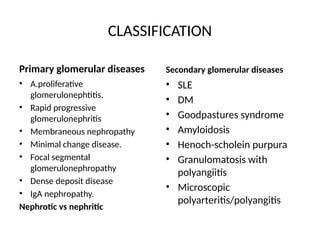
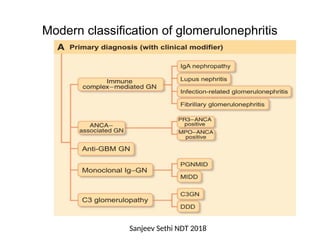
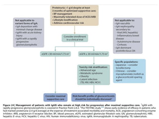
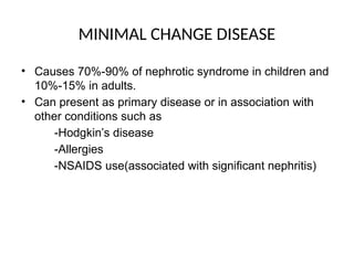
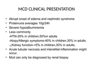
1
GLOMERULAR DISEASES
Presenter: Monica Nchimbi .Y.
Supervisor:Dr Meremo
MMED IM
Clinical nephrology
OUTLINE
• Basic anatomy of the glomerulus
• Introduction
• Classification
• Mechanism and extent of injury
• Pathogenesis
• General principle of management
BASIC ANATOMY
Richard et al CJASN 2016
INTRODUCTION
• Glomerular diseases-injury to the glomerulus due to a range of
different insults.
• Insults -immune mediated,metabolic,toxins,malignancy or hereditary.
• Glomerular cells will respond to the injuries depending on the
nature,magnitude and duration of the insults.
• Glomerular disease-one of the most common cause of irreversible kidney
damage in the young.
• Encompasses 20%-25% of existing kidney failure cases in most registries.
KDIGO glomerular 2021
Richard et al CJASN 2016
CLASSIFICATION
Primary glomerular diseases
• A.proliferative
glomerulonephtitis.
• Rapid progressive
glomerulonephritis
• Membraneous nephropathy
• Minimal change disease.
• Focal segmental
glomerulonephropathy
• Dense deposit disease
• IgA nephropathy.
Nephrotic vs nephritic
Secondary glomerular diseases
• SLE
• DM
• Goodpastures syndrome
• Amyloidosis
• Henoch-scholein purpura
• Granulomatosis with
polyangiitis
• Microscopic
polyarteritis/polyangitis
• Hereditary –Al port syndrome
-Thin basement membrane
disease
-Fabry disease.
Jefferey B et al, NIDDK
Modern classification of glomerulonephritis
Sanjeev Sethi NDT 2018
MECHANISM AND EXTENT OF INJURY
• The pathological responses of glomerulus injury involves -
hypercellurarity
-glomerular basement membrane thickening and
-hyalinosis and sclerosis.
• Hypercellularity involves -cellular proliferation
&leukocyte infiltration.
- crescent formation.
• GBM thickening-lamina densa dublication
-immune complex deposition
-increased protein content of the GBM
and electron dense materials.
• hyalinosis
-leakage of plasma proteins from the injured endothelium
or capillaries with deposition of homogeneous and
oesinophilic materials in the glomeruli.
• Sclerosis
-deposition of extracellular collageneous matrix.
Sanjeev et al, NDT 2018
• The extent of glomerular injury can be categorized as
follows:
• Diffuse-whole glomeruli of the kidney.
• Global-whole individual glomerulus.
• focal -involves some of the glomeruli
• Segmental-part of the individual glomerulus.
• Capillary or mesengial.
UPTODATE
PATHOGENESIS
• Pathogenesis involves two main pathways:
a)immune mediated glomerular diseases.
-antibody mediated.
-cell mediated.
-alternate complement system mediated.
-secondary pathogenetic mechanisms.
b)non- immune mediated glomerular diseases
• Antibody mediated involves formation of immune complexes in situ
or circulating complexes.
• Insitu- can be fixed or planted immuno- complexes
E.g –α3 NCI domain of collagen IV antigen (Goodpasture
syndrome)
-PLA2R antigen
-mesengial antigen
• Circulating immune complexes that can be exogeneous(infectious
agents or drugs) or endogeneous.(DNA,nuclear proteins or
immunoglobulins)
• Cell mediated glomerular injury is mostly seen in
immunoglobulin deficient states.
• T-lymphocytes recruits leukocytes and macrophages
which in turn
-mount antibody dependent cellular cytotoxic reactions
-promote fibrogenesis.
e.g type 3 pauci RPGN
• Alternate complement mediated injury involves
persistent C3 mediated complement activation due to
antibodies against C3 convertase.
e.g Type 2 MPGN.
• Secondary pathogenetics mechanisms involves production of
inflammatory mediators(reactive oxygen
species,cytokines,chemokines,proteases etc)
• These are released from an array of cells(platelets,NK
cells,mesangial cells and NK cells)
b)Non immune –mediated includes
• Metabolic Diabetes nephropathy
• Hemodynamic Hypertension induced
• Amyloidosis deposition
• NSAIDS
• Alport disease
• Infections (hepatitis C&B,HIV and E.Coli)
CLINICAL PRESENTATION OF
GLOMERULAR DISEASES
• Can be asymptomatic with
-proteinuria <2g/day
-microscopic hematuria(++ or +++)
-dysmorphic rbcs/hpf
• Can present with gross hematuria which will be
-non painful
-with no blood clots
• Can present with nephrotic syndrome with
-proteinuria>3.5g/d
-hypoalbuminemia
-edema
-hyperlipidemia
• Can present with nephritic syndrome with
-oliguria
-hematuria with rbcs casts
-sub-nephrotic proteinuria
-hypertension
• Can present with signs showing rapid progressing GN or
chronic GN.
Amesh Khana JMSA 2011
GENERAL PRINCIPLES OF EVALUATION AND
MANAGEMENT OF GLOMERULAR DISEASES
• Kidney biopsy
-gold standard for diagnosis of glomerular diseases.
-Mandatory for diagnosis in adults with nephrotic
syndrome when the cause is not evident.
-Should be done when benefit of information obtained
exceed risk.
• Kidney tissue should meet standard biopsy adequacy.
-mostly 8-10 glomeruli.
KDIGO GLOMERULAR 2021
KDIGO 2021 Clinical Practice
Guideline for the Management of Glomerular Diseases. Kidney Int. 2021;100(4S):S1–
S276.
Assessment of kidney function
• Achieved by measurement of proteinuria and GFR.
• Proteinuria
-24hrs urine collection should be used to determine total
protein excretion in patients with glomerular diseases
especially when immunosuppression is need.
• If can not achieved the best option is to determine PCR
on an aliquot of attempted 12-24hrs urine collection at
first presentation or first morning void.
Definition of nephrotic syndrome,nephrotic range
proteinuria and non-nephrotic range proteinuria
KDIGO GLOMERULAR 2021
• Hematuria
-Cardinal manifestation of glomerular diseases
-Initial detection-dipstick analysis very sensitive in
detection of hemoglobin.
-Abnormal dipstick test should be confirmed by
microscopy of the sediment under both low and high
magnification.
-in GN erythrocytes 50%-80% are dysmorphic and
microcytic.
-RBC casts and acanthocytes indicates inflammatory
disease.
KDIGO 2021
MANAGEMENT
MANAGEMENT OF EDEMA
MANAGEMENT OF HYPERTENSION
MANAGEMENT OF HYPERLIPIDEMIA
MANAGEMENT OF VTE
KDIGO 2021
IgA NEPHROPATHY
• Most prevalent primary glomerulonephritis globally.
• Male >female.
• Peak -2nd
and 3rd
decades.
• Incidence 2.5/100000
• More prevalent in East Asia up to 36%,a dominant cause
of kidney failure.
• Lower in North America and Europe.
• Paucity of data in Africa.
• 20%-40% of patient with IgA nephropathy progress to
kidney failure within 10-20 years.
IgA Nephropathy -pathogenesis
• Is an immune complex mediated glomerulonephritis.
• Involves diffuse mesengial IgA deposition with
hypercellulality.
• Pathogenesis driven by poor glycosylation of hinge region of
polymeric IgA1.
• The poorly galactosylated IgA1 leads to exposure of N-
acetyl-galactosamine to IgG or IgM antibodies,with formation
of immune complexes in situ when deposited or circulating.
• Galactose deficient IgA1 dodge liver catabolism.
• End product of unlimited inflammation is glomerular and
interstitial fibrosis.
IgA Nephropathy clinical presentation
• Most common-recurrent macroscopic hematuria
associated with or following URTI.
-persistent asymptomatic microscopic
hematuria.
-nephrotic syndrome is uncommon.
• Is a benign disease mostly.
• 5%-30%-complete remission
• 25%-30%-progress slowly to renal failure in 20-25 years.
• Population variations in progressive disease.
Ct..
• Risk of progression
-hypertension
-proteinuria(persistent ≥ 6months)
-absence of macroscopic hematuria.
-male sex
-older age at onset.
-renal biopsy(extensive glomerulosclerosis/interstitial
fibrosis).
IgA nephropathy diagnosis
• renal biopsy(Gold standard)
• Histological lesions include
-Diffuse proliferative glomerulonephritis
-segmental sclerosis and seldom
-segmental necrosis with cellural crescent.
• Revised Oxford MEST-C score for progression
determination.
• Assessment of secondary causes.
• No validated serum or urine biomarkers for IgAN.
IgAN Management
• Primary focus-optimization of supportive care.
• Life style modifications
-diatery sodium restriction
-smoking cessation
-weight management
-exercise
• Manage hypertension-proteinuria>0.5g/dl ACEi or ARB
• Proteinuria >0.5 with or without HTN-ACEi or ARB
• Address cardiovascular risk
• High risk of progressive CKD despite max supportive care-
6months course of glucocorticoids.
Follow up
• Target
-proteinuria of ≤200mg/l(≤400mg/1.73m2/d)
-blood pressure <90th
percentile for age,sex and height
• Continue following the patient even with complete
remission as they might relapse after a long duration.
MINIMAL CHANGE DISEASE
• Causes 70%-90% of nephrotic syndrome in children and
10%-15% in adults.
• Can present as primary disease or in association with
other conditions such as
-Hodgkin’s disease
-Allergies
-NSAIDS use(associated with significant nephritis)
MCD PATHOGENESIS
• Uncertain
• Appealing circumstantial explanation-circulating cytokine
related to T-cell response with alteration of capillary
charge and podocyte intergrity.
• Light microscopy-no obvious change,occasional
mesangial IgM deposition.
• E.microscopy-effacement of foot process supporting the
podocytes.
-weakening of slit membrane.
MCD CLINICAL PRESENTATION
• Abrupt onset of edema and nephrotic syndrome
• Proteinuria averages 10g/24h
• Severe hypoalbuminemia
• Less commonly
-HTN-30% in children,50%in adults
-Atopy/Allergic symptoms-40% in children,30% in adults.
-↓Kidney function-<5% in children,30% in adults.
• Acute tubular necrosis and interstitial inflammation might
occur.
• Mcd can only be diagnosed by renal biopsy
MCD MANAGEMENT
• High dose oral glucocorticoid is recommended
• Route and frequency can be individualized according to
the patient needs.
• Should not be given for longer than 16 weeks.
• Begin tapering 2 weeks after complete remission.
MEMBRANOUS NEPHROPATHY
• Accounts 20% of nephrotic syndrome in adults.
• Peak 30-50 years.
• Idiopathic MN rare in childhood,but most common cause
of nephrotic syndrome in the elderly.
• Male to female ratio 2:1
• Can be idiopathic or Secondary
• 20%-30% secondary associated with
-Malignancy(breast,colon and lung)
-Infections(hepatitis B,symphilis,malaria)
-Rheumatoid diseases(SLE,RA) and
-Drug exposure.
MN PATHOGENESIS
• Associated with diffuse granular subepithelial deposits of
IgG and C3 with thickening of the basement membrane.
• 70% of Idiopathic MG is associated with circulating
autoantibodies that binds withPLA2R on podocytes with
resultant insitu deposition.
• 5%-10%-autoantibodies to thrombospondin type 1
domain that binds to PLA2R.
• In secondary MG-no involvement of autoantibodies only
rarely in Hepatitis B and sarcoidosis.
MN CLINICAL PRESENTATION
• 80%-nephrotic syndrome and non selective proteinuria.
• Less commonly-microscopic hematuria
• Spontaneous remission in 20%-33%
• 30% will present with relapsing nephrotic syndrome with
preserved renal function.
• 30% will present with renal failure or die from complications ass
with nephrotic syndrome.
• Poor prognostic indicators
-Male gender
-Older age
-HTN
-Persistant proteinuria.
MG cont..
• Has the highest incident of
-Renal vein thrombosis
-Pulmonary embolism
-DVT
DIAGNOSIS
• Renal biopsy is not compulsory in diagnosis of
MG,especially in a patient with nephrotic syndrome and
positive anti-PLA2R antibody test.
• Evaluation of other conditions is highly recommended.
MN MANAGEMENT
• Optimal supportive care for all patients
• Immunosuppressive therapy-for those at risk of
progressive kidney disease.
Assessment of risk of progressive kidney
disease in MN.
TREATMENT OF MN
FOLLOW UP MN.
• Longitudinal monitoring of anti-PLA2R antibody level at
6months is recommended to determine treatment
response and to aid adjustment of medication when
required.
REFERENCES
1.Harrison’s Principles of Internal Medicine 20th
edition
2.KDIGO 2021 Clinical Practice Guideline for the
management of Gromerular Diseases.
3.Prapa et al;Core Curriculum on IgA 2021,AJKD
4.Saeed et al;Renal biopsy reports in nephrotic
syndrome.update(World Journal of Nephrology)
5.Richard et al;The players:Cells involved in Glomerular
Diseases.(CJASN 2016)
6Michelle M et al;Glomerular disease frequencies by race,sex
and region;International Kidney Biopsy Survey 2017.
7.Harsh Mohan Book of Pathology 5th
Edition.

9. GLOMhhhhhhhhhhhhhhhhhhhERULAR DISEASES.pptx

  • 1.
    1 GLOMERULAR DISEASES Presenter: MonicaNchimbi .Y. Supervisor:Dr Meremo MMED IM Clinical nephrology
  • 2.
    OUTLINE • Basic anatomyof the glomerulus • Introduction • Classification • Mechanism and extent of injury • Pathogenesis • General principle of management
  • 3.
  • 4.
    INTRODUCTION • Glomerular diseases-injuryto the glomerulus due to a range of different insults. • Insults -immune mediated,metabolic,toxins,malignancy or hereditary. • Glomerular cells will respond to the injuries depending on the nature,magnitude and duration of the insults. • Glomerular disease-one of the most common cause of irreversible kidney damage in the young. • Encompasses 20%-25% of existing kidney failure cases in most registries. KDIGO glomerular 2021 Richard et al CJASN 2016
  • 5.
    CLASSIFICATION Primary glomerular diseases •A.proliferative glomerulonephtitis. • Rapid progressive glomerulonephritis • Membraneous nephropathy • Minimal change disease. • Focal segmental glomerulonephropathy • Dense deposit disease • IgA nephropathy. Nephrotic vs nephritic Secondary glomerular diseases • SLE • DM • Goodpastures syndrome • Amyloidosis • Henoch-scholein purpura • Granulomatosis with polyangiitis • Microscopic polyarteritis/polyangitis
  • 6.
    • Hereditary –Alport syndrome -Thin basement membrane disease -Fabry disease. Jefferey B et al, NIDDK
  • 7.
    Modern classification ofglomerulonephritis Sanjeev Sethi NDT 2018
  • 8.
    MECHANISM AND EXTENTOF INJURY • The pathological responses of glomerulus injury involves - hypercellurarity -glomerular basement membrane thickening and -hyalinosis and sclerosis. • Hypercellularity involves -cellular proliferation &leukocyte infiltration. - crescent formation. • GBM thickening-lamina densa dublication -immune complex deposition -increased protein content of the GBM and electron dense materials.
  • 9.
    • hyalinosis -leakage ofplasma proteins from the injured endothelium or capillaries with deposition of homogeneous and oesinophilic materials in the glomeruli. • Sclerosis -deposition of extracellular collageneous matrix. Sanjeev et al, NDT 2018
  • 12.
    • The extentof glomerular injury can be categorized as follows: • Diffuse-whole glomeruli of the kidney. • Global-whole individual glomerulus. • focal -involves some of the glomeruli • Segmental-part of the individual glomerulus. • Capillary or mesengial. UPTODATE
  • 13.
    PATHOGENESIS • Pathogenesis involvestwo main pathways: a)immune mediated glomerular diseases. -antibody mediated. -cell mediated. -alternate complement system mediated. -secondary pathogenetic mechanisms. b)non- immune mediated glomerular diseases
  • 14.
    • Antibody mediatedinvolves formation of immune complexes in situ or circulating complexes. • Insitu- can be fixed or planted immuno- complexes E.g –α3 NCI domain of collagen IV antigen (Goodpasture syndrome) -PLA2R antigen -mesengial antigen • Circulating immune complexes that can be exogeneous(infectious agents or drugs) or endogeneous.(DNA,nuclear proteins or immunoglobulins)
  • 15.
    • Cell mediatedglomerular injury is mostly seen in immunoglobulin deficient states. • T-lymphocytes recruits leukocytes and macrophages which in turn -mount antibody dependent cellular cytotoxic reactions -promote fibrogenesis. e.g type 3 pauci RPGN • Alternate complement mediated injury involves persistent C3 mediated complement activation due to antibodies against C3 convertase. e.g Type 2 MPGN.
  • 16.
    • Secondary pathogeneticsmechanisms involves production of inflammatory mediators(reactive oxygen species,cytokines,chemokines,proteases etc) • These are released from an array of cells(platelets,NK cells,mesangial cells and NK cells) b)Non immune –mediated includes • Metabolic Diabetes nephropathy • Hemodynamic Hypertension induced • Amyloidosis deposition • NSAIDS • Alport disease • Infections (hepatitis C&B,HIV and E.Coli)
  • 17.
    CLINICAL PRESENTATION OF GLOMERULARDISEASES • Can be asymptomatic with -proteinuria <2g/day -microscopic hematuria(++ or +++) -dysmorphic rbcs/hpf • Can present with gross hematuria which will be -non painful -with no blood clots • Can present with nephrotic syndrome with -proteinuria>3.5g/d -hypoalbuminemia -edema -hyperlipidemia
  • 18.
    • Can presentwith nephritic syndrome with -oliguria -hematuria with rbcs casts -sub-nephrotic proteinuria -hypertension • Can present with signs showing rapid progressing GN or chronic GN. Amesh Khana JMSA 2011
  • 19.
    GENERAL PRINCIPLES OFEVALUATION AND MANAGEMENT OF GLOMERULAR DISEASES • Kidney biopsy -gold standard for diagnosis of glomerular diseases. -Mandatory for diagnosis in adults with nephrotic syndrome when the cause is not evident. -Should be done when benefit of information obtained exceed risk. • Kidney tissue should meet standard biopsy adequacy. -mostly 8-10 glomeruli.
  • 20.
  • 21.
    KDIGO 2021 ClinicalPractice Guideline for the Management of Glomerular Diseases. Kidney Int. 2021;100(4S):S1– S276.
  • 22.
    Assessment of kidneyfunction • Achieved by measurement of proteinuria and GFR. • Proteinuria -24hrs urine collection should be used to determine total protein excretion in patients with glomerular diseases especially when immunosuppression is need. • If can not achieved the best option is to determine PCR on an aliquot of attempted 12-24hrs urine collection at first presentation or first morning void.
  • 24.
    Definition of nephroticsyndrome,nephrotic range proteinuria and non-nephrotic range proteinuria KDIGO GLOMERULAR 2021
  • 25.
    • Hematuria -Cardinal manifestationof glomerular diseases -Initial detection-dipstick analysis very sensitive in detection of hemoglobin. -Abnormal dipstick test should be confirmed by microscopy of the sediment under both low and high magnification. -in GN erythrocytes 50%-80% are dysmorphic and microcytic. -RBC casts and acanthocytes indicates inflammatory disease. KDIGO 2021
  • 26.
  • 27.
  • 28.
  • 29.
  • 31.
  • 32.
    IgA NEPHROPATHY • Mostprevalent primary glomerulonephritis globally. • Male >female. • Peak -2nd and 3rd decades. • Incidence 2.5/100000 • More prevalent in East Asia up to 36%,a dominant cause of kidney failure. • Lower in North America and Europe. • Paucity of data in Africa. • 20%-40% of patient with IgA nephropathy progress to kidney failure within 10-20 years.
  • 33.
    IgA Nephropathy -pathogenesis •Is an immune complex mediated glomerulonephritis. • Involves diffuse mesengial IgA deposition with hypercellulality. • Pathogenesis driven by poor glycosylation of hinge region of polymeric IgA1. • The poorly galactosylated IgA1 leads to exposure of N- acetyl-galactosamine to IgG or IgM antibodies,with formation of immune complexes in situ when deposited or circulating. • Galactose deficient IgA1 dodge liver catabolism. • End product of unlimited inflammation is glomerular and interstitial fibrosis.
  • 34.
    IgA Nephropathy clinicalpresentation • Most common-recurrent macroscopic hematuria associated with or following URTI. -persistent asymptomatic microscopic hematuria. -nephrotic syndrome is uncommon. • Is a benign disease mostly. • 5%-30%-complete remission • 25%-30%-progress slowly to renal failure in 20-25 years. • Population variations in progressive disease.
  • 35.
    Ct.. • Risk ofprogression -hypertension -proteinuria(persistent ≥ 6months) -absence of macroscopic hematuria. -male sex -older age at onset. -renal biopsy(extensive glomerulosclerosis/interstitial fibrosis).
  • 36.
    IgA nephropathy diagnosis •renal biopsy(Gold standard) • Histological lesions include -Diffuse proliferative glomerulonephritis -segmental sclerosis and seldom -segmental necrosis with cellural crescent. • Revised Oxford MEST-C score for progression determination. • Assessment of secondary causes. • No validated serum or urine biomarkers for IgAN.
  • 37.
    IgAN Management • Primaryfocus-optimization of supportive care. • Life style modifications -diatery sodium restriction -smoking cessation -weight management -exercise • Manage hypertension-proteinuria>0.5g/dl ACEi or ARB • Proteinuria >0.5 with or without HTN-ACEi or ARB • Address cardiovascular risk • High risk of progressive CKD despite max supportive care- 6months course of glucocorticoids.
  • 39.
    Follow up • Target -proteinuriaof ≤200mg/l(≤400mg/1.73m2/d) -blood pressure <90th percentile for age,sex and height • Continue following the patient even with complete remission as they might relapse after a long duration.
  • 40.
    MINIMAL CHANGE DISEASE •Causes 70%-90% of nephrotic syndrome in children and 10%-15% in adults. • Can present as primary disease or in association with other conditions such as -Hodgkin’s disease -Allergies -NSAIDS use(associated with significant nephritis)
  • 41.
    MCD PATHOGENESIS • Uncertain •Appealing circumstantial explanation-circulating cytokine related to T-cell response with alteration of capillary charge and podocyte intergrity. • Light microscopy-no obvious change,occasional mesangial IgM deposition. • E.microscopy-effacement of foot process supporting the podocytes. -weakening of slit membrane.
  • 42.
    MCD CLINICAL PRESENTATION •Abrupt onset of edema and nephrotic syndrome • Proteinuria averages 10g/24h • Severe hypoalbuminemia • Less commonly -HTN-30% in children,50%in adults -Atopy/Allergic symptoms-40% in children,30% in adults. -↓Kidney function-<5% in children,30% in adults. • Acute tubular necrosis and interstitial inflammation might occur. • Mcd can only be diagnosed by renal biopsy
  • 43.
    MCD MANAGEMENT • Highdose oral glucocorticoid is recommended • Route and frequency can be individualized according to the patient needs. • Should not be given for longer than 16 weeks. • Begin tapering 2 weeks after complete remission.
  • 46.
    MEMBRANOUS NEPHROPATHY • Accounts20% of nephrotic syndrome in adults. • Peak 30-50 years. • Idiopathic MN rare in childhood,but most common cause of nephrotic syndrome in the elderly. • Male to female ratio 2:1 • Can be idiopathic or Secondary • 20%-30% secondary associated with -Malignancy(breast,colon and lung) -Infections(hepatitis B,symphilis,malaria) -Rheumatoid diseases(SLE,RA) and -Drug exposure.
  • 47.
    MN PATHOGENESIS • Associatedwith diffuse granular subepithelial deposits of IgG and C3 with thickening of the basement membrane. • 70% of Idiopathic MG is associated with circulating autoantibodies that binds withPLA2R on podocytes with resultant insitu deposition. • 5%-10%-autoantibodies to thrombospondin type 1 domain that binds to PLA2R. • In secondary MG-no involvement of autoantibodies only rarely in Hepatitis B and sarcoidosis.
  • 48.
    MN CLINICAL PRESENTATION •80%-nephrotic syndrome and non selective proteinuria. • Less commonly-microscopic hematuria • Spontaneous remission in 20%-33% • 30% will present with relapsing nephrotic syndrome with preserved renal function. • 30% will present with renal failure or die from complications ass with nephrotic syndrome. • Poor prognostic indicators -Male gender -Older age -HTN -Persistant proteinuria.
  • 49.
    MG cont.. • Hasthe highest incident of -Renal vein thrombosis -Pulmonary embolism -DVT
  • 50.
    DIAGNOSIS • Renal biopsyis not compulsory in diagnosis of MG,especially in a patient with nephrotic syndrome and positive anti-PLA2R antibody test. • Evaluation of other conditions is highly recommended.
  • 52.
    MN MANAGEMENT • Optimalsupportive care for all patients • Immunosuppressive therapy-for those at risk of progressive kidney disease.
  • 53.
    Assessment of riskof progressive kidney disease in MN.
  • 54.
  • 55.
    FOLLOW UP MN. •Longitudinal monitoring of anti-PLA2R antibody level at 6months is recommended to determine treatment response and to aid adjustment of medication when required.
  • 56.
    REFERENCES 1.Harrison’s Principles ofInternal Medicine 20th edition 2.KDIGO 2021 Clinical Practice Guideline for the management of Gromerular Diseases. 3.Prapa et al;Core Curriculum on IgA 2021,AJKD 4.Saeed et al;Renal biopsy reports in nephrotic syndrome.update(World Journal of Nephrology) 5.Richard et al;The players:Cells involved in Glomerular Diseases.(CJASN 2016) 6Michelle M et al;Glomerular disease frequencies by race,sex and region;International Kidney Biopsy Survey 2017. 7.Harsh Mohan Book of Pathology 5th Edition.

Editor's Notes