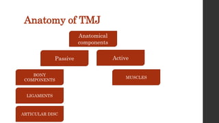
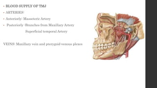
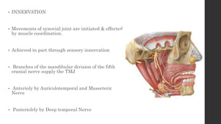
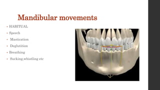
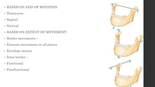
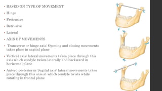
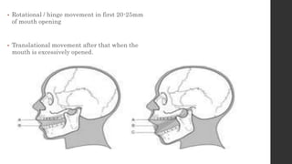
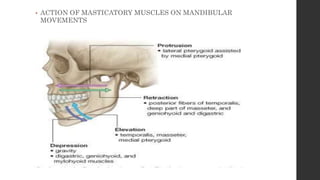
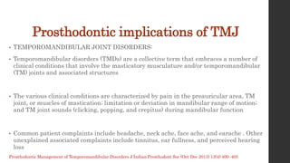
2. Contents
• Introduction
• Anatomy of the joint and its components
• Muscle and ligaments
• Blood supply
• Nerve supply
• Mandibular movements
• Prosthodontic implications of TMJ
3. Introduction
• The articulation between the temporal bone and the mandible. It is a
bilateral diarthrodial, bilateral ginglymoid joint – GPT 9
• It provides hinging movement in one plane therefore it considered as
ginglymoid joint however at the same time it provides gliding
movements which classifies it as arthrodial joint therefore it is
technically called as ginglymoarthrodial joint
• Temporomandibular joint is • Compound • Diarthrodial •
Ginglymoid • Multiaxial • Secondary • Sqaumo-dentary Joint •
Cranio–mandibular articulation
4. Anatomy of TMJ
Passive Active
Anatomical
components
BONY
COMPONENTS
LIGAMENTS
ARTICULAR DISC
MUSCLES
6. • GLENOID FOSSA
• The squamous portion of the temporal bone (concave )
• Measure approx 23 mm (both Antero-Posteriorly and
Medio-Laterally)
• Anterior : a convex bony prominence ( tubercle ) is
called as articular eminence
• Posterior : Post Glenoid Plane Prevents forced
posterior displacement of condyle
7. • The posterior roof is thin not designed to
sustain heavy force
• The articular eminence consists of thick
dense bone to tolerate such forces.
• The steepness of the articular eminence
surface dictates the pathway of the condyle
CONDYLAR GUIDANCE
9. • The articular surface lies on its
anterosuperior aspect, thus facing the
posterior slope of the articular eminence of
the temporal bone.
• If the long axes of two condyles are extended
posteriorly and medially, they meet approximately
at the anterior limit of the foramen magnum,
forming an angle that opens toward the front
ranging from 145° to 160°
10. • Transverse bony bar that forms the
root of zygomatic process
• This articular surface have dense bone
and is most heavily traversed by
condyle and disk during forward and
backward movements of jaw
11. • ARTICULAR DISC
• The articular disc is the most important
anatomic structure of the TMJ
• It is a biconcave fibrocartilaginous structure
located between the mandibular condyle and
the temporal bone component of the joint.
• Its functions to accommodate a hinging action
as well as the gliding actions between the
temporal and mandibular articular bone.
12. • The articular disc is composed of dense
fibrous connective tissue for the most of part
devoid of any blood vessel or nerve fibers
• The extreme periphery of the disc however is
slightly innervated
• In sagittal plane it can be divided in to three
regions according to thickness
• anterior band = 2 mm in thickness,
• posterior band = 3 mm thick,
• thin in the center intermediate band of 1
mm thickness.
13. • The articular disc is attached to the capsular
ligament not only anteriorly and posteriorly but also
medially and laterally This divides the joint into two
distinct compartments.
• The upper or superior compartment is bordered by
the mandibular fossa and the superior surface of the
disc. Volume of the compartment is 1.2 ml
• The lower or inferior compartment is bordered by
the mandibular condyle and the inferior surface of
the disc. Volume of the compartment is 0.9 ml
• Hinging movements take place in the lower
compartment and gliding movements take place in
the upper compartment.
14. • The internal surfaces of the cavities are surrounded by
specialized endothelial cells that form a synovial lining.
• This lining, along with a specialized synovial fringe
located at the anterior border of the retrodiscal tissues,
produces synovial fluid, which fills both joint cavities.
• Thus the TMJ is referred to as a synovial joint. This
synovial fluid serves two purposes
• Because the articular surfaces of the joint are
nonvascular, the synovial fluid acts as a medium for
providing metabolic requirements to these tissues.
• Free and rapid exchange exists between the vessels of
the capsule, the synovial fluid, and the articular
tissues
15. • The synovial fluid also serves as a lubricant between articular
surfaces during function
• Synovial fluid lubricates the articular surfaces by two mechanisms –
• Boundary lubrication: which occurs when the joint is moved and the
synovial fluid is forced from one area of the cavity in to another .
Boundary lubrication prevents friction
• Weeping lubrication : this refers to the ability of the articular
surfaces to absorbs a small amount of synovial fluid . Weeping
lubrication helps eliminate friction in the compressed but not moving
joint
16.
17. • ATTACHMENTS OF DISC
• Anteriorly :
• Anterior region of the disc is attached to the
capsular ligament
• Anterio-Superior : anterior margin of the
articular surface of the temporal bone
• Anterio-Inferior : anterior margin of the articular
surface of the condyle
18. • POSTERIORLY:
• RETRODISCAL TISSUE
• It is a loose connective tissue region that is
highly vascularized and innervated.
• SUPERIOR : Upper lamina (CONTAINS
MAINLY ELASTIC FIBERS) It attaches the
disc posteriorly to the tympanic plate &
prevents slipping of the disc while yawning.
• INFERIOR : lower lamina ( COMPOSED
CHIEFLY COLLAGENOUS FIBERS )
• It prevents excessive rotation of the disc over
the condyle.
19. TMJ LIGAMENTS
Functional ligaments Accessory ligament
1. Collateral / Discal
ligament
2. Capsular ligament
3. Temporomandibular/
Lateral ligament
1. Sphenomandibular
2. Stylomandibular
Act predominantly as restraint to
motion of the condyle and the disk
Serves as passive restraints on
mandibular motion
20. • COLLATERAL / DISCAL LIGAMENTS :
• From medial and lateral borders of the disc to the
poles of the condyle Medial discal ligament
Lateral discal ligament
• Dividing the joint mediolaterally into superior
and inferior joint cavities
21. • FUNCTIONS :
• Allow the disc to move passively with the condyle as it glides A - P
• Permit the disc to be rotated A-P on the articular surface of the
condyle
• These ligaments are RESPONSIBLE FOR THE HINGING
MOVEMENT BETWEEN THE CONDYLE AND THE ARTICULAR
DISC
• They have a vascular supply and are innervated
22. • CAPSULAR LIGAMENT
• CAPSULAR LIGAMENT/ FIBROUS CAPSULE THIN
SLEEVE OF FIBROUS TISSUE surrounding the
entire TMJ
• Superior attachment the borders of the articular
surface of the glenoid fossa and anterior edge of
preglenoid plane of articular tubercle
• Inferior attachment periphery of neck of mandibular
condyle
• This capsule is reinforced more laterally by an external
Temporomandibular ligament, which also limits the
distraction and the posterior movement of the condyle.
23. • Anteriorly, the capsule has an orifice through which the tendon of
lateral pterygoid muscle passes.
• This area of relative weakness in the capsular lining becomes a
source of possible herniation of intra-articular tissues
• May allow forward displacement of the disk.
• FUNCTIONS
• To resist any lateral or downward forces that tends to separate or
dislocate the articular surface
• To retain the synovial fluid
• Proprioception
24. • TEMPOROMANDIBULAR LIGAMENT :
• It lies at the lateral aspect of the capsular
ligament Attached above to articular tubercle
• Below attached to lateral and posterior surface
of neck of condyle
• Inner horizontal fibers
• Outer oblique fibers
• Reinforces the capsular ligament
• OOF – prevents excessive drooping of the
condyle / limits the extent of mouth opening.
• IHF – limits posterior movement of the condyle
and disk
25. • ACCESSORY LIGAMENTS -
• SPHENOMANDIBULAR LIGAMENT:
• Arises from the angular spine of the sphenoid and
petro tympanic fissure.
• Runs downward and outward.
• Insert on the lingula of the mandible.
• This ligament is passive during jaw movements,
maintaining relatively the same degree of tension
during both opening and closing of the mouth.
26. • STYLOMANDIBULAR LIGAMENT:
• This is a specialized dense, local concentration of
deep cervical fascia extending from the apex and
being adjacent to the anterior aspect of the styloid
process and the stylohyoid ligament to the
mandible’s angle and posterior border
• This ligament becomes tense only in extreme
protrusive movements. Thus, it can be considered
only as an accessory ligament of uncertain function
30. • BLOOD SUPPLY OF TMJ
• ARTERIES:
• Anteriorly: Masseteric Artery
• Posteriorly :Branches from Maxillary Artery
Superficial temporal Artery
VEINS: Maxillary vein and pterygoid venous plexes
31. • INNERVATION
• Movements of synovial joint are initiated & effected
by muscle coordination.
• Achieved in part through sensory innervation
• Branches of the mandibular division of the fifth
cranial nerve supply the TMJ
• Anterioly by Auriculotemporal and Masseteric
Nerve
• Posteriolrly by Deep temporal Nerve
33. • BASED ON AXIS OF ROTATION
• Transverse
• Sagital
• Vertical
• BASED ON EXTENT OF MOVEMENT
• Border movements –
• Extreme movements in all planes
• Envelope motion
• Intra border –
• Functional
• Parafunctional
34. • BASED ON TYPE OF MOVEMENT
• Hinge
• Protrusive
• Retrusive
• Lateral
• AXIS OF MOVEMENTS
• Transverse or hinge axis: Opening and closing movements
takes place in sagital plane
• Vertical axis: lateral movements takes place through this
axis which condyle twists laterally and backward in
horizontal plane
• Antero-posterior or Sagital axis: lateral movements takes
place through this axis at which condyle twists while
rotating in frontal plane
35.
36. • Rotational / hinge movement in first 20-25mm
of mouth opening
• Translational movement after that when the
mouth is excessively opened.
37. • ACTION OF MASTICATORY MUSCLES ON MANDIBULAR
MOVEMENTS
38. Prosthodontic implications of TMJ
• TEMPOROMANDIBULAR JOINT DISORDERS:
• Temporomandibular disorders (TMDs) are a collective term that embraces a number of
clinical conditions that involve the masticatory musculature and/or temporomandibular
(TM) joints and associated structures
• The various clinical conditions are characterized by pain in the preauricular area, TM
joint, or muscles of mastication; limitation or deviation in mandibular range of motion;
and TM joint sounds (clicking, popping, and crepitus) during mandibular function
• Common patient complaints include headache, neck ache, face ache, and earache . Other
unexplained associated complaints include tinnitus, ear fullness, and perceived hearing
loss
Prosthodontic Management of Temporomandibular Disorders J Indian Prosthodont Soc (Oct-Dec 2013) 13(4):400–405
39. • ETIOLOGIC FACTORS
• 1. Predisposing.
• 2. Initiating (precipitating).
• 3. Perpetuating to emphasize their role in the progression of TMD.
• A. Predisposing Factors Include Structural, metabolic, and/or psychologic conditions
that adversely affect the masticatory system sufficiently to increase the risk of
developing TMD
• It has been reported that an extreme anterior open bite, overjet greater than 6–7 mm
discrepancy between the retruded contact position and five or more missing posterior
teeth, and unilateral maxillary posterior lingual crossbite in children may be
associated with TMD
40. • B. Initiating Factors
• Those leads to the onset of symptoms are primarily related to trauma or repetitive
adverse loading of the masticatory system
• Overt trauma producing injury to the head, neck, or jaw can result from an impact injury,
possibly a flexion–extension injury, and an injury while eating, yawning, or even from
prolonged mouth opening during long dental appointments.
• A second form of trauma is associated with the sustained and repetitious adverse loading
of the masticatory system as a result of parafunction
41. • C. Perpetuating Factors
• Such as Para function, hormonal factors, or psychosocial factors, maybe associated with
any predisposing or initiating factor and can sustain the patient’s disorder, complicating
management of it.
• Diagnostic Classification of TMDs
• Congenital or developmental disorders - Aplasia – Hypoplasia – Hyperplasia – Neoplasia
• Disk derangement disorders - Disk displacement with reduction - Disk displacement
without reduction Joint dislocation
• Inflammatory conditions - Capsulitis/synovitis - Polyarthritides
• Noninflammatory (Osteoarthrosis) - Osteoarthritis: primary - Osteoarthritis: secondary
• Ankylosis – Fibrous – Bony
• Fracture (Condylar process)
42. • ASSESMENT OF TMD’S:
• The collection of baseline records and indicated diagnostic tests is fundamental to the
proper management of TMD
• Screening for TMD is recommended as an essential part of all routine dental and/or
orofacial pain examinations which includes questionnaires
• The comprehensive physical examination consists of a general inspection of the head and
neck, including a visual inspection and palpation; a comprehensive orthopaedic evaluation
of the TM joint
• an intraoral evaluation including an occlusal analysis
43.
44.
45. • MANAGEMENT :
• The majority of TMD patients achieve good relief of symptoms with a conservative model of
non-invasive management
• A multidisciplinary model that includes patient education and self-care, cognitive
behavioural intervention, Pharmacotherapy, physical therapy, and orthopaedic appliance
therapy (interocclusal splints) is endorsed for the management of nearly all TMD patients.
• Patient Education and Self-care
• The success of a self-care program is often enough to control an uncomplicated TMD
problem. Instruction in a self-care routine should include the following: rest of the
masticatory system through voluntary reduction of mandibular function, habit awareness
and modification, and a home physiotherapeutic program
• . A home physiotherapeutic programme of moist heat and/or ice to the affected areas,
massage of the affected muscles, and gentle range of motion exercises can reduce pain and
increase range of motion.
46. • Cognitive Behavioural Intervention
• Cognitive behavioural intervention is an important part of the overall biopsychosocial
treatment programme for TMD
• Behavioural strategies involving a combination of EMG biofeedback, relaxation techniques,
and self-directed lifestyle changes are more effective than any single behavioural treatment
procedure.
• Pharmacotherapy
• The indicated classes of pharmacologic agents include analgesics, anti inflammatory
agents, corticosteroids, anxiolytics, muscle relaxants, and low-dose antidepressants
• The non-opiate analgesics are effective for mild to moderate acute pain associated with
TMD pain, and the opioid narcotics should only be used short-term for controlling acute
severe pain
47. • Nonsteroidal, anti-inflammatory drugs (NSAIDs) are effective analgesics and anti-
inflammatory agents and are prescribed for painful articular disorders
• Intra-articular TM joint injection of corticosteroids has been recommended on a limited
basis in cases of severe joint pain when other conservative treatment has been
unsuccessful.
• The benzodiazepines are classified as sedative-hypnotic drugs and are most commonly
prescribed for their antianxiety effects
• Muscle relaxants, with the possible exception of cyclobenzaprine hydrochloride (Flexeril),
derive their therapeutic action from their sedative effect only, but can be useful for acute
muscle pain
48. • Orthopaedic Appliance Therapy
• Orthopaedic appliances, also referred to as intraoral appliances, occlusal splints, orthotics,
night guards, or bruxism appliances, have a reported 70–90 % rate of clinical success
• there are a large variety of appliances, two major types of appliances are commonly used
for TMD, namely, stabilization appliances and anterior positioning appliances
• Stabilization appliances are designed to provide stabilization of the joint, redistribution of
forces at the tooth and/or joint level, relaxation of the elevator muscles (at least short
term), and/ or protection of the teeth from the effects of bruxism.
• Anterior positioning appliances, also termed mandibular orthopaedic repositioning
appliances (MORAs), are indicated for acute joint pain, painful joint noise and closedlock,
and associated secondary muscle symptoms from articular inflammation and pain [1
49. • OCCLUSAL SPLINTS
• An occlusal splint is a removable device made of hard
acrylic resin creating precise occlusal contact with the
teeth of the opposing arch.
• PRIMARY FUNCTIONS OF OCCLUSAL SPLINTS
• Temporarily provide an orthopedically musculoskeletal
stable joint position.
• Introduces an optimum occlusal condition that prevents
the muscular hyperactivity.
• Used to protect teeth from excessive tooth wear.
• SECONDARY FUNCTION OF OCCLUSAL SPLINTS
• Stablization of week teeth
• Distribution of occlusal forces
• Stablization of unopposed teeth
50. • TYPES OF OCCLUSAL SPLINTS
• THE TWO MOST COMMONLY USED ARE:
• 1. The Permissive Split
• 2. The Directive Splint
• INDICATIONS PERMISSIVE SPLINT
• Stabilizing appliance are generally used to treat muscle pain
disorders. (also called as Muscle De-programmer)
• DIRECTIVE SPLINT
• Anterior positioning appliance are used to position or align the
condyle disk assembly
51. • Instructions
• The patient is instructed how to properly seat the appliance and the
final seating is done by biting.
• Patients are instructed to wear it in night for bruxism and in day
time for disc problems.
• IMPORTANT POINTS IN SPLINT MANAGEMENT
• A splint should be checked at least once during the 1 st week after
delivery and adjustments are done if required
• Patients with occlusal splint should preferably be recalled after 3
months months
52. • Occlusal Therapy
• There are many dental conditions that require treatment of the occlusion, that is, lack of
intra-inter-arch tooth stability, tooth mobility, fremitus, tooth or restoration fracture, tooth
sensitivity, compromised function, and/or a compromised periodontal health requiring a
redistribution of forces to minimize the effects of adverse loading
• Usually, it is wise to maintain the functional equilibrium established by the TMD
management program, especially when the intercuspal position (ICP) and the vertical
dimension of the occlusion (VDO) are acceptable.
• On the other hand, if a functional equilibrium has not been established, or if the ICP and/or
VDO are unacceptable or need to be altered to perform necessary dental treatment, the
occlusion may need to be re-established.
• When an occlusal scheme has to be re-established, a treatment reference position must be
established, namely centric relation, to allow the clinician to design treatment from a
known starting point and to evaluate the progress and the outcome of the treatment on the
basis of that starting point.
53. • The specific treatment objectives that are desired from an optimum structural and
functional reestablishment standpoint for all patients including TMD patients, are as
follows:
• (1) maximum symmetrical distribution of intercuspal contacts in the predetermined jaw
relationship;
• (2) axial or near axial loading of the teeth;
• (3) an acceptable occlusal plane;
• (4) guidance contacts that allow freedom during closing, incursive and excursive gliding
mandibular movements without deflection of the mandible or teeth; and
• (5) an acceptable vertical dimension of occlusion and interocclusal resting range
54. • Surgery
• Temporomandibular surgery is the indicated treatment for a very small percentage of TMD
patients and only those with specific TMD articular disorders
• Surgical management may vary from closed surgical procedures (arthrocentesis
andarthroscopy) or open surgical procedures (arthrotomy) to subcondylar osteotomies
(condylotomy).
• The promising effectiveness of arthrocentesis, lavage through import and export needles
with and without steroid injection and subsequent mobilization, is rapidly replacing
arthroscopic procedures
• Open joint surgical procedures may range from discoplasty, disk repositioning and repair,
discectomy (meniscectomy) with or without replacement, to arthroplasty including high
condylectomy
55.
56. REFERENCES
• Textbook of human anatomy volume 3 BD chaurasia
• Textbook of management of temporomandibular disorders and
occlusion Jeffery p Okeson
• Kaur H, Datta K. Prosthodontic management of temporomandibular
disorders. The Journal of Indian Prosthodontic Society. 2013 Dec
1;13(4):400-5.