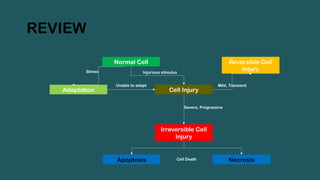
REVIEW
Normal Cell
Cell Injury
IrreversibleCell
Injury
Adaptation
Unable to adapt
Stress Injurious stimulus
Reversible Cell
Injury
Mild, Transient
Severe, Progressive
Apoptosis Necrosis
Cell Death
3.
INTRODUCTION
Apoptosis is atype of cell death that is induced by
a tightly regulated suicide program in which cells
destined to die activate intrinsic enzymes that
degrade the cells’ genomic DNA and nuclear and
cytoplasmic proteins.
• Causes of apoptosis:
i. Physiological Processes
ii. Pathophysiologic Mechanism
4.
Physiological Processes
• Deathby apoptosis is a normal phenomenon that
serves to eliminate cells that are no longer needed,
or as a mechanism to maintain constant number of
various cells in the body.
1. The removal of supernumerary cell during
development.
2. Involution of hormone dependent tissue on
hormonal withdrawal
5.
Physiological Processes
3. Cellturnover in proliferating cell population.
4. Elimination of potentially harmful self-reactive
lymphocytes.
5. Death of cells that have served their purpose.
6.
PATHOPHYSIOLOGICAL PROCESS
• Apoptosisremoves cells that are injured beyond
repair.
1. DNA damage
2. Accumulation of misfolded protein
3. Infections: HIV, Adenovirus, Viral hepatitis
7.
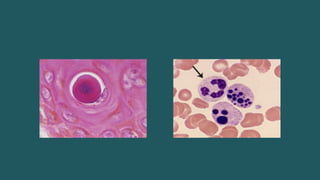
MORPHOLOGICAL CHANGES
• Cellshrinkage
• Chromatin condensation
• Formation of cytoplasmic blebs and apoptotic
bodies
• Phagocytosis of apoptotic cells/ cell bodies,
usually by macrophages.
9.
MECHANISM OF APOPTOSIS
•Apoptosis results from the activation of proteins
called caspases.
• As other proteases they exist as inactive
proenzyme and must undergo enzymatic cleavage
to become active.
• The entire process can be divided into two parts:
i. Initiation phase
ii. Execution phase
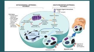
10.
MECHANISM OF APOPTOSIS
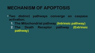
•Two distinct pathways converge on caspase
activation:
1. The Mitochondrial pathway (Intrinsic pathway)
2. The Death Receptor pathway (Extrinsic
pathway)
11.
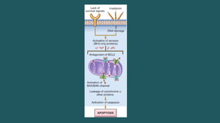
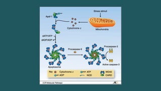
MITOCHONDRIAL PATHWAY
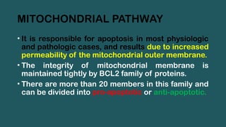
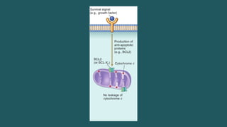
• Itis responsible for apoptosis in most physiologic
and pathologic cases, and results due to increased
permeability of the mitochondrial outer membrane.
• The integrity of mitochondrial membrane is
maintained tightly by BCL2 family of proteins.
• There are more than 20 members in this family and
can be divided into pro-apoptotic or anti-apoptotic.
12.
MITOCHONDRIAL PATHWAY
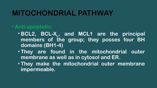
• Anti-apoptotic:
•BCL2, BCL-XL, and MCL1 are the principal
members of the group; they posses four BH
domains (BH1-4)
• They are found in the mitochondrial outer
membrane as well as in cytosol and ER.
• They make the mitochondrial outer membrane
impermeable.
14.
MITOCHONDRIAL PATHWAY
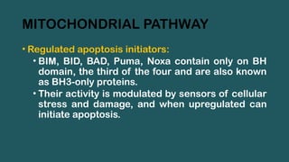
• Regulatedapoptosis initiators:
• BIM, BID, BAD, Puma, Noxa contain only on BH
domain, the third of the four and are also known
as BH3-only proteins.
• Their activity is modulated by sensors of cellular
stress and damage, and when upregulated can
initiate apoptosis.
15.
MITOCHONDRIAL PATHWAY
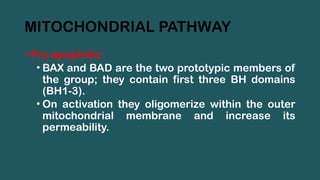
• Pro-apoptotic:
•BAX and BAD are the two prototypic members of
the group; they contain first three BH domains
(BH1-3).
• On activation they oligomerize within the outer
mitochondrial membrane and increase its
permeability.
17.
MITOCHONDRIAL PATHWAY
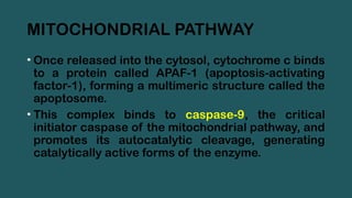
• Oncereleased into the cytosol, cytochrome c binds
to a protein called APAF-1 (apoptosis-activating
factor-1), forming a multimeric structure called the
apoptosome.
• This complex binds to caspase-9, the critical
initiator caspase of the mitochondrial pathway, and
promotes its autocatalytic cleavage, generating
catalytically active forms of the enzyme.
19.
MITOCHONDRIAL PATHWAY
• Othermitochondrial proteins with arcane names
like Smac/DIABLO enter the cytoplasm, where they
bind to and neutralize cytoplasmic proteins that
function as physiologic inhibitors of apoptosis
(IAPs).
20.
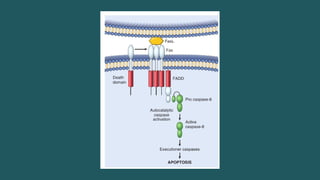
DEATH RECEPTOR PATHWAY
•This pathway is initiated by engagement of plasma
membrane death receptors.
• Death receptors are a member of tumor necrosis
factor (TNF) receptor family that contain a
cytoplasmic domain involved in protein-protein
interaction.
• Type 1 TNF receptor (TNFR1) and related protein
Fas (CD95) are the best known death receptors
21.
DEATH RECEPTOR PATHWAY
•When FasL binds to Fas, three or more molecules
of Fas are brought together, and their cytoplasmic
death domains form a binding site for an adaptor
protein called FADD (Fas-associated death
domain).
• Once attached to this complex, FADD binds
inactive caspase-8 (or caspase-10), bringing
together multiple caspase molecules and leading
to autocatalytic cleavage and generation of active
caspase-8.
23.
DEATH RECEPTOR PATHWAY
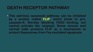
•This extrinsic apoptosis pathway can be inhibited
by a protein called FLIP, which binds to pro-
caspase-8, thereby blocking FADD binding, but
cannot activate the caspase. Some viruses and
normal cells produce FLIP as a mechanism to
protect themselves from Fas mediated apoptosis.
24.
THE EXECUTION PHASE
•The intrinsic mitochondrial pathway activates the
initiator caspase-9, whereas the extrinsic death
receptor pathway activates caspase-8 and
caspase-10.
• The active forms of these caspases trigger the
rapid and sequential activation of the executioner
caspases, such as caspase-3 and caspase-6,
which then act on many cellular components.
25.
THE EXECUTION PHASE
•Once activated these caspases cleave an inhibitor
of Dnase, making Dnase enzymatically active.
• Caspases also proteolyze structural component of
nuclear matrix thus promoting fragmentation of
nuclei.
• Other steps are ill defined; we still do not know how
membrane blebs and apoptotic bodies are formed.
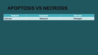
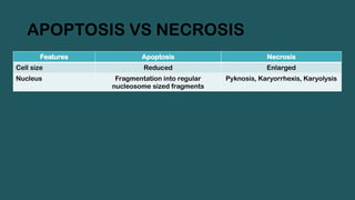
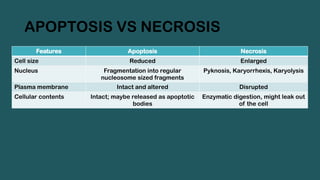
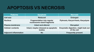
APOPTOSIS VS NECROSIS
FeaturesApoptosis Necrosis
Cell size Reduced Enlarged
Nucleus Fragmentation into regular
nucleosome sized fragments
Pyknosis, Karyorrhexis, Karyolysis
29.
APOPTOSIS VS NECROSIS
FeaturesApoptosis Necrosis
Cell size Reduced Enlarged
Nucleus Fragmentation into regular
nucleosome sized fragments
Pyknosis, Karyorrhexis, Karyolysis
Plasma membrane Intact and altered Disrupted
30.
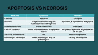
APOPTOSIS VS NECROSIS
FeaturesApoptosis Necrosis
Cell size Reduced Enlarged
Nucleus Fragmentation into regular
nucleosome sized fragments
Pyknosis, Karyorrhexis, Karyolysis
Plasma membrane Intact and altered Disrupted
Cellular contents Intact; maybe released as apoptotic
bodies
Enzymatic digestion, might leak out
of the cell
31.
APOPTOSIS VS NECROSIS
FeaturesApoptosis Necrosis
Cell size Reduced Enlarged
Nucleus Fragmentation into regular
nucleosome sized fragments
Pyknosis, Karyorrhexis, Karyolysis
Plasma membrane Intact and altered Disrupted
Cellular contents Intact; maybe released as apoptotic
bodies
Enzymatic digestion, might leak out
of the cell
Adjacent inflammation No Frequently present
32.
APOPTOSIS VS NECROSIS
FeaturesApoptosis Necrosis
Cell size Reduced Enlarged
Nucleus Fragmentation into regular
nucleosome sized fragments
Pyknosis, Karyorrhexis, Karyolysis
Plasma membrane Intact and altered Disrupted
Cellular contents Intact; maybe released as apoptotic
bodies
Enzymatic digestion, might leak out
of the cell
Adjacent inflammation No Frequently present
Physiologic/ Pathologic Often physiologic, may be
pathological
Usually pathological
#9 Caspase: Contain cysteine and cleave proteins after Aspartate residues
Humans have a total of 10 caspases
The presence of active caspases is therefore a marker of cell undergoing apoptosis
#11 Mitochondria are cellular organelle that are responsible for generating energy and contains enzymes such as cytochrome-C.
#12 Transcription of these factors depends upon survival signals.
#20 Cytoplasmic death domain is essential for delivering apoptotic signal. Some TNF receptor family do not have said domain and act in inflammatory process and not apoptosis.
#21 FasL is expressed on T cells that recognize self antigens (an functions to eliminate self-reactive lymphocytes that also express receptor Fas upon recognition of self antigens)
Also expressed by Cytotoxic T-cells that kill virus infected and tumor cells.