antepartum haemorrhage by shubham kumbare
•Download as PPTX, PDF•
1 like•294 views

This document discusses antepartum haemorrhage (APH), specifically placenta previa and abruptio placenta as causes of APH. Placenta previa occurs when the placenta implants over or near the cervical os, potentially causing bleeding as the cervix dilates. Risk factors include multiparity, advanced maternal age, prior placenta previa, and placenta abnormalities. Abruptio placenta involves premature separation of a normally implanted placenta, potentially causing concealed or revealed bleeding. Both conditions require careful monitoring and management of bleeding, with delivery by caesarean section often needed to prevent complications.

Recommended
More Related Content
What's hot
What's hot (20)
Similar to antepartum haemorrhage by shubham kumbare
Similar to antepartum haemorrhage by shubham kumbare (20)
Recently uploaded
Recently uploaded (20)
antepartum haemorrhage by shubham kumbare
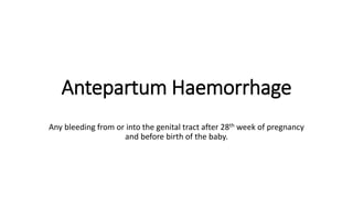
- 1. Antepartum Haemorrhage Any bleeding from or into the genital tract after 28th week of pregnancy and before birth of the baby.
- 2. causes
- 3. Placenta previa • When the placenta is implanted partially or completely over the lower uterine segment (over or adjacent to internal os). • Incidence= 0.5 to 1% among hospital deliveries • Aetiology- postulated theories- 1. dropping down theory 2. persistence of chorionic activity Defective decidua Big surface of placenta
- 4. High risk factors for placenta previa • Multiparity • Maternal age >35 • Asian women • Presence of uterine scar • Multiple pregnancy • Prior placenta previa • Placenta size and abnormality • smoking
- 5. Type I ( low lying) Type II (Marginal)
- 6. Type III (partial central) Type IV (central)
- 7. Dangerous placenta previa it is type II posteriors placenta previa
- 8. Causes of bleeding • Lower segment progressively dilates, the inelastic placenta is sheered off the wall of the lower segment. This leads to the opening of sinuses and episodes of bleeding.
- 9. Placental migration • With progressive increase in the length of lower uterine segment, the lower placental edge relocates away from the cervical os. • Due to trophotrophism.
- 10. Clinical features • Symptom- sudden painless vaginal bleeding which is apparently causeless and recurrent. • Signs- general condition and anaemia are proportionate to the visible blood loss.
- 11. Confirmation of diagnosis • Painless and recurrent vaginal bleeding in second half of pregnancy.
- 12. placentography • Sonography- 1. transabdominal USG 2. transvaginal USG 3.color Doppler flow study • MRI
- 13. Clinical confirmation • Double setup vaginal examination it is done in OT under anaesthesia, keeping everything ready for C-section.
- 14. Complications- • Maternal: during pregnancy 1. APH 2. malpresentation 3. premature labour • Maternal: During labour 1. Early rupture of membranes. 2. cord prolapse 3. slow dilatation of cervix 4. IPH 5. increased incidence of operative interference 6. PPH 7. Retained placenta • Maternal: During pueperium 1. sepsis 2. subinvolution 3. embolism • Fetal complications 1.LBW 2. FGR 3. Asphyxia 4. IUD 5. Birth injuries 6. congenital malformation 7 maternal fetal morbidity and mortality
- 15. Management-
- 16. prevention • Regular anc • Antenatal diagnosis • Significance of warning signs • Colour Doppler usg
- 17. At home • Put to bed • To assess blood loss • Quick but gentle abdominal examination • Vaginal examination must not done
- 18. Immediate attention • Amount of blood loss • Blood sample are taken • A large bore IV cannula is sited and infusion of normal saline • Gentle abdominal palpation • Inspection of vulva • Confirmation of diagnosis
- 19. Expectant management • Vital pre-requires:- 1)blood for transfusion 2)facilities for caesarean section should be available throughtout 24hr Selection of cases:- 1)HB more than equal 10gm%,haematocrit >30% 2)pregnancy should be<37weeks 3)active vaginal bleeding 4) foetal well being is assured Termination of expectant pregnancy is carried upto 37 wks
- 20. Active management • Caesarean delivery done in women where placenta edge is within 2cm from the internal os • Vaginal delivery is considered when placental edge is 2-3cm away from internal os
- 21. Practical approach to lower segment CS for placenta privia • To make infra-umbilical longitudinal incision • To tackle the engorged vessels • To tackle the placenta lying underneath the incision • B-lynch suture or tight intrauterine packing is done by temponade
- 22. Practical guide to LSCS for placenta privia accreta • Women with anterior placenta privia implanted at the site of prior cs, there is an increase risk of placenta accrete. This may need hysterectomy • Incision made away from the placenta • Any attempt of placental separation in placenta accreta may cause massive bleeding • In presence of bleeding
- 23. Abruptio placenta • It is one of the APH where bleeding occurs due to premature separation of normally situated placenta
- 26. Type-3 • Mixed
- 27. Incidence • PNM 15-20% • MM 2-5%
- 28. pathogenesis • premature placental separation is initiated by haemorrhage into decidua basalis • Absence of uterine contraction lead to concealed type • Thrombin generated following decidual haemorrhage triggers the action of MMP,cytokines,coagulation cascade
- 29. Couvelaire uterus Can be diagnosed by laprotomy
- 30. Aetiology • Genetic factors • HTP in pregnancy • Trauma • Sudden uterine decompression • Short cord • Placental anomalies • Sick placenta • Uterine factors • Folic acid def. • Torsion of uterus • Cocaine abuse • Thrombophilia • Prior abruption
- 31. Clinical classification • Grade 0:asymptomatic • Grade1: maternal BP and fibrinogen level are unaffected • Grade2: 45%, shock is absent and foetal distress or foetal death may occur • grade3: coagulation defect or anuria
- 33. Complications MATERNAL :- revealed type : maternal risk is proportionate to visible blood loss and maternal death is rear Concealed type : haemorrhage , shock , blood coagulation dis , oliguria , anuria , PPH , puerperal sepsis • FETAL:- • revealed type : fetal death is 25-30% • Concealed : fetal death is 50-100%, the death are due to prematurity and anoxia due to placental separation
- 34. management