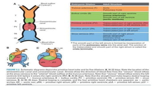
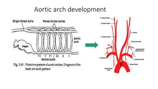
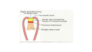
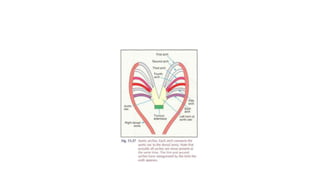
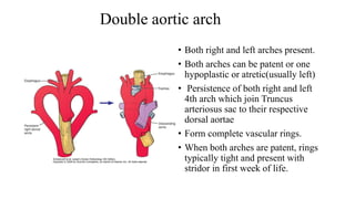
The document summarizes the development of the aortic arch, thoracic and abdominal aorta, and venous system during embryogenesis. It describes how the paired dorsal aortas fuse to form the descending aorta and how the aortic arches give rise to major arteries like the carotid and subclavian arteries. Common aortic arch anomalies are discussed along with the regression of embryonic veins and formation of major veins like the inferior vena cava from segments including the vitelline, umbilical, and cardinal veins. Rare anomalies of the venous system and arterial variations are also mentioned.
Join live classes, download study aids, sell your documents, join or host your own classes online, get tutoring, tutor students, take practices tests and more at Examville.com
Echocardiography, Class II, Introduction to Echocardiography - Anatomy of the heart, cardiac hemodynamic concepts, coronary arteries, coronary artery branches, coronary distribution, 17 segment model, coronary perfusion, the pathway of the heart, cardiovascular blood flow, the cardiac cycle, semilunar valve function, cardiac intrinsic function, electrophysiology of the heart, electrocardiogram, phases of the cardiac cycle (chart), cardiac output, stroke volume, preload & afterload of the heart, calculation of target heart rate
How many patients does case series should have In comparison to case reports.pdfpubrica101
Pubrica’s team of researchers and writers create scientific and medical research articles, which may be important resources for authors and practitioners. Pubrica medical writers assist you in creating and revising the introduction by alerting the reader to gaps in the chosen study subject. Our professionals understand the order in which the hypothesis topic is followed by the broad subject, the issue, and the backdrop.
https://pubrica.com/academy/case-study-or-series/how-many-patients-does-case-series-should-have-in-comparison-to-case-reports/
Navigating Challenges: Mental Health, Legislation, and the Prison System in B...Guillermo Rivera
This conference will delve into the intricate intersections between mental health, legal frameworks, and the prison system in Bolivia. It aims to provide a comprehensive overview of the current challenges faced by mental health professionals working within the legislative and correctional landscapes. Topics of discussion will include the prevalence and impact of mental health issues among the incarcerated population, the effectiveness of existing mental health policies and legislation, and potential reforms to enhance the mental health support system within prisons.
Defecation
Normal defecation begins with movement in the left colon, moving stool toward the anus. When stool reaches the rectum, the distention causes relaxation of the internal sphincter and an awareness of the need to defecate. At the time of defecation, the external sphincter relaxes, and abdominal muscles contract, increasing intrarectal pressure and forcing the stool out
The Valsalva maneuver exerts pressure to expel faeces through a voluntary contraction of the abdominal muscles while maintaining forced expiration against a closed airway. Patients with cardiovascular disease, glaucoma, increased intracranial pressure, or a new surgical wound are at greater risk for cardiac dysrhythmias and elevated blood pressure with the Valsalva maneuver and need to avoid straining to pass the stool.
Normal defecation is painless, resulting in passage of soft, formed stool
CONSTIPATION
Constipation is a symptom, not a disease. Improper diet, reduced fluid intake, lack of exercise, and certain medications can cause constipation. For example, patients receiving opiates for pain after surgery often require a stool softener or laxative to prevent constipation. The signs of constipation include infrequent bowel movements (less than every 3 days), difficulty passing stools, excessive straining, inability to defecate at will, and hard feaces
IMPACTION
Fecal impaction results from unrelieved constipation. It is a collection of hardened feces wedged in the rectum that a person cannot expel. In cases of severe impaction the mass extends up into the sigmoid colon.
DIARRHEA
Diarrhea is an increase in the number of stools and the passage of liquid, unformed feces. It is associated with disorders affecting digestion, absorption, and secretion in the GI tract. Intestinal contents pass through the small and large intestine too quickly to allow for the usual absorption of fluid and nutrients. Irritation within the colon results in increased mucus secretion. As a result, feces become watery, and the patient is unable to control the urge to defecate. Normally an anal bag is safe and effective in long-term treatment of patients with fecal incontinence at home, in hospice, or in the hospital. Fecal incontinence is expensive and a potentially dangerous condition in terms of contamination and risk of skin ulceration
HEMORRHOIDS
Hemorrhoids are dilated, engorged veins in the lining of the rectum. They are either external or internal.
FLATULENCE
As gas accumulates in the lumen of the intestines, the bowel wall stretches and distends (flatulence). It is a common cause of abdominal fullness, pain, and cramping. Normally intestinal gas escapes through the mouth (belching) or the anus (passing of flatus)
FECAL INCONTINENCE
Fecal incontinence is the inability to control passage of feces and gas from the anus. Incontinence harms a patient’s body image
PREPARATION AND GIVING OF LAXATIVESACCORDING TO POTTER AND PERRY,
An enema is the instillation of a solution into the rectum and sig
Telehealth Psychology Building Trust with Clients.pptxThe Harvest Clinic
Telehealth psychology is a digital approach that offers psychological services and mental health care to clients remotely, using technologies like video conferencing, phone calls, text messaging, and mobile apps for communication.
The Importance of Community Nursing Care.pdfAD Healthcare
NDIS and Community 24/7 Nursing Care is a specific type of support that may be provided under the NDIS for individuals with complex medical needs who require ongoing nursing care in a community setting, such as their home or a supported accommodation facility.
Antibiotic Stewardship by Anushri Srivastava.pptxAnushriSrivastav
Stewardship is the act of taking good care of something.
Antimicrobial stewardship is a coordinated program that promotes the appropriate use of antimicrobials (including antibiotics), improves patient outcomes, reduces microbial resistance, and decreases the spread of infections caused by multidrug-resistant organisms.
WHO launched the Global Antimicrobial Resistance and Use Surveillance System (GLASS) in 2015 to fill knowledge gaps and inform strategies at all levels.
ACCORDING TO apic.org,
Antimicrobial stewardship is a coordinated program that promotes the appropriate use of antimicrobials (including antibiotics), improves patient outcomes, reduces microbial resistance, and decreases the spread of infections caused by multidrug-resistant organisms.
ACCORDING TO pewtrusts.org,
Antibiotic stewardship refers to efforts in doctors’ offices, hospitals, long term care facilities, and other health care settings to ensure that antibiotics are used only when necessary and appropriate
According to WHO,
Antimicrobial stewardship is a systematic approach to educate and support health care professionals to follow evidence-based guidelines for prescribing and administering antimicrobials
In 1996, John McGowan and Dale Gerding first applied the term antimicrobial stewardship, where they suggested a causal association between antimicrobial agent use and resistance. They also focused on the urgency of large-scale controlled trials of antimicrobial-use regulation employing sophisticated epidemiologic methods, molecular typing, and precise resistance mechanism analysis.
Antimicrobial Stewardship(AMS) refers to the optimal selection, dosing, and duration of antimicrobial treatment resulting in the best clinical outcome with minimal side effects to the patients and minimal impact on subsequent resistance.
According to the 2019 report, in the US, more than 2.8 million antibiotic-resistant infections occur each year, and more than 35000 people die. In addition to this, it also mentioned that 223,900 cases of Clostridoides difficile occurred in 2017, of which 12800 people died. The report did not include viruses or parasites
VISION
Being proactive
Supporting optimal animal and human health
Exploring ways to reduce overall use of antimicrobials
Using the drugs that prevent and treat disease by killing microscopic organisms in a responsible way
GOAL
to prevent the generation and spread of antimicrobial resistance (AMR). Doing so will preserve the effectiveness of these drugs in animals and humans for years to come.
being to preserve human and animal health and the effectiveness of antimicrobial medications.
to implement a multidisciplinary approach in assembling a stewardship team to include an infectious disease physician, a clinical pharmacist with infectious diseases training, infection preventionist, and a close collaboration with the staff in the clinical microbiology laboratory
to prevent antimicrobial overuse, misuse and abuse.
to minimize the developme
Empowering ACOs: Leveraging Quality Management Tools for MIPS and BeyondHealth Catalyst
Join us as we delve into the crucial realm of quality reporting for MSSP (Medicare Shared Savings Program) Accountable Care Organizations (ACOs).
In this session, we will explore how a robust quality management solution can empower your organization to meet regulatory requirements and improve processes for MIPS reporting and internal quality programs. Learn how our MeasureAble application enables compliance and fosters continuous improvement.
India Clinical Trials Market: Industry Size and Growth Trends [2030] Analyzed...Kumar Satyam
According to TechSci Research report, "India Clinical Trials Market- By Region, Competition, Forecast & Opportunities, 2030F," the India Clinical Trials Market was valued at USD 2.05 billion in 2024 and is projected to grow at a compound annual growth rate (CAGR) of 8.64% through 2030. The market is driven by a variety of factors, making India an attractive destination for pharmaceutical companies and researchers. India's vast and diverse patient population, cost-effective operational environment, and a large pool of skilled medical professionals contribute significantly to the market's growth. Additionally, increasing government support in streamlining regulations and the growing prevalence of lifestyle diseases further propel the clinical trials market.
Growing Prevalence of Lifestyle Diseases
The rising incidence of lifestyle diseases such as diabetes, cardiovascular diseases, and cancer is a major trend driving the clinical trials market in India. These conditions necessitate the development and testing of new treatment methods, creating a robust demand for clinical trials. The increasing burden of these diseases highlights the need for innovative therapies and underscores the importance of India as a key player in global clinical research.
CHAPTER 1 SEMESTER V PREVENTIVE-PEDIATRICS.pdfSachin Sharma
This content provides an overview of preventive pediatrics. It defines preventive pediatrics as preventing disease and promoting children's physical, mental, and social well-being to achieve positive health. It discusses antenatal, postnatal, and social preventive pediatrics. It also covers various child health programs like immunization, breastfeeding, ICDS, and the roles of organizations like WHO, UNICEF, and nurses in preventive pediatrics.
CHAPTER 1 SEMESTER V - ROLE OF PEADIATRIC NURSE.pdfSachin Sharma
Pediatric nurses play a vital role in the health and well-being of children. Their responsibilities are wide-ranging, and their objectives can be categorized into several key areas:
1. Direct Patient Care:
Objective: Provide comprehensive and compassionate care to infants, children, and adolescents in various healthcare settings (hospitals, clinics, etc.).
This includes tasks like:
Monitoring vital signs and physical condition.
Administering medications and treatments.
Performing procedures as directed by doctors.
Assisting with daily living activities (bathing, feeding).
Providing emotional support and pain management.
2. Health Promotion and Education:
Objective: Promote healthy behaviors and educate children, families, and communities about preventive healthcare.
This includes tasks like:
Administering vaccinations.
Providing education on nutrition, hygiene, and development.
Offering breastfeeding and childbirth support.
Counseling families on safety and injury prevention.
3. Collaboration and Advocacy:
Objective: Collaborate effectively with doctors, social workers, therapists, and other healthcare professionals to ensure coordinated care for children.
Objective: Advocate for the rights and best interests of their patients, especially when children cannot speak for themselves.
This includes tasks like:
Communicating effectively with healthcare teams.
Identifying and addressing potential risks to child welfare.
Educating families about their child's condition and treatment options.
4. Professional Development and Research:
Objective: Stay up-to-date on the latest advancements in pediatric healthcare through continuing education and research.
Objective: Contribute to improving the quality of care for children by participating in research initiatives.
This includes tasks like:
Attending workshops and conferences on pediatric nursing.
Participating in clinical trials related to child health.
Implementing evidence-based practices into their daily routines.
By fulfilling these objectives, pediatric nurses play a crucial role in ensuring the optimal health and well-being of children throughout all stages of their development.
4. • Once endothelial cells are established as vascular elements, they
begin to sprout and bud, forming simple capillary networks.
• vascular endothelial growth factor (VEGF)- Stable vascular network
5. Basics of embryology of heart
• Heart is first seen in the form of two
endothelial heart tubes- 18th day of
foetal life
• Fusion results in a single tube with a
series of dilatations (Bulbus cordis ,
Ventricle , Atrium ,Sinus venosus) and
begins to beat by 22nd day
8. Parts of Primitive Aorta
• 1. Ventral aorta-Ventral to foregut
• 2. Arched portion-connected to first pharyngeal arch
• 3. Dorsal aorta-Dorsal to foregut
9.
10.
11. 1st and 2nd arches (maxillary and stapedial)
• 1st pair of aortic arches hidden
in the mandibular arch and
participates in formation of
the maxillary artery
• 2nd pair of aortic arches give
rise to stapedial and hyoid
arteries.
12. 3rd arch(carotids)
• 3rd pair of aortic arches make
their appearance at the end of
week 4. They give rise to
the common
carotids and proximal portions
of the internal carotid arteries.
• Portion of dorsal aorta b/w 3rd
and 4th (ductus
caroticus)disappear
13. First, second and fifth arches disappear
The paired dorsal aortas fuse at the level of the seventh
cervical vertebra to become the thoracic and abdominal aorta.
The dorsal aorta between the third and fourth arches involutes
so that blood entering the third arch perfuses the head only.
The third arch and its branches become the carotid system. The right dorsal aorta distal
to the right seventh intersegmental artery involutes so that blood entering the right
fourth arch perfuses the right upper limb. This system becomes the right subclavian
artery.
The distal part of the right sixth arch involutes, but the distal
part of the left sixth arch persists and becomes the ductus
arteriosus.
The right fourth arch, the dorsal aorta distal to the right fourth
arch+ right seventh intersegmental artery - Right subclavian
artery
Left fourth arch+ left dorsal aorta distal to it become part of
the Aortic archLeft seventh intersegmental artery - left subclavian artery
Pulmonary arteries form from the sixth arch with the ductus
arteriosus present
Patent ductus arteriosus becomes ligamentum arteriosum
later on
14.
15. Aortic Arch Anomalies
• BOVINE ARCH
• DOUBLE ARCH
• RIGHT ARCH
• PATENT DUCTUS ARTERIOSIS
• ABERRANT RIGHT SUBCLAVIAN ARTERY
• INTERRUPTED AORTIC ARCH
• COARCTATION
• ANOMALOUS ORIGIN OF PULMONARYARTERY
• ABNORMAL LEFT ARCH
• ABNORMAL RIGHT ARCH
• CERVICALARCH
16. BOVINE ARCH
• Most common anomaly -8% in
whites and 25% in blacks
• Found in African descents
17.
18. Double aortic arch
• Both right and left arches present.
• Both arches can be patent or one
hypoplastic or atretic(usually left)
• Persistence of both right and left
4th arch which join Truncus
arteriosus sac to their respective
dorsal aortae
• Form complete vascular rings.
• When both arches are patent, rings
typically tight and present with
stridor in first week of life.
19. PATENT DUCTUS ARTERIOSIS
• The ductus arteriosus, which is
normally occluded soon after
birth, may remain patent
• Communication between the
pulmonary artery and the aorta
20. TYPES OF PDA
• Type A - conical duct with well
defined aortic ampulla and constriction
near the pulmonary artery end.
• Type B - large duct with window like
structure which is very short in length.
• Type C - tubular duct without any
constriction.
• Type D - complex duct with multiple
constrictions.
• Type E - elongated duct with
constriction remote from the edge of
the trachea (as viewed on lateral
angiography)
21. COARCTATION OF THE AORTA
• Aorta may show a localized
narrowing of its lumen
• May be distal to the attachment
of the ductus (postductal)
• proximal to the attachment
(preductal)
23. RIGHT AORTIC ARCH-MIRROR IMAGE
TYPE
• Sequence of arch vessels-left
innominate,right carotid.
• Ligamentum left sided.
• No vascular ring.
• Almost always associated with
CHD (48% TOF)
24. RETRO ESOPHAGEAL RIGHT
SUBCLAVIAN ARTERY
• The right subclavian artery
originates distal to the left SCA
and reaches the right upper limb
• The right fourth arch and the
dorsal aorta distal to the right
fourth arch abnormally involute,
and therefore cannot contribute
to formation of the RT. SCA
25. DEVELOPMENT OF DTAAND
ABDOMINALAORTA
• During the fourth week, the
paired dorsal aortas caudal to the
level of the seventh cervical
somite fuse to become the
descending (thoracic and
abdominal) aorta.
• Dorsal, Ventral, and Lateral
branches
• Dorsal branches- dorsal branches
supply the developing neural
tube and epimere
• ventral branches- superior
thoracic artery, internal thoracic
artery, and the superior epigastric
arteries.
26. • At the lumbar level, the dorsal intersegmental branches become
lumbar arteries, with the fifth lumbar pair remaining as the common
iliac arteries.
• Lumbar ventral branches form Allantoic and Vitelline vessels.
• As the gastrointestinal tract develops, the Vitelline arteries become its
blood supply and fuse to become the three unpaired gut vessels: the
Celiac artery supplying the foregut, the Superior mesenteric artery
supplying the Midgut, and the Inferior mesenteric artery supplying the
Hindgut.
Lateral segmental arteries
• The lateral segmental arteries supply the primitive urogenital ridge,
which is the source of the gonads and kidney, adrenal and gonadal
arteries
27. Development of the venous system
3 pairs of major veins can be distinguished:
• (a) The vitelline veins (omphalomesenteric
veins), carrying blood from the yolk sac to the sinus venosus.
• (b) The umbilical veins, originating in the chorionic villi and
carrying oxygenated blood to the embryo.
• (c) The cardinal veins, draining the body of the embryo proper.
28.
29. Vitelline veins
• 2 vitelline veins : Right and Left
• Each one divided into distal, middle and proximal parts by the
developing liver
• The distal parts of 2 veins anastomosis around the duodenum to
form the portal vein
• The middle part of 2 veins forming the liver sinusoids
• The proximal part of the left vein disappear
• The proximal part of the right above the liver forming the
Hepato-cardiac part of the IVC
30.
31. At first 2 umbilical veins are present (right and left)The right vein completely disappearsThe proximal part of the left vein disappearsThe distal part of the left vein carries blood from the placenta
to the liver
32. Cardinal Veins
• The cardinal veins form the main
venous drainage system of the
embryo.
• This system consists of:
The anterior cardinal
veins
The posterior cardinal
veins, of the embryo.
The short common
cardinal veins.
33. In fifth to the seventh week, posterior cardinal system on both side
regressed:
• (a) the supracardinal veins, which drain the body wall by way of
the intercostal veins
• (b) the subcardinal veins, which mainly drain the kidneys
• (c) the sacrocardinal veins, which drain the lower extremities
34.
35. • 2 posterior cardinal veins, with the
development the greater part of posterior
cardinal veins disappear in both sides and the
supracardinal veins take their places
• The right supracardinal vein with a small
portion of the right posterior cardinal vein
forming the azygos vein
• The left supracardinal vein forms the
hemiazygos veins which drains in the
azygos vein
posterior cardinal veins and the
supracardinal veins
36. subcardinal veins
• 2 subcardinal veins and anastomosis in between the left
subcardinal vein disappears, and only its distal portion
remains as the left gonadalvein
• The anastomosis between the subcardinal veins forms the left
renal vein.
• The right subcardinal vein develops into the renal segment
of the inferior vena cava and a small distal portion forms the
right gonadal vein which drain into the inferior vena cava
37. Sacrocardinal veins
• The anastomosis between the sacrocardinal veins forms the left
common iliac vein.
• The right sacrocardinal vein becomes the sacrocardinal
segment of the inferior vena cava.
• Renal segment of the inferior vena cava connects with the
hepatic segment, which is derived from the right vitelline vein,
• The inferior vena cava, consisting of hepatic, renal, and
sacrocardinal segments, is complete.
38. Anomalies of venous system
• Double vena cava
• Left sided vena cava
• IVC duplication
• Retro aortic left renal vein
• Left Circum-aortic renal
vein
40. Persistent sciatic artery
• If the iliofemoral artery fails to
develop, the sciatic artery may
persist as the dominant vessel
supplying blood to the thigh.
• This is a rare anomaly, with an
incidence of 0.05%
41. Popliteal entrapment syndrome
• Popliteal entrapment syndrome is
caused by displacement of the
popliteal artery medial to the
medial head of the
gastrocnemius.
• It results from migration of the
medial head of the
gastrocnemius that forces the
vessels across the popliteal fossa
and entraps them against the
medial condyle of the femur