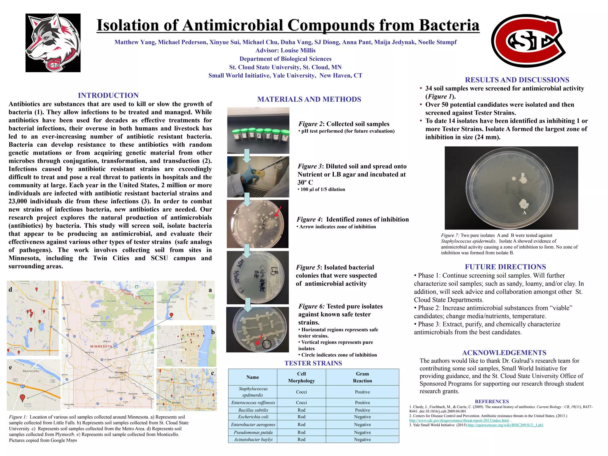
Isolation of Antimicrobial Compounds from Bacteria
Matthew Yang, Michael Pederson, Xinyue Sui, Michael Chu, Duha Vang, SJ Diong, Anna Pant, Maija Jedynak, Noelle Stumpf
Advisor: Louise Millis
Department of Biological Sciences
St. Cloud State University, St. Cloud, MN
Small World Initiative, Yale University, New Haven, CT
RESULTS AND DISCUSSIONS
INTRODUCTION
Antibiotics are substances that are used to kill or slow the growth of
bacteria (1). They allow infections to be treated and managed. While
antibiotics have been used for decades as effective treatments for
bacterial infections, their overuse in both humans and livestock has
led to an ever-increasing number of antibiotic resistant bacteria.
Bacteria can develop resistance to these antibiotics with random
genetic mutations or from acquiring genetic material from other
microbes through conjugation, transformation, and transduction (2).
Infections caused by antibiotic resistant strains are exceedingly
difficult to treat and pose a real threat to patients in hospitals and the
community at large. Each year in the United States, 2 million or more
individuals are infected with antibiotic resistant bacterial strains and
23,000 individuals die from these infections (3). In order to combat
new strains of infectious bacteria, new antibiotics are needed. Our
research project explores the natural production of antimicrobials
(antibiotics) by bacteria. This study will screen soil, isolate bacteria
that appear to be producing an antimicrobial, and evaluate their
effectiveness against various other types of tester strains (safe analogs
of pathogens). The work involves collecting soil from sites in
Minnesota, including the Twin Cities and SCSU campus and
surrounding areas. FUTURE DIRECTIONS
• Phase 1: Continue screening soil samples. Will further
characterize soil samples; such as sandy, loamy, and/or clay. In
addition, will seek advice and collaboration amongst other St.
Cloud State Departments.
• Phase 2: Increase antimicrobial substances from “viable”
candidates; change media/nutrients, temperature.
• Phase 3: Extract, purify, and chemically characterize
antimicrobials from the best candidates.
REFERENCES
1. Clardy, J., Fischbach, M., & Currie, C. (2009). The natural history of antibiotics. Current Biology : CB, 19(11), R437–
R441. doi:10.1016/j.cub.2009.04.001
2. Centers for Disease Control and Prevention. Antibiotic resistance threats in the United States, (2013.)
http://www.cdc.gov/drugresistance/threat-report-2013/index.html. .
3. Yale Small World Initiative (2015) http://openwetware.org/wiki/BISC209/S12:_Lab1
ACKNOWLEDGEMENTS
The authors would like to thank Dr. Gulrud’s research team for
contributing some soil samples, Small World Initiative for
providing guidance, and the St. Cloud State University Office of
Sponsored Programs for supporting our research through student
research grants.
Figure 1: Location of various soil samples collected around Minnesota. a) Represents soil
sample collected from Little Falls. b) Represents soil samples collected from St. Cloud State
University. c) Represents soil samples collected from the Metro Area. d) Represents soil
samples collected from Plymouth. e) Represents soil sample collected from Monticello.
Pictures copied from Google Maps
A
B
A
12
Figure 1
Figure 2
Figure 3
Figure 4
Figure 5
Figure 5
TESTER STRAINS
Figure 7: Two pure isolates A and B were tested against
Staphylococcus epidermidis. Isolate A showed evidence of
antimicrobial activity causing a zone of inhibition to form. No zone of
inhibition was formed from isolate B.
Figure 2: Collected soil samples
• pH test performed (for future evaluation)
Figure 3: Diluted soil and spread onto
Nutrient or LB agar and incubated at
30º C
• 100 µl of 1/5 dilution
Figure 4: Identified zones of inhibition
• Arrow indicates zone of inhibition
Figure 5: Isolated bacterial
colonies that were suspected
of antimicrobial activity
Figure 6: Tested pure isolates
against known safe tester
strains.
• Horizontal regions represents safe
tester strains.
• Vertical regions represents pure
isolates
• Circle indicates zone of inhibition
MATERIALS AND METHODS
A
B
• 34 soil samples were screened for antimicrobial activity
(Figure 1).
• Over 50 potential candidates were isolated and then
screened against Tester Strains.
• To date 14 isolates have been identified as inhibiting 1 or
more Tester Strains. Isolate A formed the largest zone of
inhibition in size (24 mm).
a
b
c
d
e
Name
Cell
Morphology
Gram
Reaction
Staphylococcus
epdimerdis
Cocci Positive
Enterococcus raffinosis Cocci Positive
Bacillus subtilis Rod Positive
Escherichia coli Rod Negative
Enterobacter aerogenes Rod Negative
Pseudomonas putida Rod Negative
Acinetobacter baylyi Rod Negative

Antibiotic Research Poster (2)

  • 1.
    Isolation of AntimicrobialCompounds from Bacteria Matthew Yang, Michael Pederson, Xinyue Sui, Michael Chu, Duha Vang, SJ Diong, Anna Pant, Maija Jedynak, Noelle Stumpf Advisor: Louise Millis Department of Biological Sciences St. Cloud State University, St. Cloud, MN Small World Initiative, Yale University, New Haven, CT RESULTS AND DISCUSSIONS INTRODUCTION Antibiotics are substances that are used to kill or slow the growth of bacteria (1). They allow infections to be treated and managed. While antibiotics have been used for decades as effective treatments for bacterial infections, their overuse in both humans and livestock has led to an ever-increasing number of antibiotic resistant bacteria. Bacteria can develop resistance to these antibiotics with random genetic mutations or from acquiring genetic material from other microbes through conjugation, transformation, and transduction (2). Infections caused by antibiotic resistant strains are exceedingly difficult to treat and pose a real threat to patients in hospitals and the community at large. Each year in the United States, 2 million or more individuals are infected with antibiotic resistant bacterial strains and 23,000 individuals die from these infections (3). In order to combat new strains of infectious bacteria, new antibiotics are needed. Our research project explores the natural production of antimicrobials (antibiotics) by bacteria. This study will screen soil, isolate bacteria that appear to be producing an antimicrobial, and evaluate their effectiveness against various other types of tester strains (safe analogs of pathogens). The work involves collecting soil from sites in Minnesota, including the Twin Cities and SCSU campus and surrounding areas. FUTURE DIRECTIONS • Phase 1: Continue screening soil samples. Will further characterize soil samples; such as sandy, loamy, and/or clay. In addition, will seek advice and collaboration amongst other St. Cloud State Departments. • Phase 2: Increase antimicrobial substances from “viable” candidates; change media/nutrients, temperature. • Phase 3: Extract, purify, and chemically characterize antimicrobials from the best candidates. REFERENCES 1. Clardy, J., Fischbach, M., & Currie, C. (2009). The natural history of antibiotics. Current Biology : CB, 19(11), R437– R441. doi:10.1016/j.cub.2009.04.001 2. Centers for Disease Control and Prevention. Antibiotic resistance threats in the United States, (2013.) http://www.cdc.gov/drugresistance/threat-report-2013/index.html. . 3. Yale Small World Initiative (2015) http://openwetware.org/wiki/BISC209/S12:_Lab1 ACKNOWLEDGEMENTS The authors would like to thank Dr. Gulrud’s research team for contributing some soil samples, Small World Initiative for providing guidance, and the St. Cloud State University Office of Sponsored Programs for supporting our research through student research grants. Figure 1: Location of various soil samples collected around Minnesota. a) Represents soil sample collected from Little Falls. b) Represents soil samples collected from St. Cloud State University. c) Represents soil samples collected from the Metro Area. d) Represents soil samples collected from Plymouth. e) Represents soil sample collected from Monticello. Pictures copied from Google Maps A B A 12 Figure 1 Figure 2 Figure 3 Figure 4 Figure 5 Figure 5 TESTER STRAINS Figure 7: Two pure isolates A and B were tested against Staphylococcus epidermidis. Isolate A showed evidence of antimicrobial activity causing a zone of inhibition to form. No zone of inhibition was formed from isolate B. Figure 2: Collected soil samples • pH test performed (for future evaluation) Figure 3: Diluted soil and spread onto Nutrient or LB agar and incubated at 30º C • 100 µl of 1/5 dilution Figure 4: Identified zones of inhibition • Arrow indicates zone of inhibition Figure 5: Isolated bacterial colonies that were suspected of antimicrobial activity Figure 6: Tested pure isolates against known safe tester strains. • Horizontal regions represents safe tester strains. • Vertical regions represents pure isolates • Circle indicates zone of inhibition MATERIALS AND METHODS A B • 34 soil samples were screened for antimicrobial activity (Figure 1). • Over 50 potential candidates were isolated and then screened against Tester Strains. • To date 14 isolates have been identified as inhibiting 1 or more Tester Strains. Isolate A formed the largest zone of inhibition in size (24 mm). a b c d e Name Cell Morphology Gram Reaction Staphylococcus epdimerdis Cocci Positive Enterococcus raffinosis Cocci Positive Bacillus subtilis Rod Positive Escherichia coli Rod Negative Enterobacter aerogenes Rod Negative Pseudomonas putida Rod Negative Acinetobacter baylyi Rod Negative