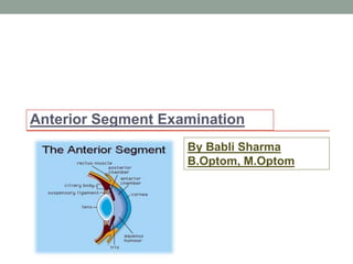
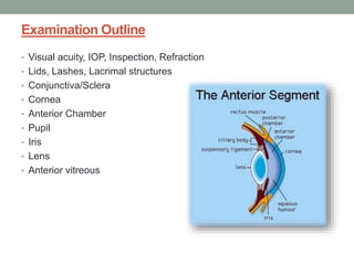
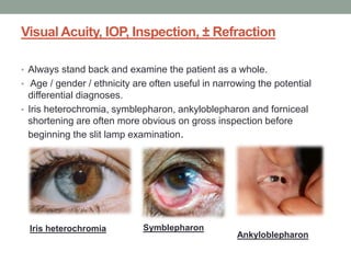
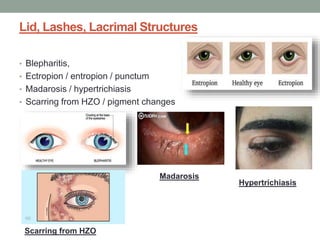
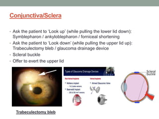
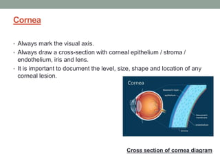
1. Visual Acuity, IOP, Inspection, ± Refraction
Always stand back and examine the patient as a whole. Age / gender / ethnicity are often useful in narrowing the potential differential diagnoses. Iris heterochromia, symblephara, ankyloblepharon and forniceal shortening are often more obvious on gross inspection before beginning the slit lamp examination.
2. Lid, Lashes, Lacrimal Structures
Blepharitis,
Ectropion / entropion / punctum
Madarosis / hypertrichiasis
Scarring from HZO / pigment changes
3. Conjunctiva/Sclera
Ask the patient to ‘Look up’ (while pulling the lower lid down): Symblepharon / ankyloblepharon / forniceal shortening
Ask the patient to ‘Look down’ (while pulling the upper lid up): Trabeculectomy bleb / glaucoma drainage device
Scleral buckle
Offer to evert the upper lid
4. Cornea
Always mark the visual axis.
Always draw a cross-section with corneal epithelium / stroma / endothelium, iris and lens (Figure 1.1.1). It is important to document the level, size, shape and location of any corneal lesion.
Note:
Shape: e.g. Keratoconus
Size: Measure the corneal diameter with a handheld ruler if suspicious of congenital glaucoma. Greater than 12mm in an adult is abnormal, Microcornea is defined as corneal diameter <10mm
Thinning: Estimate and document the remaining thickness as a % of normal
Scars, wounds
Corneal graft
i. Type (penetrating vs. anterior lamellar vs. endothelial)? Size? Clarity?
ii. Sutures? (interrupted, continuous, broken / loose?)
iii. Pay special attention to the graft-host junction
iv. Neovascularisation?
v. Look at the other eye for clues as to why the patient had a graft
± Tear film / marginal tear strip
In addition, look at each corneal layer:
Epithelium: Epithelial defects, punctate epithelial erosions, superficial punctate keratitis, dystrophies, microcystic oedema, Fleischer ring, limbal stem cell failure (conjunctivalisation of the cornea)
Stroma: Dystrophies, stromal oedema, vessels (superficial or deep?), prominent corneal nerves
Endothelium: Guttata, pigment dispersion, keratic precipitates (KP), Descemet’s membrane folds
The presence of one sign (e.g. a conical cornea) should prompt you to look for other associated signs of that condition (e.g. Down syndrome, vernal kerato-conjunctivitis, Fleischer ring- best seen with cobalt blue light, prominent corneal nerves, hydrops / corneal scarring and Vogt striae of keratoconus).
If an ocular surface mass is present (e.g. Ocular surface squamous neoplasia), you must exclude orbital invasion. Check for:
Lymphadenopathy
Corneal sensation
Ophthalmoplegia
Intraocular examination including gonioscopy for angle involvement
5. Anterior Chamber
Cells / flare
Hypopyon, hyphaema
Anterior chamber depth (narrow angle?). Von Herrick’s test can estimate angle depth
If an intraocular mass is present (e.g. iris melanoma), then check for:
Hyphaema
Nodular vs diffuse configuration
Ectropion uveae – seen in iris melanoma
Refractory glaucoma - raised IOP
Gonioscopy
3. Examination Structure
• Attention should be given to the wording of the question which often
directs the candidate where to look:
“Examines the”…
External Eye – Periorbital tissue,
• Eyelid, margins,
• Conjunctiva,
• Fornices,
• Limbus and Corneal surface
• Anterior Segment –
• Eyelids,
• Conjunctiva,
• Cornea (all layers),
• Anterior chamber,
• Iris and Lens
4. Visual Acuity, IOP, Inspection, ± Refraction
• Always stand back and examine the patient as a whole.
• Age / gender / ethnicity are often useful in narrowing the potential
differential diagnoses.
• Iris heterochromia, symblepharon, ankyloblepharon and forniceal
shortening are often more obvious on gross inspection before
beginning the slit lamp examination.
Iris heterochromia Symblepharon
Ankyloblepharon
6. Conjunctiva/Sclera
• Ask the patient to ‘Look up’ (while pulling the lower lid down):
Symblepharon / ankyloblepharon / forniceal shortening
• Ask the patient to ‘Look down’ (while pulling the upper lid up):
Trabeculectomy bleb / glaucoma drainage device
• Scleral buckle
• Offer to evert the upper lid
Trabeculectomy bleb
7. Cornea
• Always mark the visual axis.
• Always draw a cross-section with corneal epithelium / stroma /
endothelium, iris and lens.
• It is important to document the level, size, shape and location of any
corneal lesion.
Cross section of cornea diagram
8. Note:
• Shape: e.g. Keratoconus
• Size: Measure the corneal diameter with a handheld ruler if
suspicious of congenital glaucoma. Greater than 12mm in an adult is
abnormal, Microcornea is defined as corneal diameter <10mm
• Thinning: Estimate and document the remaining thickness as a % of
normal
• Scars, wounds
Keratoconus Microcornea
Megalocornea:
suspicious of
congenital glaucoma
9. Contd..
• Corneal graft
i. Type (penetrating vs. anterior lamellar vs. endothelial)? Size?
Clarity?
ii. Sutures? (interrupted, continuous, broken / loose?)
iii. Pay special attention to the graft-host junction
iv. Neovascularisation?
v. Look at the other eye for clues as to why the patient had a graft
• ± Tear film / marginal tear strip
Sutures Neovascularisation
10. In addition, look at each corneal layer:
Epithelium:
• Epithelial defects,
• punctate epithelial erosions,
• superficial punctate keratitis,
• dystrophies,
• microcystic oedema,
• Fleischer ring,
• limbal stem cell failure (conjunctivalisation of the cornea)
Epithelial defect Punctate epithelial
erosions
Superficial punctate
keratitis
Dystrophies
12. Contd..
• The presence of one sign (e.g. a conical cornea)
• Should prompt you to look for other associated signs of that condition:
e.g.
• Down syndrome,
• Vernal kerato-conjunctivitis,
• Fleischer ring- best seen with cobalt blue light,
• Prominent corneal nerves,
• Hydrops / corneal scarring and Vogt striae of keratoconus).
VKC Hydrops Vogt striae
Down syndrome
13. Contd…
• If an ocular surface mass is present (e.g. Ocular surface squamous
neoplasia : OSSN),
• We must exclude orbital invasion.
Check for:
• Lymphadenopathy
• Corneal sensation
• Ophthalmoplegia
• Intraocular examination including gonioscopy for angle involvement
OSSN
Ophthalmoplegia
14. Anterior Chamber
• Cells / flare
• Hypopyon, hyphaema
• Anterior chamber depth (narrow angle?). Von Herrick’s test can
estimate angle depth
If an intraocular mass is present (e.g. iris melanoma), then check for:
• Hyphaema
• Nodular vs diffuse configuration
• Ectropion uveae – seen in iris melanoma
• Refractory glaucoma - raised IOP
• Gonioscopy for angle involvement
• Transillumination – more likely to be a cyst
• Subjacent ciliary body involvement – will need eye to be dilated
• Dilated fundus examination for extension posteriorly
Hypopyon Hyphaema
15. Iris
• Posterior bow (pigment dispersion syndrome PDS)
• Rubeosis (neovascular vessels run parallel to pupil rather than
radially and are uncuffed)
• Transillumination defects: Peripheral iridotomy (PI), PXF, PDS, HZO,
trauma (IOL, ACG)
Rubeosis Peripheral iridotomy PXF
16. Lens
• Cataract: What type? Anterior cortical / Nuclear sclerotic / Posterior
subcapsular
• Phacodonesis? (ask the patient to ‘look up’ then ‘look straight’).
Anterior or posterior lenticonus
• IOL (PCIOL in bag / sulcus, open-loop ACIOL, iris clipped, scleral
fixed- sutured vs. tunnelled)
• Posterior capsular opacification / YAG capsulotomy
• PXF, posterior synechiae, glaucomflecken
Cataract PCIOL
Posterior
Synechiae