posterior curciate liagment injury, machanisum of injury, type of injury, special test, associated injuries ti PCL injury, physiotherapy treatment
posteior sag test, posterior drawer test, abduction stress test, adduction stress test, day wie trsetment
General talk about Anterior Cruciate Ligament tear.
it presented during my orthopedic rotation in KFUH.
under supervision of Dr. Balwi "sport injuries consultant"
MCL. LCL.ALL injuries
To understand the relevant anatomy of the side ligaments of the knee
To study the mechanism of injury of each ligament and how to diagnose such injury
To highlight the different treatment options in acute or chronic situations
Arthroscopic ACL Reconstruction By Dr Shekhar ShrivastavDelhiArthroscopy
Arthroscopic Acl Reconstruction By Dr Shekhar Shrivastav.
HOW NORMAL KNEE WORKS ?
The knee is the largest joint in the body, and one of the most easily injured. It is made up of the lower end of the thigh bone(femur), the upper end of the shin bone (tibia), and the knee cap (patella), which slides in a groove on the end of the femur. Four bands of tissue, the anterior and posterior cruciate ligaments, and the medial and lateral collateral ligaments connect the femur and the tibia and provide joint stability. The surfaces where the femur, tibia and patella touch are covered with articular cartilage, a smooth substance that cushions the bones and enables them to glide freely. Semicircular rings of tough fibrous-cartilage tissue called the lateral and medial menisci act as shock absorbers and stabilizers.
WHAT IS THE ROLE OF ACL ?
ACL along with other ligaments of the knee joint and meniscus provides stability to the knee joint.
WHAT IS LIGAMENT RECONSTRUCTION ( ACL ) ?
Ligament reconstruction involves replacing the torn ligament with a tendon (graft) from your knee and fixing the graft in place with screws. This procedure is performed with the use of the arthroscope. The anterior cruciate ligament (ACL) is the most common ligament requiring reconstruction procedures. The torn ligament is excised arthroscopically and new ligament is prepared by ligament grafts taken from your own body. Bony tunnels are prepared in femur and tibia using specialized instruments through which the new ligament is passed and fixed with special screws. This procedure requires relative rest or leave from your work or studies for about 2-3 weeks after which you will be allowed normal day to day activities.
WHEN CAN THE PATIENT BE AMBULATED AFTER SURGERY ?
The patient can walk from the same evening of the surgery. Initially the patient is advised to walk with a brace and a walking cane. Strengthening and range of motion exercises for the knee are started from the next day. The patient is discharged from the hospital 2nd or 3rd day after surgery. The patient can walk without support by 10-14 days depending on muscle strengthening. Slow Jogging and other strenuous activities are permitted after 3 months and the patient can return to active sports only 8-9 months after surgery.
Torn ACL Reconstructed ACL
For Further Queries contact your Orthopedic Surgeon at
+ 91 9971192233
Collapse of medial longitudinal arch, with the entire sole of the foot coming into complete or near-complete contact with the ground.
Books Refered :
Text Book Of ANATOMY - Vishram Singh
Joint Structure And Function – Cynthia Norkin
Therapeutic Exrercise – Carolyn Kisner
Orthopaedic Physical Assessment – Magee
Orthopaedic Medicine – L. Ombregt
Campbell’s Operative Orthopaedics
Can read freely here
https://sethiortho.blogspot.com/
Examination of Knee Joint Ligaments
SethiNet Presentations
Introduction
Proper use of the examination techniques requires
An understanding of the anatomy
Pathophysiology of knee ligament injuries
Advanced imaging - Augment a history and examination when necessary
Imaging should not replace a thorough history and physical examination
History taking
A description of the mechanism of injury
The patient should be queried about previous injuries
The current injury may be the sequela of a previous injury
Common ligament Injuries
Anterior Cruciate Ligament
Anatomy
The ACL originates at posteromedial aspect of the lateral femoral condyle
Wide tibial insertion at the lateral aspect of the anterior tibial spine
The ACL has two fiber bundles
The anteromedial
Posterolateral bundles
Which provide varying tension from flexion through extension
Functions
Primary restraint against anterior tibial translation
Provides rotational stability, especially in extension
ACL - Mechanism of Injury
Injury to the knee ligaments is typically the result of
A non contact change in direction
Twisting injury
Landing from a jump.
The patient often describes a “pop” that is felt or heard at injury
The appearance of swelling (hemarthrosis) within a few hours
ACL -Examinations
Examinations
The Anterior drawer test
The Lachman Test
The Pivot Shift Test
Novel Tests
ACL - Anterior drawer test
Patient with patient supine position
The hip flexed at 45° / knee flexed at 90°
The foot is fixed to the table - often by sitting on it
The clinician applies an anterior force to the proximal tibia, palpating the joint line for anterior translation.
Increased anterior translation indicates ACL insufficiency.
Sensitivity – only 50% with the patient under anesthesia
because the posterior horn of the medial meniscus may act as a so-called doorstop that prevents anterior translation, even in the presence of a torn ACL.
ACL - Lachman Test
It was designed to overcome three identified limitations of the anterior drawer test
Acute effusion that often precludes flexion to 90°
Protective spasm of the hamstring muscles that can prevent anterior translation of the tibia
The articulation of the relatively acute convexity of the posterior medial femoral condyle and the posterior horn of the medial meniscus that buttresses and prevents anterior translation of the tibia.
These limitations can lead to false-negative findings
The Lachman test is typically done with the knee flexed 20° to 30°.
The examiner places one hand laterally on the patient’s thigh to stabilize the femur
while the other hand grasps the proximal and more subcutaneous medial tibia and applies anterior stres
The test is positive
In the presence of anterior translation
A soft or mushy end point.
When the ACL is intact, the end point is hard
ACL - Grading - Lachman test
Over the past decade, technology and research have greatly expanded the functionality and aesthetics of prosthetic feet. Today, amputees have a wide array of feet from which to choose. Various models are designed for activities ranging from walking, dancing and running to cycling, golfing, swimming and even snow skiing.
Knee pain treatment approaches will vary, relying upon what precisely is inflicting your knee ache. Your physician can also additionally prescribe medicinal drugs to assist relieve aches and dealing with the situations inflicting your knee ache, inclusive of rheumatoid arthritis or gout. Find more info here: https://philaholisticclinic.com/knee-pain-treatment/
The knee is prone to injuries like ACL tears, meniscus tears, and patellar tendinitis. ACL tears often require surgery and extensive rehabilitation, while meniscus tears can be treated with physical therapy or arthroscopic surgery. Patellar tendinitis is typically managed with rest, physical therapy, and anti-inflammatory medications. Knee clinics offer specialized care for these injuries, with knee specialists providing accurate diagnosis and personalized treatment plans to facilitate prompt recovery and long-term joint health.
Ligament Injuries - Types, Symptoms and TreatmentHealth Quest
Ligament injuries occur when a ligament is stretched beyond its normal range. Ligaments are tough band of fibrous tissues that connects bone to bone or bone to cartilage and support, stabilize and strengthen joints. The primary function of ligaments is to keep the bones in proper alignment and prevent abnormal joint movements. Leading pain management centers in Brooklyn, NYC offer effective treatment options for ligament injuries to help patients attain better mobility, balance, and strength.
For more information about knee sprains, watch this presentation. Here you will find four ligaments in the knee joint, any of which can be sprained.
URL:- www.metrophysio.co.uk
Description of sports injuries in footballers. Read about prevention and treatment. Treatment of sports injuries is possible with Stem cells and PRP in Chennai.
TEST BANK for Operations Management, 14th Edition by William J. Stevenson, Ve...kevinkariuki227
TEST BANK for Operations Management, 14th Edition by William J. Stevenson, Verified Chapters 1 - 19, Complete Newest Version.pdf
Pulmonary Thromboembolism - etilogy, types, medical- Surgical and nursing man...VarunMahajani
Disruption of blood supply to lung alveoli due to blockage of one or more pulmonary blood vessels is called as Pulmonary thromboembolism. In this presentation we will discuss its causes, types and its management in depth.
Explore natural remedies for syphilis treatment in Singapore. Discover alternative therapies, herbal remedies, and lifestyle changes that may complement conventional treatments. Learn about holistic approaches to managing syphilis symptoms and supporting overall health.
Report Back from SGO 2024: What’s the Latest in Cervical Cancer?bkling
Are you curious about what’s new in cervical cancer research or unsure what the findings mean? Join Dr. Emily Ko, a gynecologic oncologist at Penn Medicine, to learn about the latest updates from the Society of Gynecologic Oncology (SGO) 2024 Annual Meeting on Women’s Cancer. Dr. Ko will discuss what the research presented at the conference means for you and answer your questions about the new developments.
Couples presenting to the infertility clinic- Do they really have infertility...Sujoy Dasgupta
Dr Sujoy Dasgupta presented the study on "Couples presenting to the infertility clinic- Do they really have infertility? – The unexplored stories of non-consummation" in the 13th Congress of the Asia Pacific Initiative on Reproduction (ASPIRE 2024) at Manila on 24 May, 2024.
The prostate is an exocrine gland of the male mammalian reproductive system
It is a walnut-sized gland that forms part of the male reproductive system and is located in front of the rectum and just below the urinary bladder
Function is to store and secrete a clear, slightly alkaline fluid that constitutes 10-30% of the volume of the seminal fluid that along with the spermatozoa, constitutes semen
A healthy human prostate measures (4cm-vertical, by 3cm-horizontal, 2cm ant-post ).
It surrounds the urethra just below the urinary bladder. It has anterior, median, posterior and two lateral lobes
It’s work is regulated by androgens which are responsible for male sex characteristics
Generalised disease of the prostate due to hormonal derangement which leads to non malignant enlargement of the gland (increase in the number of epithelial cells and stromal tissue)to cause compression of the urethra leading to symptoms (LUTS
- Video recording of this lecture in English language: https://youtu.be/lK81BzxMqdo
- Video recording of this lecture in Arabic language: https://youtu.be/Ve4P0COk9OI
- Link to download the book free: https://nephrotube.blogspot.com/p/nephrotube-nephrology-books.html
- Link to NephroTube website: www.NephroTube.com
- Link to NephroTube social media accounts: https://nephrotube.blogspot.com/p/join-nephrotube-on-social-media.html
micro teaching on communication m.sc nursing.pdfAnurag Sharma
Microteaching is a unique model of practice teaching. It is a viable instrument for the. desired change in the teaching behavior or the behavior potential which, in specified types of real. classroom situations, tends to facilitate the achievement of specified types of objectives.
Factory Supply Best Quality Pmk Oil CAS 28578–16–7 PMK Powder in Stockrebeccabio
Factory Supply Best Quality Pmk Oil CAS 28578–16–7 PMK Powder in Stock
Telegram: bmksupplier
signal: +85264872720
threema: TUD4A6YC
You can contact me on Telegram or Threema
Communicate promptly and reply
Free of customs clearance, Double Clearance 100% pass delivery to USA, Canada, Spain, Germany, Netherland, Poland, Italy, Sweden, UK, Czech Republic, Australia, Mexico, Russia, Ukraine, Kazakhstan.Door to door service
Hot Selling Organic intermediates
ARTIFICIAL INTELLIGENCE IN HEALTHCARE.pdfAnujkumaranit
Artificial intelligence (AI) refers to the simulation of human intelligence processes by machines, especially computer systems. It encompasses tasks such as learning, reasoning, problem-solving, perception, and language understanding. AI technologies are revolutionizing various fields, from healthcare to finance, by enabling machines to perform tasks that typically require human intelligence.
Prix Galien International 2024 Forum ProgramLevi Shapiro
June 20, 2024, Prix Galien International and Jerusalem Ethics Forum in ROME. Detailed agenda including panels:
- ADVANCES IN CARDIOLOGY: A NEW PARADIGM IS COMING
- WOMEN’S HEALTH: FERTILITY PRESERVATION
- WHAT’S NEW IN THE TREATMENT OF INFECTIOUS,
ONCOLOGICAL AND INFLAMMATORY SKIN DISEASES?
- ARTIFICIAL INTELLIGENCE AND ETHICS
- GENE THERAPY
- BEYOND BORDERS: GLOBAL INITIATIVES FOR DEMOCRATIZING LIFE SCIENCE TECHNOLOGIES AND PROMOTING ACCESS TO HEALTHCARE
- ETHICAL CHALLENGES IN LIFE SCIENCES
- Prix Galien International Awards Ceremony
Evaluation of antidepressant activity of clitoris ternatea in animals
Anterior cruciate-ligament
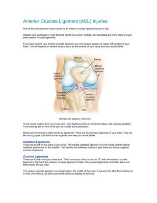
1. Anterior Cruciate Ligament (ACL) Injuries
One of the most common knee injuries is an anterior cruciate ligament sprain or tear.
Athletes who participate in high demand sports like soccer, football, and basketball are more likely to injure
their anterior cruciate ligaments.
If you have injured your anterior cruciate ligament, you may require surgery to regain full function of your
knee. This will depend on several factors, such as the severity of your injury and your activity level.
Normal knee anatomy, front view
Three bones meet to form your knee joint: your thighbone (femur), shinbone (tibia), and kneecap (patella).
Your kneecap sits in front of the joint to provide some protection.
Bones are connected to other bones by ligaments. There are four primary ligaments in your knee. They act
like strong ropes to hold the bones together and keep your knee stable.
Collateral Ligaments
These are found on the sides of your knee. The medial collateral ligament is on the inside and the lateral
collateral ligament is on the outside. They control the sideways motion of your knee and brace it against
unusual movement.
Cruciate Ligaments
These are found inside your knee joint. They cross each other to form an "X" with the anterior cruciate
ligament in front and the posterior cruciate ligament in back. The cruciate ligaments control the back and
forth motion of your knee.
The anterior cruciate ligament runs diagonally in the middle of the knee. It prevents the tibia from sliding out
in front of the femur, as well as provides rotational stability to the knee.
2. Description
About half of all injuries to the anterior cruciate ligament occur along with damage to other structures in the
knee, such as articular cartilage, meniscus, or other ligaments.
Injured ligaments are considered "sprains" and are graded on a severity scale.
Grade 1 Sprains. The ligament is mildly damaged in a Grade 1 Sprain. It has been slightly stretched, but is
still able to help keep the knee joint stable.
Grade 2 Sprains. A Grade 2 Sprain stretches the ligament to the point where it becomes loose. This is often
referred to as a partial tear of the ligament.
Grade 3 Sprains. This type of sprain is most commonly referred to as a complete tear of the ligament. The
ligament has been split into two pieces, and the knee joint is unstable.
Partial tears of the anterior cruciate ligament are rare; most ACL injuries are complete or near complete
tears.
Cause
The anterior cruciate ligament can be injured in several ways:
• Changing direction rapidly
• Stopping suddenly
• Slowing down while running
• Landing from a jump incorrectly
• Direct contact or collision, such as a football tackle
Several studies have shown that female athletes have a higher incidence of ACL injury than male athletes in
certain sports. It has been proposed that this is due to differences in physical conditioning, muscular
3. strength, and neuromuscular control. Other suggested causes include differences in pelvis and lower
extremity (leg) alignment, increased looseness in ligaments, and the effects of estrogen on ligament
properties.
Symptoms
When you injure your anterior cruciate ligament, you might hear a "popping" noise and you may feel your
knee give out from under you. Other typical symptoms include:
• Pain with swelling. Within 24 hours, your knee will swell. If ignored, the swelling and pain may
resolve on its own. However, if you attempt to return to sports, your knee will probably be unstable
and you risk causing further damage to the cushioning cartilage (meniscus) of your knee.
• Loss of full range of motion
• Tenderness along the joint line
• Discomfort while walking
Doctor Examination
Physical Examination and Patient History
During your first visit, your doctor will talk to you about your symptoms and medical history.
During the physical examination, your doctor will check all the structures of your injured knee, and compare
them to your non-injured knee. Most ligament injuries can be diagnosed with a thorough physical
examination of the knee.
Imaging Tests
Other tests which may help your doctor confirm your diagnosis include:
X-rays. Although they will not show any injury to your anterior cruciate ligament, x-rays can show whether
the injury is associated with a broken bone.
Magnetic resonance imaging (MRI) scan. This study creates better images of soft tissues like the anterior
cruciate ligament. However, an MRI is usually not required to make the diagnosis of a torn ACL.
Treatment
Treatment for an ACL tear will vary depending upon the patient's individual needs. For example, the young
athlete involved in agility sports will most likely require surgery to safely return to sports. The less active,
usually older, individual may be able to return to a quieter lifestyle without surgery.
Nonsurgical Treatment
A torn ACL will not heal without surgery. But nonsurgical treatment may be effective for patients who are
elderly or have a very low activity level. If the overall stability of the knee is intact, your doctor may
recommend simple, nonsurgical options.
Bracing. Your doctor may recommend a brace to protect your knee from instability. To further protect your
knee, you may be given crutches to keep you from putting weight on your leg.
Physical therapy. As the swelling goes down, a careful rehabilitation program is started. Specific exercises
will restore function to your knee and strengthen the leg muscles that support it.
Surgical Treatment
4. Rebuilding the ligament. Most ACL tears cannot be sutured (stitched) back together. To surgically repair
the ACL and restore knee stability, the ligament must be reconstructed. Your doctor will replace your torn
ligament with a tissue graft. This graft acts as a scaffolding for a new ligament to grow on.
Grafts can be obtained from several sources. Often they are taken from the patellar tendon, which runs
between the kneecap and the shinbone. Hamstring tendons at the back of the thigh are a common source of
grafts. Sometimes a quadriceps tendon, which runs from the kneecap into the thigh, is used. Finally,
cadaver graft (allograft) can be used.
There are advantages and disadvantages to all graft sources. You should discuss graft choices with your
own orthopaedic surgeon to help determine which is best for you.
Because the regrowth takes time, it may be six months or more before an athlete can return to sports after
surgery.
Procedure. Surgery to rebuild an anterior cruciate ligament is done with an arthroscope using small
incisions. Arthroscopic surgery is less invasive. The benefits of less invasive techniques include less pain
from surgery, less time spent in the hospital, and quicker recovery times.
Animation courtesy Visual Health Solutions, Inc.
Unless ACL reconstruction is treatment for a combined ligament injury, it is usually not done right away. This
delay gives the inflammation a chance to resolve, and allows a return of motion before surgery. Performing
an ACL reconstruction too early greatly increases the risk of arthrofibrosis, or scar forming in the joint, which
would risk a loss of knee motion.
Rehabilitation
Whether your treatment involves surgery or not, rehabilitation plays a vital role in getting you back to your
daily activities. A physical therapy program will help you regain knee strength and motion.
If you have surgery, physical therapy first focuses on returning motion to the joint and surrounding muscles.
This is followed by a strengthening program designed to protect the new ligament. This strengthening
gradually increases the stress across the ligament. The final phase of rehabilitation is aimed at a functional
return tailored for the athlete's sport.