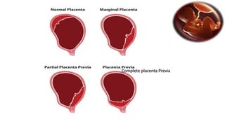
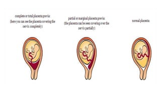
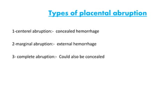
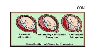
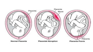
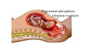
Placental abruption and placenta previa are two common causes of bleeding in late pregnancy. Placental abruption occurs when the placenta separates from the inner wall of the uterus before delivery. It can cause pain and vaginal bleeding. Placenta previa is when the placenta lies low in the uterus, covering all or part of the cervical opening. It typically causes painless vaginal bleeding. Both conditions can lead to complications for the mother like hemorrhage, shock, and infection or complications for the baby like low birth weight or stillbirth. Treatment depends on gestational age and severity of bleeding but may involve bed rest, monitoring, blood transfusions, or delivery via c-section
Uterine Rupture
Deepa Mishra
Assistant Professor (OBG)
Introduction
Uterine rupture is when the muscular wall of the uterus tears during pregnancy or childbirth
Symptoms while classically including increased pain, vaginal bleeding, or a change in contractions are not always present.
Disability or death of the mother or baby may result.
Definition
Uterine rupture is giving way of gravid uterus or dissolution in the continuity of uterine wall anytime after 28 weeks of gestation with or without expulsion of the fetus.
Incidence
Rates of uterine rupture during vaginal birth following one previous C-section, done by the typical technique, are estimated at 0.9%
Rates are greater among those who have had multiple prior C-sections or an atypical type of C-section.
In those who do have uterine scarring, the risk during a vaginal birth is about 1 per 12,000
Risk of death of the baby is about 6%
Etiology
Risk Factors
Previous cesarean section
Myomectomy
Dysfunctional labor
Labor augmentation by oxytocin or prostaglandins
High parity
First pregnancy- very rare
Types of uterine rupture
Complete Rupture
All the layers including peritoneum are torn and the uterine contents escape into the peritoneal cavity.
Usually results in death
Incomplete Rupture
Visceral peritoneum is intact and usually the fetus remains in the uterine cavity
Sign & Symptoms
Uterine dehiscence and abdominal pain and vaginal bleeding
Deterioration of fetal heart rate
Loss of fetal station on manual vaginal exam
Hypovolemic shock due to intrabdominal bleeding
Chest pain between the scapulae, pain during inspiration due to irritation of blood below the perineum
Cessation of uterine contractions
Palpation of fetus outside the uterus
Signs of abdominal pregnancy
Post term pregnancy
Diagnosis
Signs of obstructed labor with dehydration, exhaustion, tachycardia raised temperature tonic contraction , pathological retraction ring
Absent fetal heart sound
On PV hot, dry vagina with a large caput over the presenting part
Prevention
Early diagnosis and management of CPD mal presentation and obstructed labor
Proper selection of cases for vaginal delivery
Carefull monitoring of oxytocin infusion specially in multipara
Avoid intra uterine manipulation no version in single fetus
Instrumental delivery after cervical dilatation
Immediate CS in obstructed labor
Hospital delivery for high risk cases
ECV should be avoided during general anaesthesia
Careful manual removal of placenta
Treatment
Resuscitation with adequate hydration and blood transfusion
Laprotomy
Hysterectomy
Repair
Complication
Rupture uterus with haemorrhage, shock and sepsis
Fetal loss is high in spontaneous and traumatic rupture
Mortality is low in LSCS scar rupture
Gestetional hypertension, Preeclampsia and Eclampsiasunil kumar daha
Please find the power point on Gestetional hypertension, Preeclampsia and Eclampsia . I tried to present it on understandable way and all the contents are reviewed by experts and from very reliable references. Thank you
Uterine rupture - All you need to know.Sandeep Das
This presentation gives the detailed information about uterine rupture - definition, epidemiology, classification, signs and symptoms, prevention and management.
Labour induction
Induction of labour
Guidelines on induction of labour
Guidelines on labour induction
induction of labour is not risk free
prostaglandins for induction of labour
Bishop score
Cervical ripening techniques
mechanical and pharmacological induction of labour
Post dates induction
options for cervical ripening
oral vs. vaginal misoprostol
advantages diadvantages and techniques for induction of labour
gynecology & obstetrics
Labour induction methods
review of guidelines for labour induction
Placenta previa is a condition in which the placenta lies very low in the uterus and covers all or part of the cervix. The cervix is the opening to the uterus that sits at the top of the vagina. Placenta previa happens in about 1 in 200 pregnancies.
Placenta praevia risk factors include a previous delivery, age older than 35 and a history of previous surgeries, such as a caesarean section (C-section) or uterine fibroid removal.
The main symptom is bright red vaginal bleeding without pain during the second-half of pregnancy. The condition can also cause severe bleeding before or during delivery.
Limited physical activity is recommended. A C-section is often required in severe cases.
Bleeding from the genital tract in the late pregnancy, after 20th weeks of gestation and before the onset of labor.
This may place the life of the mother and fetus at risk.
Placental abruption is premature separation of placenta from the uterus/ in other words separates before childbirth.
It occurs most commonly around 25 weeks of pregnancy characterized by vaginal bleeding, lower abdominal pain, and dangerously low blood pressure
Prix Galien International 2024 Forum ProgramLevi Shapiro
June 20, 2024, Prix Galien International and Jerusalem Ethics Forum in ROME. Detailed agenda including panels:
- ADVANCES IN CARDIOLOGY: A NEW PARADIGM IS COMING
- WOMEN’S HEALTH: FERTILITY PRESERVATION
- WHAT’S NEW IN THE TREATMENT OF INFECTIOUS,
ONCOLOGICAL AND INFLAMMATORY SKIN DISEASES?
- ARTIFICIAL INTELLIGENCE AND ETHICS
- GENE THERAPY
- BEYOND BORDERS: GLOBAL INITIATIVES FOR DEMOCRATIZING LIFE SCIENCE TECHNOLOGIES AND PROMOTING ACCESS TO HEALTHCARE
- ETHICAL CHALLENGES IN LIFE SCIENCES
- Prix Galien International Awards Ceremony
TEST BANK for Operations Management, 14th Edition by William J. Stevenson, Ve...kevinkariuki227
TEST BANK for Operations Management, 14th Edition by William J. Stevenson, Verified Chapters 1 - 19, Complete Newest Version.pdf
Ethanol (CH3CH2OH), or beverage alcohol, is a two-carbon alcohol
that is rapidly distributed in the body and brain. Ethanol alters many
neurochemical systems and has rewarding and addictive properties. It
is the oldest recreational drug and likely contributes to more morbidity,
mortality, and public health costs than all illicit drugs combined. The
5th edition of the Diagnostic and Statistical Manual of Mental Disorders
(DSM-5) integrates alcohol abuse and alcohol dependence into a single
disorder called alcohol use disorder (AUD), with mild, moderate,
and severe subclassifications (American Psychiatric Association, 2013).
In the DSM-5, all types of substance abuse and dependence have been
combined into a single substance use disorder (SUD) on a continuum
from mild to severe. A diagnosis of AUD requires that at least two of
the 11 DSM-5 behaviors be present within a 12-month period (mild
AUD: 2–3 criteria; moderate AUD: 4–5 criteria; severe AUD: 6–11 criteria).
The four main behavioral effects of AUD are impaired control over
drinking, negative social consequences, risky use, and altered physiological
effects (tolerance, withdrawal). This chapter presents an overview
of the prevalence and harmful consequences of AUD in the U.S.,
the systemic nature of the disease, neurocircuitry and stages of AUD,
comorbidities, fetal alcohol spectrum disorders, genetic risk factors, and
pharmacotherapies for AUD.
Ozempic: Preoperative Management of Patients on GLP-1 Receptor Agonists Saeid Safari
Preoperative Management of Patients on GLP-1 Receptor Agonists like Ozempic and Semiglutide
ASA GUIDELINE
NYSORA Guideline
2 Case Reports of Gastric Ultrasound
Couples presenting to the infertility clinic- Do they really have infertility...Sujoy Dasgupta
Dr Sujoy Dasgupta presented the study on "Couples presenting to the infertility clinic- Do they really have infertility? – The unexplored stories of non-consummation" in the 13th Congress of the Asia Pacific Initiative on Reproduction (ASPIRE 2024) at Manila on 24 May, 2024.
Title: Sense of Taste
Presenter: Dr. Faiza, Assistant Professor of Physiology
Qualifications:
MBBS (Best Graduate, AIMC Lahore)
FCPS Physiology
ICMT, CHPE, DHPE (STMU)
MPH (GC University, Faisalabad)
MBA (Virtual University of Pakistan)
Learning Objectives:
Describe the structure and function of taste buds.
Describe the relationship between the taste threshold and taste index of common substances.
Explain the chemical basis and signal transduction of taste perception for each type of primary taste sensation.
Recognize different abnormalities of taste perception and their causes.
Key Topics:
Significance of Taste Sensation:
Differentiation between pleasant and harmful food
Influence on behavior
Selection of food based on metabolic needs
Receptors of Taste:
Taste buds on the tongue
Influence of sense of smell, texture of food, and pain stimulation (e.g., by pepper)
Primary and Secondary Taste Sensations:
Primary taste sensations: Sweet, Sour, Salty, Bitter, Umami
Chemical basis and signal transduction mechanisms for each taste
Taste Threshold and Index:
Taste threshold values for Sweet (sucrose), Salty (NaCl), Sour (HCl), and Bitter (Quinine)
Taste index relationship: Inversely proportional to taste threshold
Taste Blindness:
Inability to taste certain substances, particularly thiourea compounds
Example: Phenylthiocarbamide
Structure and Function of Taste Buds:
Composition: Epithelial cells, Sustentacular/Supporting cells, Taste cells, Basal cells
Features: Taste pores, Taste hairs/microvilli, and Taste nerve fibers
Location of Taste Buds:
Found in papillae of the tongue (Fungiform, Circumvallate, Foliate)
Also present on the palate, tonsillar pillars, epiglottis, and proximal esophagus
Mechanism of Taste Stimulation:
Interaction of taste substances with receptors on microvilli
Signal transduction pathways for Umami, Sweet, Bitter, Sour, and Salty tastes
Taste Sensitivity and Adaptation:
Decrease in sensitivity with age
Rapid adaptation of taste sensation
Role of Saliva in Taste:
Dissolution of tastants to reach receptors
Washing away the stimulus
Taste Preferences and Aversions:
Mechanisms behind taste preference and aversion
Influence of receptors and neural pathways
Impact of Sensory Nerve Damage:
Degeneration of taste buds if the sensory nerve fiber is cut
Abnormalities of Taste Detection:
Conditions: Ageusia, Hypogeusia, Dysgeusia (parageusia)
Causes: Nerve damage, neurological disorders, infections, poor oral hygiene, adverse drug effects, deficiencies, aging, tobacco use, altered neurotransmitter levels
Neurotransmitters and Taste Threshold:
Effects of serotonin (5-HT) and norepinephrine (NE) on taste sensitivity
Supertasters:
25% of the population with heightened sensitivity to taste, especially bitterness
Increased number of fungiform papillae
Title: Sense of Smell
Presenter: Dr. Faiza, Assistant Professor of Physiology
Qualifications:
MBBS (Best Graduate, AIMC Lahore)
FCPS Physiology
ICMT, CHPE, DHPE (STMU)
MPH (GC University, Faisalabad)
MBA (Virtual University of Pakistan)
Learning Objectives:
Describe the primary categories of smells and the concept of odor blindness.
Explain the structure and location of the olfactory membrane and mucosa, including the types and roles of cells involved in olfaction.
Describe the pathway and mechanisms of olfactory signal transmission from the olfactory receptors to the brain.
Illustrate the biochemical cascade triggered by odorant binding to olfactory receptors, including the role of G-proteins and second messengers in generating an action potential.
Identify different types of olfactory disorders such as anosmia, hyposmia, hyperosmia, and dysosmia, including their potential causes.
Key Topics:
Olfactory Genes:
3% of the human genome accounts for olfactory genes.
400 genes for odorant receptors.
Olfactory Membrane:
Located in the superior part of the nasal cavity.
Medially: Folds downward along the superior septum.
Laterally: Folds over the superior turbinate and upper surface of the middle turbinate.
Total surface area: 5-10 square centimeters.
Olfactory Mucosa:
Olfactory Cells: Bipolar nerve cells derived from the CNS (100 million), with 4-25 olfactory cilia per cell.
Sustentacular Cells: Produce mucus and maintain ionic and molecular environment.
Basal Cells: Replace worn-out olfactory cells with an average lifespan of 1-2 months.
Bowman’s Gland: Secretes mucus.
Stimulation of Olfactory Cells:
Odorant dissolves in mucus and attaches to receptors on olfactory cilia.
Involves a cascade effect through G-proteins and second messengers, leading to depolarization and action potential generation in the olfactory nerve.
Quality of a Good Odorant:
Small (3-20 Carbon atoms), volatile, water-soluble, and lipid-soluble.
Facilitated by odorant-binding proteins in mucus.
Membrane Potential and Action Potential:
Resting membrane potential: -55mV.
Action potential frequency in the olfactory nerve increases with odorant strength.
Adaptation Towards the Sense of Smell:
Rapid adaptation within the first second, with further slow adaptation.
Psychological adaptation greater than receptor adaptation, involving feedback inhibition from the central nervous system.
Primary Sensations of Smell:
Camphoraceous, Musky, Floral, Pepperminty, Ethereal, Pungent, Putrid.
Odor Detection Threshold:
Examples: Hydrogen sulfide (0.0005 ppm), Methyl-mercaptan (0.002 ppm).
Some toxic substances are odorless at lethal concentrations.
Characteristics of Smell:
Odor blindness for single substances due to lack of appropriate receptor protein.
Behavioral and emotional influences of smell.
Transmission of Olfactory Signals:
From olfactory cells to glomeruli in the olfactory bulb, involving lateral inhibition.
Primitive, less old, and new olfactory systems with different path
Factory Supply Best Quality Pmk Oil CAS 28578–16–7 PMK Powder in Stockrebeccabio
Factory Supply Best Quality Pmk Oil CAS 28578–16–7 PMK Powder in Stock
Telegram: bmksupplier
signal: +85264872720
threema: TUD4A6YC
You can contact me on Telegram or Threema
Communicate promptly and reply
Free of customs clearance, Double Clearance 100% pass delivery to USA, Canada, Spain, Germany, Netherland, Poland, Italy, Sweden, UK, Czech Republic, Australia, Mexico, Russia, Ukraine, Kazakhstan.Door to door service
Hot Selling Organic intermediates
These lecture slides, by Dr Sidra Arshad, offer a quick overview of physiological basis of a normal electrocardiogram.
Learning objectives:
1. Define an electrocardiogram (ECG) and electrocardiography
2. Describe how dipoles generated by the heart produce the waveforms of the ECG
3. Describe the components of a normal electrocardiogram of a typical bipolar leads (limb II)
4. Differentiate between intervals and segments
5. Enlist some common indications for obtaining an ECG
Study Resources:
1. Chapter 11, Guyton and Hall Textbook of Medical Physiology, 14th edition
2. Chapter 9, Human Physiology - From Cells to Systems, Lauralee Sherwood, 9th edition
3. Chapter 29, Ganong’s Review of Medical Physiology, 26th edition
4. Electrocardiogram, StatPearls - https://www.ncbi.nlm.nih.gov/books/NBK549803/
5. ECG in Medical Practice by ABM Abdullah, 4th edition
6. ECG Basics, http://www.nataliescasebook.com/tag/e-c-g-basics
Ocular injury ppt Upendra pal optometrist upums saifai etawah
Bleeding in late pregnancy
1. Bleeding in Late Pregnancy
Under supervisor Dr:- Ragaa.
Dr:- Tereza.
Student name :- Mostafa Mosleh Shakshak.
Thorya Abd Elaty
2. Outline
• Introduction
• Definition
• Causes and high risk
• Clinical manifestation S,S and investigation.
• Complication maternal & fetus.
• Management medical, surgical and nursing.
3. Introduction
• The causes of bleeding in the second half of pregnancy are different from those in
early pregnancy. Common conditions causing minor bleeding include inflammation
of, or growths on, the cervix. At times sexual intercourse may irritate the cervix and
cause bleeding.
• Bleeding can also be serious and pose a threat to the health of the women or the
fetus. It may require treatment in a hospital. Heavy vaginal bleeding usually
involves a problem with the placenta, The two most common causes are placental
abruption and placenta previa. Preterm labor can also cause vaginal bleeding.
4. General causes for bleeding
• Placenta previa
• Placenta abruption
• Vasa previa
• Direct truma
• Cancer cervix
• cervities
5. Normal placenta
Def.:-
- Fetomaternal organ involved in nutrition, waste
elimination and gas exchange between the
developing fetus and mother
- The placenta is usually attached to the upper
part of the uterus, away from the cervix, the
opening which the baby passes through during
delivery.
6. Definition of placenta Previa
The term placenta previa refers to a placenta that overlies
to the internal os of the cervix. The placenta normally
implants in the upper uterine segment. In placenta previa,
the placenta either
totally or partially lies within the lower
uterine segment.
7. Placenta Previa has been categorized
into 3 type
1- Complete placenta Previa, where the placenta completely
covers the internal os.
Symptoms of placenta Previa include:-
• Sudden, painless vaginal bleeding that ranges from slight to
heavy. The blood is often bright red. Bleeding can occur as early
as the 20th week of pregnancy.
• Symptoms of preterm labor, such as regular, menstrual-like
cramps, or a feeling of pressure in lower abdomen. The
bleeding from placenta Previa can cause the uterus to contract.
8. Following types of placenta
2- Partial placenta Previa, where the
placenta partially covers the internal os. As a
result, this situation occurs only when the
internal os is dilated to some degree.
9. Following types of placenta
• 3- Marginal placenta Previa, OR Low-lying
placenta which extends (SPREAD) into the
lower uterine segment but does not reach the
internal os.
12. Causes and high risk
• The cause of placenta previa is unknown, but it is
associated with certain conditions including the
following
• Chronic hypertension
• Multiparity
• Multiple gestations
• Older age
• Previous cesarean delivery
13. Following causes .
• Tobacco use
• Uterine curettage D&C
• Abnormally shaped uterus
• Scarring on the lining of the uterus, due to history of
surgery, c-section, previous pregnancy, or abortion
14. Clinical manifestation
• S&S
Painless, bright red vaginal bleeding during the second half of pregnancy is the
main sign of placenta Previa. The bleeding often starts near the end of the second
trimester or beginning of the third trimester. Labor sometimes starts within several
days of heavy bleeding.
• Investigation
Placenta Previa is diagnosed through abdominal ultrasound and transvaginal
ultrasound
15. complication
• Maternal
- shock and death of the mother if the bleeding is excessive
- hysterectomy
- antepartum bleeding
- infection and formation of blood clots
- septicemia
- blood loss requiring transfusion
• Fetus
- slow fetal growth due to insufficient blood supply
- fetal death are intra uterine death from hypoxia due to placental separation and
maternal anemia
16. Following complication
• Placenta accrete
• the placenta is attached not on the wall of the uterus, but deeply in it. It
can sometimes go all the way through the uterine wall and attach itself
to a nearby organ as well. A placenta that is so deeply attached in the
uterine wall is not easily expelled after the baby is born. Manual
intervention is necessary, and in most cases, surgery.
17. The uterus is made up of four layers:
• The endometrium or the innermost layer – the lining of the uterine
wall
• The myometrium comes next; this is the muscle layer of the uterus
• then there is the layer of connective tissue around the uterus
called the parametrium
• and lastly, there is the perimetrium which is the outermost layer
18. Following complication
• Vasa Previa
This is an even rarer complication. It happens when the blood vessels
from the umbilical cord run through the membranes covering
the cervix . Because these membranes aren't protected by the
umbilical cord, they can easily tear and cause bleeding.
19. Management
• Medical & Surgical management
- maternal stabilization and fetal monitoring.
- Control of blood loss ,blood replacement .
- With fetus of less than 36 weeks gestation carefully observation
to determine safety of pregnancy or need for preterm delivery.
- Hospitalization with complete bed rest until 36 weeks gestation
with complete placenta Previa.
- Possible vaginal delivery with minimal bleeding .
- Corticosteroid for lung maturity
- No vaginal exams
20. Management
Nursing assessment
1. Determine the amount and type of bleeding and any history of
bleeding throughout any pregnancy.
2. Record maternal and fetal vital signs .
3. Palpate for the presence of uterine contractions.
4. Evaluate laboratory data on hemoglobin and hematocrit status.
5. Assess fetal status with continuous fetal monitoring.
21. Management
Nursing diagnosis
1- Ineffective tissue perfusion, placental, related to excessive bleeding
causing fetal compromise .
2- Deficient fluid volume related to excessive bleeding.
3- Risk for infection related to excessive blood loss and open vessels near
cervix .
4- Anxiety excessive bleeding, procedures, and possible maternal – fetal
complications.
22. Management
Nursing intervention
1- If continuation of the pregnancy is thought safe for patient and fetus administer magnesium
sulfate as ordered for premature labor
2- Obtain blood samples for complete blood count and blood type and cross matching
3- complete bed rest
4- If the patient and placenta Previa is experiencing active bleeding, continuously monitor her
blood pressure, pulse rate, respiration, central venous pressure, intake and output, and amount
of vaginal bleeding as well as the fetal heart rate and rhythm
23. Following Nursing management
5- Administer prescribed IV fluids and blood products.
6- Provide information about labor progress and the condition of the fetus.
7- Prepare the patient and her family for a possible caesarian delivery
8- Assure the patient that frequent monitoring and prompt management greatly reduce the risk of neonatal death.
9- Emotional support
10- Monitor VS for elevated temperature, pulse, and blood pressure, monitor laboratory results for elevated WBC
count,check for vaginal discharge to detect early signs of infection resulting from exposure of placental tissue.
24. Following Nursing management
11- Provide or teach perineal hygiene to decrease the risk of ascending infection.
12- Observe for abnormal fetal heart rate
13- Position the patient in side lying position
14- Assess fetal movement to evaluate for possible fetal hypoxia.
15- Teach woman to monitor fetal movement to evaluate well being
16- Administer oxygen as ordered to increase oxygenation to mother and fetus.
27. definition
• Abruption placenta is separation of the placenta from its
implantation site before delivery.
28. Types of placental abruption
1-centerel abruption:- concealed hemorrhage
2-marginal abruption:- external hemorrhage
3- complete abruption:- Could also be concealed
32. Causes placental abruption
cause of placental abruption is unknown. It is, however,
associated with certain conditions, including the following:-
• Maternal age .
• multiple pregnancy.
• hypertension in pregnancy.
• Preterm premature rupture of membranes.
• direct trauma.
• cigarette smoking and drugs use .
33. • vaginal bleeding
symptoms of placental abruption:
• Shock in the mother (hypovolemic shock
• Decreased perfusion to the kidneys during massive blood loss May cause oliguria
• abdominal pain and tenderness or rigid
34. Following symptoms
• uterine contractions that do not relax
• blood in amniotic fluid
• nausea
• thirst
• decreased fetal movements
35. Complication:-
Maternal :-
1.preterm birth
2.sever anemia related to hemorrhage 3.postpartum (after delivery) hemorrhage
3. Acute renal failure
4.Hypovolemia shock
5.DIC
6.embolism during the placental separation
7.death.
37. Medical management
1-depend on condition of mother and fetus.
2-monitor lab investigation(CBC,ANTICOAGULANT)
3-cross matching and RH.
4-i.v fluid to correct hypovolemic shock.
5-blood transfusion.
38. 6- Assessment of fetus
7- Termination of pregnancy: CS or Vaginal delivery
8- anti coagulant drug as physician's describe.
Cont…
39. Nursing assessment
1- Determine the amount and type of bleeding and the presence or absence of
pain.
2. Monitor maternal and fetal vital signs ,especially maternal Bp ,pulse ,fetal
heart rate.
3. Palpate the abdomen .
4. Measure and record fundal height to evaluate the presence of concealed
bleeding.
5. Prepare for possible delivery.
40. Nursing Diagnosis:
_Ineffective tissue perfusion: placental related to excessive
bleeding,hypotention,and decreased cardiac output, causing fetal
compromise.
_ Deficient fluid volume related to excessive bleeding.
_Fear related to excessive bleeding,procedures,and unknown
outcome
41. Nursing Intervention
1. Maintaining tissue perfusion by: Evaluate amount of bleeding by weighing all pads,monitor CBC
and v/s.
2- Position in left lateral position,with the head elevated to enhance placenta perfusion.
3- Maintain oxygen saturation level above 90% by using pulse oximetry monitoring.
4-Evaluate fetal status with continuous external fetal monitoring.
42. Cont..
5-Encourage relaxation techniques.
6-Prepare for possible cesarean delivery if maternal or compromise is evident.
7. Maintaining fluid volume by :Maintain large –bore I.V line for fluids and blood
products, Evaluate coagulation studies, Monitor maternal v/s and contractions,
Monitor vaginal bleeding.
43. investigation
1-US:
is essential on diagnosis . Usually TVS.
will determined on going pregnancy, failing pregnancy and rule out
ectopic and trophoplastic disease.
2-pregnancy test:
by urinary or serum HCG to distinguish in early complete miscarriage.
3-blood group and RH typing.