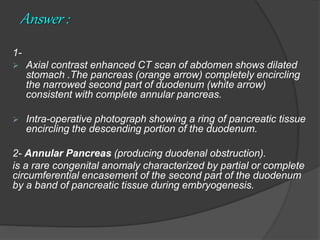
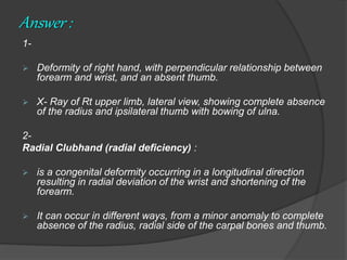
Dr Karrar Adil
Team E
Question 1
a 22-year-old male presented with a history of recurrent attacks of epigastric
pain, nausea and vomiting.
The symptoms had been present for more than 2 years, but had become more
frequent in the last few months.
1- Describe the images.
2- What is the diagnosis?
3- What is the pathogenesis?
4- Mention the associated anomalies.
5- How to treat this patient?
Answer :
1-
 Axial contrast enhanced CT scan of abdomen shows dilated
stomach .The pancreas (orange arrow) completely encircling
the narrowed second part of duodenum (white arrow)
consistent with complete annular pancreas.
 Intra-operative photograph showing a ring of pancreatic tissue
encircling the descending portion of the duodenum.
2- Annular Pancreas (producing duodenal obstruction).
is a rare congenital anomaly characterized by partial or complete
circumferential encasement of the second part of the duodenum
by a band of pancreatic tissue during embryogenesis.
3- Pathogenesis :
The pancreas is normally formed from the fusion of the dorsal and ventral
pancreatic buds between the first 4–8 weeks of embryonic life.
Annular pancreas results due to failure of the ventral bud to rotate.
instead, it elongates to encircle the upper part of the duodenum. This
failure can be explained by any of the following:
• Adherence of the ventral bud to the duodenal wall prior to rotation.
• Persistence and enlargement of the left ventral bud .
• Fusion of the ventral and dorsal buds before rotation.
4- Associated congenital anomalies :
 In infants: Down syndrome, tracheoesophageal fistula, esophageal
atresia, duodenal atresia, renal anomalies, anorectal malformations,
and congenital heart disease.
 in adults: pancreatic divisum, malrotation, duodenal webs, and
Schatzki ring.
.
5- Treatment :
 The treatment of symptomatic, obstructing
annular pancreas has classically been surgical.
 The preferred treatment is a bypass operation
such as gastro- or duodenojejunostomy.
 resection or division of the annulus results in
complications of duodenal leak, pancreatitis, and
pancreatic fistula, which is why it is safer to
bypass the annular constriction.
Question 2
1- Describe the images.
2- What is the diagnosis?
3- What are the associated syndromes?
4- What is the classification of this anomaly?
Answer :
1-
 Deformity of right hand, with perpendicular relationship between
forearm and wrist, and an absent thumb.
 X- Ray of Rt upper limb, lateral view, showing complete absence
of the radius and ipsilateral thumb with bowing of ulna.
2-
Radial Clubhand (radial deficiency) :
 is a congenital deformity occurring in a longitudinal direction
resulting in radial deviation of the wrist and shortening of the
forearm.
 It can occur in different ways, from a minor anomaly to complete
absence of the radius, radial side of the carpal bones and thumb.
3-
Radial Clubhand is associated with :
 TAR (Thrombocytopenia-absent radius) syndrome.
 Fanconi's anemia.
 Holt-Oram syndrome.
 VACTERL Syndrome:
(vertebral anomalies, anal atresia, cardiac abnormalities,
tracheoesophageal fistula, renal agenesis, and limb defects).
 VATER Syndrome:
vertebral anomalies, anal atresia, tracheoesophageal fistula,
esophageal atresia, renal agenesis).
4- Bayne and Klug Classification :
Type I :
Deficient distal radial epiphysis.
Type II :
Deficient distal and proximal radial epiphyses.
Type III :
Present proximally (partial aplasia).
Type IV :
Completely absent (total aplasia - most common).
Question 3
This is a photograph of right axillary lymph node dissection
performed through mastectomy wound.
1- Name the structures marked by colored arrows.
2- What are the indications of axillary L.N. dissection?
3- What structure is most vulnerable to iatrogenic injury?
4- Mention the other complications of this procedure.
Answer :
1-
• pectoralis major muscle.
• pectoralis minor muscle.
• axillary vein.
• intercostobrachial nerve.
• long thoracic nerve.
• thoracodorsal pedicle.
2- Indications :
 Clinically positive axillary lymph nodes that have been proven
with biopsy in a patient that is not planning on undergoing
neoadjuvant chemotherapy.
 Patients with inflammatory breast cancer.
 Patients with three or more axillary sentinel nodes that are
positive for cancer.
 Persistently positive lymph nodes after neoadjuvant therapy.
 Axillas with failed radiotracer and/or lymphazurin/methylene
blue during sentinel lymph node biopsy.
 Axillary lymph node recurrence after breast cancer treatment.
3- The most likely injured structure is the intercostobrachial nerve. When
this is transected, it causes paresthesias to the medial upper arm, axilla,
and occasionally lateral chest wall.
4- Complications :
 Infection.
 Seroma or hematoma.
 temporarily decreased range of motion of the shoulder.
 Lymphedema
 axillary vein thrombosis.
 Vascular injury.
 nerves injury :
• intercostobrachial nerve.
• long thoracic nerve injury (leads to paralysis of serratus anterior)
causes winging of the scapula.
• Damage to the thoracodorsal nerve leads to paresis or paralysis of
latissimus dorsi.
Question 4
A 19-year-old male presented to the emergency department with a
5-day history of fever, sore throat, inability to talk, odynophagia,
and trismus.
On examination, the patient appeared ill, pale, dehydrated, and
anxious.
1- What is the diagnosis?
2- What are the causative agents?
3- What are the potential complications?
4- How to treat this patient ?
Answer :
1- Bilateral peritonsillar abscess (quinsy) :
is defined as accumulation of pus inside the peritonsillar
space (between the palatine tonsillar capsule medially and
the superior pharyngeal constrictor muscle laterally).
2-
 Streptococcus pyogenes is the most common aerobic
organism associated with peritonsillar abscess followed
by streptococcus viridans.
 The most common anaerobic species include
fusobacterium necrophorum,prevotella species, and
bacteroides
3- Complications :
 Parapharyngeal / Retropharyngeal abscess.
 Laryngeal edema leading to airway compromise.
 Aspiration pneumonitis.
 Lung abscess (following rupture).
 Life-threatening hemorrhage (following erosion or
septic necrosis into the carotid sheath of the neck).
 Meningitis.
 Sepsis.
 Dehydration.
4- Treatment :
 Intravenous fluids, as the patient is usually dehydrated.
 Intravenous antibiotic (ampicillin/amoxicillin with
metronidazole).
 Analgesics and antipyretics.
 Intravenous dexamethasone (single dose).
 Drainage is a must along with medical management. options
include:
• needle aspiration.
• incision and drainage.
• quinsy tonsillectomy (for recurring abscess).
Question 5
1- Name the procedure.
2- Mention the landmark and the structures targeted by this procedure.
3- What are the preferred agents to use?
4- What are the indications and contraindications of this procedure?
Answer :
1- Transversus Abdominis Plane (TAP)
block :
The TAP is a potential anatomical space that
lies between transversus abdominis and
internal oblique muscles, where local
anesthetic can be deposited, creating a non-
dermatomal “field block”.
2-
 The T6–L1 segmental nerves enter the triangle of Petit just
medial to the anterior axillary line. Injection of local
anaesthetic into the TAP allows a block of all these nerves.
 These include the intercostal nerves (T7-T11), the subcostal
nerve (T12), the iliohypogastric and ilioinguinal nerves (L1).
3- Anesthetic agent: ropivacaine or bupivacaine without epinephrine.
4-
 Common indications for TAP blocks include any major or lower
abdominal surgery (appendectomy, hernia repair, caesarean
section, hysterectomy and prostatectomy).
 Bilateral blocks can be given for midline or transverse incisions.
Contraindications to TAP blocks include the following:
 Patient refusal.
 Active infection over the site of injection.
 Those with known allergies.
 Practice caution in patients taking anticoagulation, pregnant patient,
and in patients where anatomical landmarks are indistinguishable.
Question 6
A young female with history of
school drop-out due to mental
issues, presented with the
complaint of abdominal pain,
vomiting, and a non-tender
abdominal mass.
1- Describe the image.
2- What is the diagnosis?
3- What is the etiology?
4- What is the treatment?
Answer :
1-
Contrast enhanced CT Scan of abdomen (coronal
view) showing well-circumscribed large
heterogenous intragastric mass, seen completely
separable from the gastric wall and extending to
the duodenum.
2-
Trichobezoar “Rapunzel syndrome” :
 is a rare condition consisting of a hair ball
in the proximal gastrointestinal tract,
which may cause obstruction.
 It is almost exclusively affects young
women.
 When parts of the tail of trichobezoar
break off through the pylorus and migrate
to the small intestine, this condition is
called Rapunzel syndrome.
3-
 Most patients with trichobezoars suffer from associated underlying
compulsive psychiatric disorders including trichotillomania (pulling out
of their own hair), trichophagia (eating of hair), and pica.
 Hair strands are resistant to digestion. they escape peristalsis
because of their slippery surface, and are retained in the folds of the
gastric mucosa.
 When more hair accumulates, peristalsis causes it to be enmeshed
into a ball which gets too large to leave the stomach, becomes even
more matted together and assumes the shape of the stomach,
usually as a single solid mass.
4- Treatment :
 Surgical removal (accomplished by gastrotomy).
 Endoscopic or laparoscopic therapy are unlikely to be useful except
for small sized bezoars.
 Parental counseling and psychological/psychiatric follow-up.
Thank you…

Problem solving : a quiz in general surgery .pptx

  • 1.
  • 2.
  • 3.
    a 22-year-old malepresented with a history of recurrent attacks of epigastric pain, nausea and vomiting. The symptoms had been present for more than 2 years, but had become more frequent in the last few months. 1- Describe the images. 2- What is the diagnosis? 3- What is the pathogenesis? 4- Mention the associated anomalies. 5- How to treat this patient?
  • 4.
    Answer : 1-  Axialcontrast enhanced CT scan of abdomen shows dilated stomach .The pancreas (orange arrow) completely encircling the narrowed second part of duodenum (white arrow) consistent with complete annular pancreas.  Intra-operative photograph showing a ring of pancreatic tissue encircling the descending portion of the duodenum. 2- Annular Pancreas (producing duodenal obstruction). is a rare congenital anomaly characterized by partial or complete circumferential encasement of the second part of the duodenum by a band of pancreatic tissue during embryogenesis.
  • 5.
    3- Pathogenesis : Thepancreas is normally formed from the fusion of the dorsal and ventral pancreatic buds between the first 4–8 weeks of embryonic life. Annular pancreas results due to failure of the ventral bud to rotate. instead, it elongates to encircle the upper part of the duodenum. This failure can be explained by any of the following: • Adherence of the ventral bud to the duodenal wall prior to rotation. • Persistence and enlargement of the left ventral bud . • Fusion of the ventral and dorsal buds before rotation. 4- Associated congenital anomalies :  In infants: Down syndrome, tracheoesophageal fistula, esophageal atresia, duodenal atresia, renal anomalies, anorectal malformations, and congenital heart disease.  in adults: pancreatic divisum, malrotation, duodenal webs, and Schatzki ring. .
  • 6.
    5- Treatment : The treatment of symptomatic, obstructing annular pancreas has classically been surgical.  The preferred treatment is a bypass operation such as gastro- or duodenojejunostomy.  resection or division of the annulus results in complications of duodenal leak, pancreatitis, and pancreatic fistula, which is why it is safer to bypass the annular constriction.
  • 7.
  • 8.
    1- Describe theimages. 2- What is the diagnosis? 3- What are the associated syndromes? 4- What is the classification of this anomaly?
  • 9.
    Answer : 1-  Deformityof right hand, with perpendicular relationship between forearm and wrist, and an absent thumb.  X- Ray of Rt upper limb, lateral view, showing complete absence of the radius and ipsilateral thumb with bowing of ulna. 2- Radial Clubhand (radial deficiency) :  is a congenital deformity occurring in a longitudinal direction resulting in radial deviation of the wrist and shortening of the forearm.  It can occur in different ways, from a minor anomaly to complete absence of the radius, radial side of the carpal bones and thumb.
  • 10.
    3- Radial Clubhand isassociated with :  TAR (Thrombocytopenia-absent radius) syndrome.  Fanconi's anemia.  Holt-Oram syndrome.  VACTERL Syndrome: (vertebral anomalies, anal atresia, cardiac abnormalities, tracheoesophageal fistula, renal agenesis, and limb defects).  VATER Syndrome: vertebral anomalies, anal atresia, tracheoesophageal fistula, esophageal atresia, renal agenesis).
  • 11.
    4- Bayne andKlug Classification : Type I : Deficient distal radial epiphysis. Type II : Deficient distal and proximal radial epiphyses. Type III : Present proximally (partial aplasia). Type IV : Completely absent (total aplasia - most common).
  • 12.
  • 13.
    This is aphotograph of right axillary lymph node dissection performed through mastectomy wound. 1- Name the structures marked by colored arrows. 2- What are the indications of axillary L.N. dissection? 3- What structure is most vulnerable to iatrogenic injury? 4- Mention the other complications of this procedure.
  • 14.
    Answer : 1- • pectoralismajor muscle. • pectoralis minor muscle. • axillary vein. • intercostobrachial nerve. • long thoracic nerve. • thoracodorsal pedicle.
  • 15.
    2- Indications : Clinically positive axillary lymph nodes that have been proven with biopsy in a patient that is not planning on undergoing neoadjuvant chemotherapy.  Patients with inflammatory breast cancer.  Patients with three or more axillary sentinel nodes that are positive for cancer.  Persistently positive lymph nodes after neoadjuvant therapy.  Axillas with failed radiotracer and/or lymphazurin/methylene blue during sentinel lymph node biopsy.  Axillary lymph node recurrence after breast cancer treatment.
  • 16.
    3- The mostlikely injured structure is the intercostobrachial nerve. When this is transected, it causes paresthesias to the medial upper arm, axilla, and occasionally lateral chest wall. 4- Complications :  Infection.  Seroma or hematoma.  temporarily decreased range of motion of the shoulder.  Lymphedema  axillary vein thrombosis.  Vascular injury.  nerves injury : • intercostobrachial nerve. • long thoracic nerve injury (leads to paralysis of serratus anterior) causes winging of the scapula. • Damage to the thoracodorsal nerve leads to paresis or paralysis of latissimus dorsi.
  • 17.
  • 18.
    A 19-year-old malepresented to the emergency department with a 5-day history of fever, sore throat, inability to talk, odynophagia, and trismus. On examination, the patient appeared ill, pale, dehydrated, and anxious. 1- What is the diagnosis? 2- What are the causative agents? 3- What are the potential complications? 4- How to treat this patient ?
  • 19.
    Answer : 1- Bilateralperitonsillar abscess (quinsy) : is defined as accumulation of pus inside the peritonsillar space (between the palatine tonsillar capsule medially and the superior pharyngeal constrictor muscle laterally). 2-  Streptococcus pyogenes is the most common aerobic organism associated with peritonsillar abscess followed by streptococcus viridans.  The most common anaerobic species include fusobacterium necrophorum,prevotella species, and bacteroides
  • 20.
    3- Complications : Parapharyngeal / Retropharyngeal abscess.  Laryngeal edema leading to airway compromise.  Aspiration pneumonitis.  Lung abscess (following rupture).  Life-threatening hemorrhage (following erosion or septic necrosis into the carotid sheath of the neck).  Meningitis.  Sepsis.  Dehydration.
  • 21.
    4- Treatment : Intravenous fluids, as the patient is usually dehydrated.  Intravenous antibiotic (ampicillin/amoxicillin with metronidazole).  Analgesics and antipyretics.  Intravenous dexamethasone (single dose).  Drainage is a must along with medical management. options include: • needle aspiration. • incision and drainage. • quinsy tonsillectomy (for recurring abscess).
  • 22.
  • 23.
    1- Name theprocedure. 2- Mention the landmark and the structures targeted by this procedure. 3- What are the preferred agents to use? 4- What are the indications and contraindications of this procedure?
  • 24.
    Answer : 1- TransversusAbdominis Plane (TAP) block : The TAP is a potential anatomical space that lies between transversus abdominis and internal oblique muscles, where local anesthetic can be deposited, creating a non- dermatomal “field block”.
  • 25.
    2-  The T6–L1segmental nerves enter the triangle of Petit just medial to the anterior axillary line. Injection of local anaesthetic into the TAP allows a block of all these nerves.  These include the intercostal nerves (T7-T11), the subcostal nerve (T12), the iliohypogastric and ilioinguinal nerves (L1).
  • 26.
    3- Anesthetic agent:ropivacaine or bupivacaine without epinephrine. 4-  Common indications for TAP blocks include any major or lower abdominal surgery (appendectomy, hernia repair, caesarean section, hysterectomy and prostatectomy).  Bilateral blocks can be given for midline or transverse incisions. Contraindications to TAP blocks include the following:  Patient refusal.  Active infection over the site of injection.  Those with known allergies.  Practice caution in patients taking anticoagulation, pregnant patient, and in patients where anatomical landmarks are indistinguishable.
  • 27.
  • 28.
    A young femalewith history of school drop-out due to mental issues, presented with the complaint of abdominal pain, vomiting, and a non-tender abdominal mass. 1- Describe the image. 2- What is the diagnosis? 3- What is the etiology? 4- What is the treatment?
  • 29.
    Answer : 1- Contrast enhancedCT Scan of abdomen (coronal view) showing well-circumscribed large heterogenous intragastric mass, seen completely separable from the gastric wall and extending to the duodenum.
  • 30.
    2- Trichobezoar “Rapunzel syndrome”:  is a rare condition consisting of a hair ball in the proximal gastrointestinal tract, which may cause obstruction.  It is almost exclusively affects young women.  When parts of the tail of trichobezoar break off through the pylorus and migrate to the small intestine, this condition is called Rapunzel syndrome.
  • 31.
    3-  Most patientswith trichobezoars suffer from associated underlying compulsive psychiatric disorders including trichotillomania (pulling out of their own hair), trichophagia (eating of hair), and pica.  Hair strands are resistant to digestion. they escape peristalsis because of their slippery surface, and are retained in the folds of the gastric mucosa.  When more hair accumulates, peristalsis causes it to be enmeshed into a ball which gets too large to leave the stomach, becomes even more matted together and assumes the shape of the stomach, usually as a single solid mass. 4- Treatment :  Surgical removal (accomplished by gastrotomy).  Endoscopic or laparoscopic therapy are unlikely to be useful except for small sized bezoars.  Parental counseling and psychological/psychiatric follow-up.
  • 32.