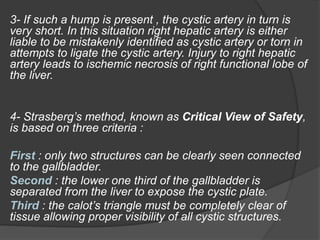
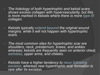
Dr Karrar Adil
Team E
Question 1
This is an intra operative image of laparoscopic
cholecystectomy.
1- Identify the structures (A,B,C,D,and E).
2- Describe the anomaly . What is it called ?
3- Why is it important to recognize this anomaly?
4- What are the critical view of safety principles?
Answer :
1-
A: Infundibulum of gallbladder.
B: cystic duct.
C: common bile duct.
D: RHA.
E: Cystic Artery and proximal clip ligation.
2- long tortuous right hepatic artery in front of the
origin of the cystic duct is called as Moynihan's or
caterpillar hump. It is important cause of bleeding
in cholecystectomy.
3- If such a hump is present , the cystic artery in turn is
very short. In this situation right hepatic artery is either
liable to be mistakenly identified as cystic artery or torn in
attempts to ligate the cystic artery. Injury to right hepatic
artery leads to ischemic necrosis of right functional lobe of
the liver.
4- Strasberg’s method, known as Critical View of Safety,
is based on three criteria :
First : only two structures can be clearly seen connected
to the gallbladder.
Second : the lower one third of the gallbladder is
separated from the liver to expose the cystic plate.
Third : the calot’s triangle must be completely clear of
tissue allowing proper visibility of all cystic structures.
Question 2
A 26-year-old dark skinned
female complains of swelling
on right ear lobe since 3 years
when she had got her ear
pierced.
1- What is the diagnosis ?
2- How to differentiate it from
other closely related
conditions?
3- How to treat this condition?
Answer :
1- Keloid scar.
2-
• Hypertrophic scar usually show a rapid growth
phase for up to 6 months, and then gradually
regresses over a period of a few years.
but keloid typically persist for long periods of time,
and do not regress spontaneously.
• the incidence of keloid scar formation is much
higher in black-skinned individuals than in whites.
More common in females.
• The histology of both hypertrophic and keloid scars
shows excess collagen with hypervascularity, but this
is more marked in keloids where there is more type III
collagen.
• Keloids typically extend beyond the original wound
margins. while it will not happen with hypertrophic
scars.
• The most common sites for hypertrophic scar are
shoulders, neck, presternum, knees, and ankles
whereas; keloids are frequently seen on anterior chest,
earlobes, upper arms, and cheeks.
• Keloids have a higher tendency to recur following
excision, whereas new hypertrophic scar formation is
rare after its excision.
3- Treatment :
Intra-marginal surgical excision of keloid tissue is
recommended to prevent the stimulation of additional
collagen synthesis.
Other most commonly used treatment modalities
include :
 Intra-lesional steroid injection (triamcinolone).
 Cryotherapy.
 Laser removal.
 Radiotherapy.
 Silicon gel sheeting.
Question 3
6 year old child presented with
Lt upper arm pain , swelling ,
and limitation of movement
following a minor fall.
X-ray was done.
1- What are the x-ray
findings?
2- What is the diagnosis?
3- What are the most common
sites for this pathology?
4- Mention the treatment
options.
Answer :
1-
 lucent lytic lesion at the upper humeral metaphysis
and shaft with symmetrical thinning of cortices.
 pathological fracture and a displaced bone chip in
its cavity giving fallen leaf sign.
2- Unicameral Bone Cyst (also called simple bone
cyst).
Is a non-neoplastic, serous fluid-filled bone lesion
typically presents in patients < 20 years of age with a
pathological fracture through the lesion.
Fallen leaf sign
3- location :
Most common site is the proximal humerus (60%).
other locations including proximal femur, distal tibia, calcaneus,
and occasionally metacarpals, phalanges, and distal radius
4- Treatment :
• Non-operative :
 Immobilization.
 methylprednisolone acetate injection.
• Operative :
 curettage and bone grafting +/- internal fixation based on
tumor location.
Question 4
A 40-year old farmer experienced right upper
abdominal pain for one week, radiating to the right
shoulder, associated with fever of (39 C), chills, and
malaise.
He had a history of diarrhea 3 months ago.
Abdominal CT scan was done.
1- Describe the image.
2- What is the diagnosis ?
3- Mention the risk factors of this condition.
4- What are the treatment options?
Answer :
1- Contrast enhanced abdominal CT scan
(Axial view) showing round hypodense
lesion in the Rt lobe of the liver displaying
double-target sign consisting of an inner
enhancing ring (white arrowhead) and
outer hypodense ring (black arrowhead).
2- Amoebic liver abscess.
3- Risk factors associated with amebic liver abscess:
 Immigrants from endemic areas. or short-term
travelers to endemic countries.
 Institutionalized persons.
 Crowding and poor hygiene.
 Homosexuality.
 Presence of immunosuppression :
(ex: HIV infection, malnutrition with hypoalbuminemia,
alcohol abuse, chronic infections, steroid use,
malignancy, extremes of age, and pregnancy).
4-
• Medical treatment :
 Metronidazole (Tinidazole is an alternative).
 luminal agent : is used for removing any
intraluminal cysts.
Paromomycin, diiodohydroxyquin, or diloxanide
furoate.
• Percutaneous drainage of abscess :
Indications :
 high risk of rupture (size >5 cm).
 failure to observe a clinical response to therapy within
5-7 days.
 abscess in pregnancy where drug therapy cannot be
used.
 Pyogenic superinfection.
 Can’t differentiate from a pyogenic liver abscess.
• Surgical treatment :
(open surgical approach or laparoscopic surgery)
Indications :
 for patients whom medical and percutaneous
treatment failed.
 when there are large, multilocular or dense content
abscesses.
 Left lobe abscess.
 imminence of rupture or development of serious
complications such as peritonitis.
Question 5
This is an image of a 2-week-old
female seen in the Emergency
Department.
she was born at 34 weeks
gestational age due to maternal
pre-eclampsia.
1- What is the diagnosis?
2- What are the predisposing
factors for this condition?
3- Which micro organisms cause
this condition?
4- What are the main
complications?
Answer :
1- Omphalitis.
2- predisposing factors for omphalitis include :
 Prematurity.
 Complicated delivery.
 Poor hygienic practices during the neonatal
period.
 maternal infection.
 umbilical catheterization.
 home birth.
3- causative micro organisms :
 S. aureus.
 Streptococcus pyogenes.
 Gram-negative organisms (i.e., E. coli, Klebsiella
species).
 anaerobes (Bacteroides fragilis and Clostridium).
4- main complications :
 Suppurative thrombophlebitis of the umbilical or portal
veins (resulting in portal vein thrombosis).
 Abscess.
 Abdominal wall necrotizing fasciitis.
 Peritonitis.
 Intestinal gangrene.
 Pyourachus (infection of the urachal remnant).
Question 6
1- Name the devise in the image below.
2- What do the digital screen readings stand for ?
3- What is the preferred gas for establishing
pneumoperitoneum? Why ? Mention other gases than can
be used.
Answer :
1- Laparoscopic Insufflator.
2- Quadro manometric Indicators:
are the four important readings of the insufflator.
These are :
 Preset Insufflation pressure.
 Actual Pressure.
 Gas flow rate (liter/minute).
 Volume of gas consumed.
3-
 Carbon dioxide (CO2) is the most commonly
used gas for insufflation during laparoscopic
surgery because it is colorless, inexpensive,
non-flammable, and has higher blood solubility
than air, which reduces the risk of
complications.
 Other used gases include Nitrous Oxide,
Helium and argon.
Thank you …

Problem solving : a quiz in general surgerypptx

  • 1.
  • 2.
  • 3.
    This is anintra operative image of laparoscopic cholecystectomy. 1- Identify the structures (A,B,C,D,and E). 2- Describe the anomaly . What is it called ? 3- Why is it important to recognize this anomaly? 4- What are the critical view of safety principles?
  • 4.
    Answer : 1- A: Infundibulumof gallbladder. B: cystic duct. C: common bile duct. D: RHA. E: Cystic Artery and proximal clip ligation. 2- long tortuous right hepatic artery in front of the origin of the cystic duct is called as Moynihan's or caterpillar hump. It is important cause of bleeding in cholecystectomy.
  • 5.
    3- If sucha hump is present , the cystic artery in turn is very short. In this situation right hepatic artery is either liable to be mistakenly identified as cystic artery or torn in attempts to ligate the cystic artery. Injury to right hepatic artery leads to ischemic necrosis of right functional lobe of the liver. 4- Strasberg’s method, known as Critical View of Safety, is based on three criteria : First : only two structures can be clearly seen connected to the gallbladder. Second : the lower one third of the gallbladder is separated from the liver to expose the cystic plate. Third : the calot’s triangle must be completely clear of tissue allowing proper visibility of all cystic structures.
  • 6.
  • 7.
    A 26-year-old darkskinned female complains of swelling on right ear lobe since 3 years when she had got her ear pierced. 1- What is the diagnosis ? 2- How to differentiate it from other closely related conditions? 3- How to treat this condition?
  • 8.
    Answer : 1- Keloidscar. 2- • Hypertrophic scar usually show a rapid growth phase for up to 6 months, and then gradually regresses over a period of a few years. but keloid typically persist for long periods of time, and do not regress spontaneously. • the incidence of keloid scar formation is much higher in black-skinned individuals than in whites. More common in females.
  • 9.
    • The histologyof both hypertrophic and keloid scars shows excess collagen with hypervascularity, but this is more marked in keloids where there is more type III collagen. • Keloids typically extend beyond the original wound margins. while it will not happen with hypertrophic scars. • The most common sites for hypertrophic scar are shoulders, neck, presternum, knees, and ankles whereas; keloids are frequently seen on anterior chest, earlobes, upper arms, and cheeks. • Keloids have a higher tendency to recur following excision, whereas new hypertrophic scar formation is rare after its excision.
  • 10.
    3- Treatment : Intra-marginalsurgical excision of keloid tissue is recommended to prevent the stimulation of additional collagen synthesis. Other most commonly used treatment modalities include :  Intra-lesional steroid injection (triamcinolone).  Cryotherapy.  Laser removal.  Radiotherapy.  Silicon gel sheeting.
  • 11.
  • 12.
    6 year oldchild presented with Lt upper arm pain , swelling , and limitation of movement following a minor fall. X-ray was done. 1- What are the x-ray findings? 2- What is the diagnosis? 3- What are the most common sites for this pathology? 4- Mention the treatment options.
  • 13.
    Answer : 1-  lucentlytic lesion at the upper humeral metaphysis and shaft with symmetrical thinning of cortices.  pathological fracture and a displaced bone chip in its cavity giving fallen leaf sign. 2- Unicameral Bone Cyst (also called simple bone cyst). Is a non-neoplastic, serous fluid-filled bone lesion typically presents in patients < 20 years of age with a pathological fracture through the lesion.
  • 14.
  • 15.
    3- location : Mostcommon site is the proximal humerus (60%). other locations including proximal femur, distal tibia, calcaneus, and occasionally metacarpals, phalanges, and distal radius 4- Treatment : • Non-operative :  Immobilization.  methylprednisolone acetate injection. • Operative :  curettage and bone grafting +/- internal fixation based on tumor location.
  • 16.
  • 17.
    A 40-year oldfarmer experienced right upper abdominal pain for one week, radiating to the right shoulder, associated with fever of (39 C), chills, and malaise. He had a history of diarrhea 3 months ago. Abdominal CT scan was done.
  • 18.
    1- Describe theimage. 2- What is the diagnosis ? 3- Mention the risk factors of this condition. 4- What are the treatment options?
  • 19.
    Answer : 1- Contrastenhanced abdominal CT scan (Axial view) showing round hypodense lesion in the Rt lobe of the liver displaying double-target sign consisting of an inner enhancing ring (white arrowhead) and outer hypodense ring (black arrowhead). 2- Amoebic liver abscess.
  • 20.
    3- Risk factorsassociated with amebic liver abscess:  Immigrants from endemic areas. or short-term travelers to endemic countries.  Institutionalized persons.  Crowding and poor hygiene.  Homosexuality.  Presence of immunosuppression : (ex: HIV infection, malnutrition with hypoalbuminemia, alcohol abuse, chronic infections, steroid use, malignancy, extremes of age, and pregnancy).
  • 21.
    4- • Medical treatment:  Metronidazole (Tinidazole is an alternative).  luminal agent : is used for removing any intraluminal cysts. Paromomycin, diiodohydroxyquin, or diloxanide furoate.
  • 22.
    • Percutaneous drainageof abscess : Indications :  high risk of rupture (size >5 cm).  failure to observe a clinical response to therapy within 5-7 days.  abscess in pregnancy where drug therapy cannot be used.  Pyogenic superinfection.  Can’t differentiate from a pyogenic liver abscess.
  • 23.
    • Surgical treatment: (open surgical approach or laparoscopic surgery) Indications :  for patients whom medical and percutaneous treatment failed.  when there are large, multilocular or dense content abscesses.  Left lobe abscess.  imminence of rupture or development of serious complications such as peritonitis.
  • 24.
  • 25.
    This is animage of a 2-week-old female seen in the Emergency Department. she was born at 34 weeks gestational age due to maternal pre-eclampsia. 1- What is the diagnosis? 2- What are the predisposing factors for this condition? 3- Which micro organisms cause this condition? 4- What are the main complications?
  • 26.
    Answer : 1- Omphalitis. 2-predisposing factors for omphalitis include :  Prematurity.  Complicated delivery.  Poor hygienic practices during the neonatal period.  maternal infection.  umbilical catheterization.  home birth.
  • 27.
    3- causative microorganisms :  S. aureus.  Streptococcus pyogenes.  Gram-negative organisms (i.e., E. coli, Klebsiella species).  anaerobes (Bacteroides fragilis and Clostridium). 4- main complications :  Suppurative thrombophlebitis of the umbilical or portal veins (resulting in portal vein thrombosis).  Abscess.  Abdominal wall necrotizing fasciitis.  Peritonitis.  Intestinal gangrene.  Pyourachus (infection of the urachal remnant).
  • 28.
  • 29.
    1- Name thedevise in the image below. 2- What do the digital screen readings stand for ? 3- What is the preferred gas for establishing pneumoperitoneum? Why ? Mention other gases than can be used.
  • 30.
    Answer : 1- LaparoscopicInsufflator. 2- Quadro manometric Indicators: are the four important readings of the insufflator. These are :  Preset Insufflation pressure.  Actual Pressure.  Gas flow rate (liter/minute).  Volume of gas consumed.
  • 31.
    3-  Carbon dioxide(CO2) is the most commonly used gas for insufflation during laparoscopic surgery because it is colorless, inexpensive, non-flammable, and has higher blood solubility than air, which reduces the risk of complications.  Other used gases include Nitrous Oxide, Helium and argon.
  • 32.