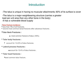
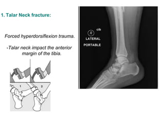
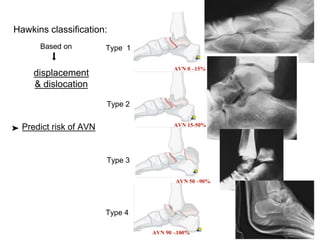
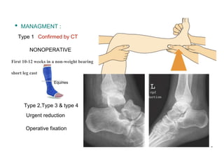
3. -Talus fracture incidence :
•less than 1% of all fractures.
• second most common tarsal fractures after calcaneus fractures .
•Talar Neck Fractures :
■ most common fracture of talus ( 50%).
• Talar body fractures :
▪ account for 13-23% of talus fractures.
• Lateral process fractures :
■account for 10.4% of talus fractures .
• Talar head fractures :
▪least common talus fracture.
Introduction
-The talus is a major weightbearing structure (carries a greater
load per unit area than any other bone in the body)
-It has a vulnerable blood supply
-The talus is unique in having no muscular attachments; 60% of its surface is covere
6. Vascularity
1. posterior tibial artery
• artery of tarsal canal : ➠
most of talar body .
• calcaneal braches:
posterior talus
• deltoid branch: ➠
body
2. anterior tibial artery
head and neck.
3. perforating peroneal arteries
artery of tarsal sinus
head and neck.
The
most
IMP
Remain
after
neck fx
7. PRESENTATION
occur in a young, active, and
mobile population
high velocity injury
Clinically
History
✔︎ Intense pain
✔︎ unable to do Wight bearing
✔︎ swelling
✔︎ echymosis
✔︎ Close deformity
✔︎ Open fracture
🔎
Fall from hight
➚
➘ MVA
9. Imaging
• X ray:
AP , LAT , oblique (ankle,foot)
Canale view( NECK)
• CT:
Congruency “ tibiotalar,
subtalar and talonavicular .
Superior details on: -Comminution
-Displacements
-Process fractures
-Associated foot
injuries .
allows for a view of the talar neck unimpeded by the calcaneus
10.
11. 1. Talar Neck fracture:
Forced hyperdorsiflexion trauma.
-Talar neck impact the anterior
margin of the tibia.
12. Type 1
Type 2
Type 3
Type 4
Hawkins classification:
displacement
& dislocation
Predict risk of AVN
AVN 0 –15%
AVN 15-50%
AVN 90 –100%
AVN 50 –90%
Based on
⬇︎
➤
13. TREATMENT
- Treatment is determined by
the Hawkins type..
Goals of treatment:
1. Early anatomic reduction of the neck fracture
2. Reduction of dislocated joints
3. minimize soft tissue compromise
4. proper rotation,length,and angulation of the neck.
5. Stable fixation
6. Avoidance of complications
14. • MANAGMENT :
NONOPERATIVE
First 10-12 weeks in a non-weight bearing
short leg cast
Type 1
Type 2,Type 3 & type 4
Urgent reduction
Operative fixation
Confirmed by CT
Equines
15. - anteromedial and anterolateral to allow accurate visualization, anatomic reduction,
Dual approach is the recommended.
Operative
16. Anteromedial approach
-anterior aspect of the medial malleolus to the dorsal aspect of the navicular
tuberosity.
-running between the anterior tibial and posterior tibial tendons
-Saphenous nerve and vein are superficial hazard.
-The dissection is carried down to the bone, just dorsal to the posterior tibial
tendon.
-Disruption of the deltoid ligament should be avoided
-Dissection of soft tissues at the level of talar neck dorsally and plantarward
should be avoided such that no further damage to the blood supply occurs
medial malleolus osteotomy can be made for
greater visualization proximally to include
the talar body.
17. Anterolateral
Superficial peroneal nerve
Protected
lateral talar neck
-The anterolateral incision is made from the anterior aspect
of the lateral malleolus toward the base of the fourth metatarsal.
- The extensor digitorum longus and peroneus tertius tendons are
retracted.
-The extensor digitorum brevis muscle is retracted dorsally.
18. Technique
• Bone graft: Potentially useful for comminuted fractures
• Fixation: 2- to 3.5-mm screws
• fully threaded titanium small fragment screws may be used.
• Since medial comminution is frequently present, lag screw fixation
is often not used. as it may redisplace the fracture into varus due to
compression of this compromised bone.
• A minimum of two screws should be placed across the fracture site.
• A hard cortical ridge of bone is usually present along the
dorsal aspect of the sinus tarsi that allows for excellent
fixation with one or two screws inserted from the lateral neck
ofthe talus across the fracture site
• In significantly comminuted fractures, a contoured plate can be
placed along the sinus tarsi with transverse screws across the neck
of the talus and anterior to posterior screws into the body of the talus
20. 2.Talar body fractures
Axial load trauma
squeezed between the tibia plafond and
Posterior facet of calcaneus.
MVA
FALL FROM HIGHT
RADIOGRAPHIC –LATERAL XRAY VIEWS
CT - congruency
-fracture geometry
✷ These fractures can occur in any plane and have a
much poorer prognosis than talar neck fractures .
◉AVN 50 %
21. ☛ Based on severity
Müller AO/OTA Classification
• Ankle joint involvement (C1)
osteochondral injury
• Subtalar joint involvement (C2)
coronal split through the body
• Ankle and subtalar joint involvement (C3)
Comminuted
23. • Before making the osteotomy, two parallel drill holes are made in the medial malleolus for osteotomy
reduction and fixation at the end of the procedure .
• The anterior ankle capsule from the axilla of the ankle joint to the anterior aspect of the deltoid ligament is
released.
• The superficial and deep posterior tibial tendon sheath is released from the level of the osteotomy to
the posterior aspect of the medial malleolus.
• A retractor is placed to protect the posterior tibial tendon when performing the osteotomy.
• The periosteum is incised approximately 5 to 10 mm superior to the ankle joint, and a narrow,
saw bladed is used to cut transversely through the tibia to the level of the medial
axilla of the ankle joint
Predrilling of the medial malleolus
prior to performing the osteotomy.
24. Anteroposterior (left) and lateral (right) drawings of an ankle, showing the different me
osteotomy of the medial malleolus
25. The femoral distractor is applied for traction to facilitate difficult fracture reduction with a pin in
calcaneus and in tibia.
26. fixed with two partially threaded, 4.0-mm, cancellous, titanium screws
27. -internal fixation can be performed for fragments of bone
and cartilage large enough to stabilize.
-Depending upon the size of the fragment, cortical screws
ranging from 2 to 4 mm in diameter can be used.
-Care should be taken to avoid prominent hardware.
Countersunk or headless screws are advantageous.
Compression screw fixation may be used in
noncomminuted fractures.
- Alternative fixation devices include Herbert screws,
Kirschner wires, and threaded wires, all of which may be
useful depending upon the size of the fragments to be
stabilized.
- Small plates can also be used to span comminuted
segments medially or laterally in some cases a portion
of the plate can be countersunk to lessen the risk of
hardware impingement.
28. Type C3
Accurate replacement of
fragments is near impossible
Long term results- bad
IN SUCH CASES TIBIOTALAR ARTHRODESIS
IS PREFFERRED.
29. axially directed loading and compression of
talar head
• Fracture without displacement
short leg cast for 6 weeks
• Displaced fractures
TALONAVICULAR ARTHRITIS
longitudianal arch support
talonavicular arthrodesis
⬇︎
⬇︎
ORIF
3.Talus head fracture
30. • VON KNOCH described v sign
• V SiGN- it is the contour of
lateral process over lateral
view xrays
• V sign positive- any
disruption in contour of V
indicating fracture lateral
processs
Snowboarder’s fracture”
Axial loading, dorsiflexion ,
external rotation and eversion of
foot
4.FRACTURE OF LATERAL PROCESS
31. FRACTURE OF LATERAL PROCESS OF TALUS
Hawkins classification
CONSERVATIVE
Unless there is articular involvement
32. 5.POSTERIOR PROCESS FRACTURES
include the medial and lateral
tubercle fractures
- severe ankle
inversion injury where posterior
talofibular ligament avulses the
lateral tubercle
• Undisplaced
Cast for 4 weeks
• Displaced
primary excision of small fragments
ORIF when entire posterior process
is fractured
☛
☛
33. -Following surgery, patients are initially splinted with the foot in
a plantigrade position until the surgical incisions have healed.
-Once the skin has healed, the patient can begin gentle rangeof-
motion exercises with the use of a removable cast brace.
-Restricted weight bearing is usually continued for approximately
3 months following the injury.
-It is worthwhile to counsel patients about the risks of osteonecrosis
and delayed union or nonunion of the talus fracture.
-Revascularization can be seen on plain radiographs with
Hawkins sign.
-MRI may be used to determine the status of the perfusion of
the talus.
-Union can be difficult to judge using plain radiographs,
and CT scanning is often helpful to define when union
has been achieved.
Postoperative Care
34. Hawkins sign.
Note the atrophy in the
subchondral area of the talus, which suggests
vascularity. (This is a good prognostic sign
for viability of the talus.)
Hawkins sign.
-Its presence signifies that osteonecrosis will not occur; its absence does not indicate that osteonecrosis
will definitely occur.
6 to 8 weeks
35.
36. complication
• Delayed union,
• Nonunion—Frank nonunion is rare
• Malunion—Varus malunion
most common leads to degenerative arthritis of the subtalar joint Clinically,
patients present and stand on the lateral border of the foot.
• Posttraumatic arthritis—Posttraumatic
arthritis occurs at the subtalar joint
(50% of cases),
• Osteonecrosis
37. Update
reduction of displaced fractures was thought to reduce the risk of osteonecrosis. Recently,
a study has shown that approximately 60% of orthopedic traumatologists find it acceptable
to operate after 8 hours, and 46% find it acceptable to operate after 24 hours.
!
38.
39.
40. • Fractures of the talus are uncommon but often represent serious injury.
• The high-energy force required to produce a talar neck fracture can
cause severe associated soft-tissue damage, including damage to the
precarious blood supply.
• Anatomic reduction and stable internal fixation of a displaced talar neck
fracture may minimize the risk of complications, but posttraumatic
sequelae may be inevitable.
• The risk of osteonecrosis is almost completely determined by the
severity of injury.
• osteonecrosis may be decreased by accurate surgical reduction as well
as meticulous surgical dissection that avoids further vascular damage.
• Osteonecrosis and posttraumatic arthritis are challenging complications
to treat.
• Talar body fractures are associated with a high rate of complications.
• Lateral process fractures of the talus can easily be overlooked and may
lead to posttraumatic sequelae.
Summary
41. REFERENCES
• Rockwood and Green’s Fractures in Adults
• Grays Anatomy for students
• ORTHOBULLET .
• AO surgery.
• Orthopaedic Knowledge Update Trauma
• Talus Fractures: Evaluation and Treatment
Paul T. Fortin, MD, and Jeffrey E. Balazsy, MD