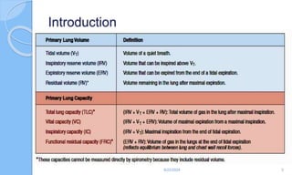
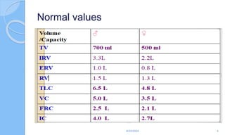
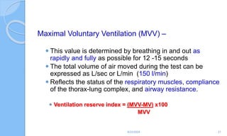
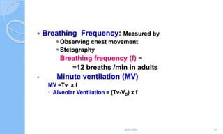
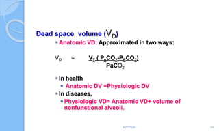
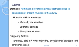
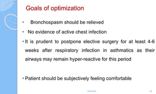
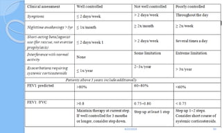
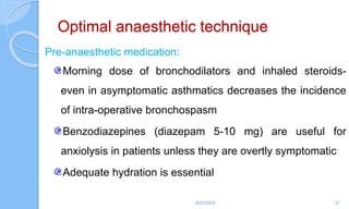
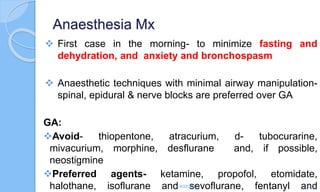
Asthma and COPD
6/22/2024 1
Objectives
 Define asthma and Explain the cause of asthma
 List symptoms and signs of asthma
 Describe the general considerations for pt with asthma
 Explain the preoperative evaluation and preparation
 Describe the perioperative medications used to treat asthma
 List drugs safe for asthmatics
 Describe anaesthesia management of asthmatic patients
 Describe management of severe bronchospasm outside OR
 Describe management of severe bronchospasm during anaesthesia
 Manage asthmatic patients during the perioperative period
 Describe the postoperative care of asthmatic patients
6/22/2024 2
Introduction
6/22/2024 3
Normal values
6/22/2024 4
 Functional resdual capacity (FRC)
◦ 40% of TLV
◦ Pulmonary variables are optimized at FRC because of
better
 Work of breathing
 Vascular resistance
 Ventilation perfusion ratio
6/22/2024 5
Factors affecting lung volumes &
capacitis
 FRC increase is caused by hyperinflation,
emphysema, asthma
  TLC is caused by restrictive pulmonary
disease, interstitial fibrosis
6/22/2024 6
Dray spirometer (vitalography)
Used to record the forced expiratory spirogram which
includes
FVC = Forced vital capacity
Total volume expired under maximal effort
FEV1= Forced expiratory volume during one sec
6/22/2024 7
◦ Vital capacity: Influenced by
 Strength of muscle
 Range of diaphragm motility
 Posture
 Compromising abdominal volume
 Chest deformity
 Pain
 Increased residual volume
 Bronchial asthma, emphysema)
 Reduction of total lung capacity
 Pneumothorax
 Pulmonary congestion
 Lung compliance
 Lung fibrosis
6/22/2024 8
Pulmonary function test (PFT)
1. Forced expiratory volume-1 (FEV1)
 Volume of air expired by the end of one second
 Fraction of vital capacity expelled in the first one second using
maximal expiratory effort
 Represents forced expiration
 It is effort dependent
 Normal value is 80% of FVC
6/22/2024 9
FEV1/FVC
◦ FEV1 Percent or (FEV1%)
◦ This number is the ratio of FEV1 to FVC
◦ It indicates what percentage of the total FVC was expelled
from the lungs during the first second of forced exhalation
◦ This number is called FEV1%, %FEV1 or FEV1/FVC
ratio.
◦ This PFT value is critically important in the diagnosis of
obstructive and restrictive diseases.
6/22/2024 10
Obstructive Pulmonary Disease
 Obstructive and restrictive diseases are the two
most common abnormal patterns as determined by
PFTs.
 Obstructive diseases include asthma, emphysema,
chronic bronchitis, cystic fibrosis, bronchiectasis,
and bronchiolitis.
 The primary characteristic of these disorders is
resistance to airflow.
6/22/2024 11
 Restrictive pulmonary diseases are characterized by
decreased lung compliance.
 Lung volumes are typically reduced, with
preservation of normal expiratory flow rates.
 Both FEV1 and FVC are reduced, but the
FEV1/FVC ratio is normal.
6/22/2024 12
 Restrictive pulmonary diseases include many acute
and chronic intrinsic pulmonary disorders, as well as
extrinsic (extrapulmonary) disorders involving the
pleura, chest wall, diaphragm, or neuromuscular
function.
 Reduced lung compliance increases the work of
breathing, resulting in a characteristic rapid but
shallow, breathing pattern
6/22/2024 13
 Elevated airway resistance and air trapping increase
the work of breathing.
 Respiratory gas exchange is impaired because of
ventilation/perfusion ( V/Q)imbalance.
 The predominance of expiratory airflow resistance
results in air trapping; residual volume and total lung
capacity (TLC) increase.
6/22/2024 14
 Wheezing is a common finding and represents
turbulent airflow.
 It is often absent with mild obstruction that may be
manifested initially only by prolonged exhalation.
 Progressive obstruction typically results first in
expiratory wheezing only, and then in both
inspiratory and expiratory wheezing.
 With marked obstruction,wheezing may be absent
when airflow has nearly ceased.
6/22/2024 15
 Obstructive lung diseases
◦ Difficulty in moving gas out of the lun
◦ There is high airway resistance
◦ Reduced FEV
◦ Reduced FEV/FVC ratio
6/22/2024 16
PFTS-OBSTRUCTIVE
PARAMETER Asthma Bronchitis Emphysem
 FVC dec dec dec
 FEV1 dec dec dec
 FEV1/FVC dec dec dec
 RV/TLC inc inc inc
6/22/2024 17
PFTS - RESTRICTIVE
PARAMETER INTRINSIC EXTRINSIC
 FVC dec dec
 FEV1 normal normal
 FEV1/FVC normal normal
 RV/TLC normal increased
6/22/2024 18
Maximal expiratory flow (MEF)
◦ Peak expiratory flow (PEF)
◦ The maximal flow rate of air during FEV1
◦ Normal value is 8 L/s
◦ It is reduced in bronchial asthma .
6/22/2024 19
Forced expiratory flow (FEF)
 The FVC expiratory curve is divided into quartiles:
therefore there is a FEF that exists for each quartile.
 The quartiles are expressed as FEF25%, FEF50%,
and FEF75% of FVC.
◦ FEF25% - Describes the volume of air (L/s) that was
forcibly expelled in the first 25% of the total FVC test.
6/22/2024 20
Maximal Voluntary Ventilation (MVV) –
 This value is determined by breathing in and out as
rapidly and fully as possible for 12 -15 seconds
 The total volume of air moved during the test can be
expressed as L/sec or L/min (150 l/min)
 Reflects the status of the respiratory muscles, compliance
of the thorax-lung complex, and airway resistance.
 Ventilation reserve index = (MVV-MV) x100
MVV
6/22/2024 21
 Breathing Frequency: Measured by
Observing chest movement
Stetography
Breathing frequency (f) =
=12 breaths /min in adults
 Minute ventilation (MV)
MV =Tv x f
 Alveolar Ventilation = (Tv-VD) x f
6/22/2024 22
Dead space volume (VD)
Anatomic VD: Approximated in two ways:
VD = VT ( PACO2-PECO2)
PaCO2
In health
 Anatomic DV =Physiologic DV
In diseases,
Physiologic VD= Anatomic VD+ volume of
nonfunctional alveoli.
6/22/2024 23
Asthma
Definition: Asthma is a reversible airflow obstruction due to
constriction of smooth muscles in the airway
Bronchial wall inflammation
- Mucus hyper secretion,
- Epithelial damage
- Airways constriction
Triggering factors:
-Exercise, cold air, viral infections, occupational exposure and
emotional stress) 6/22/2024 24
Clinical manifestations:
Wheezing
Shortness of breath
Cough and
Chest tightness
6/22/2024 25
Pathophysiology:
 The immunologic-inflammatory pathways involved in the
pathogenesis of asthma are complex and include lymphocytes,
immunoglobulin E, eosinophils, neutrophils, mast cells, leucotrienes,
and cytokines.
 These pathways are triggered and modified by extrinsic and
environmental factors such as allergens, respiratory infections,
smoke, and occupation-related exposure.
 Thus, asthma ultimately represents a dynamic interaction between
host and environmental factors
6/22/2024 26
Airway inflammation contributes to airway
hyperresponsiveness, airflow limitation, respiratory
symptoms, and disease chronicity.
In some patients, persistent changes in airway structure
occur, including sub-basement fibrosis, mucus hyper
secretion, injury to epithelial cells, smooth muscle
hypertrophy, and angiogenesis.
6/22/2024 27
Airway edema.
These include edema, inflammation, mucus hyper secretion and
the formation of mucus plugs, as well as structural changes
including hypertrophy and hyperplasia of the airway smooth
muscle.
These latter changes may not respond to usual treatment.
6/22/2024 28
Prevention of attacks
 Allergens that can be controlled by avoidance (e.g. animal
dander, dust, airborne moulds, and pollens) should be
eliminated.
 Nonspecific exacerbating factors (e.g. cigarette smoke,
odors, irritant fumes, and change in temperature,
atmospheric pressure, and humidity) should also be
investigated and avoided if possible.
6/22/2024 29
Treatments
 Drugs used to treat asthma include β-adrenergic
agonists, methylxanthines, glucocorticoids,
anticholinergics, leukotriene modifiers, and mast-
cell–stabilizing agents.
 Although devoid of any bronchodilating properties,
cromolyn sodium and nedocromil are effective in
preventing bronchospasm by blocking the
degranulation of mast cells.
6/22/2024 30
• Assessing the severity of the attack is paramount in
deciding management
◦ Bronchodilators should be used in orderly progression
◦ Decide when to start corticosteroids
6/22/2024 31
Cont.
 Treatment of the Acute Attack
 Mild acute attack: most can be managed as an outpatient
 Salbutamol aerosol (Ventolin®) two puffs every 20 minutes for
three doses is the 1st line of treatment.
 Adrenaline 1:1000 can be given in doses up to a maximum of
0.2 ml in children and 0.3ml in adults, repeated once or twice
in 20 to 30 min (if there is no hypertension or any other contra
indication). 6/22/2024 32
 If the initial treatment fails, Aminophylline 250 mg IV
diluted in dextrose in water should be given slowly over
10-15 minutes, once.
 If the patient does not respond to one dose of
aminophylline IV, then the patient is declared to have
severe asthma, and should be admitted and managed
as in-patient.
6/22/2024 33
Cont.
 In patient management
 Patients who are diagnosed to have severe and life threatening
asthma need in patient management. Some may even need
admission to ICU.
 Signs of Severity of acute asthmatic attack
1) Tachycardia HR > 120/min , Tachypnea RR >30 min
2) Presence of pulsus paradoxus
3) Use of accessory muscles of respiration
4) Cyanosis
Cont.
6) Silent chest
7) Paradoxical movement of the chest and the abdomen
8) Presence of complications: Pneumothorax, atelectasis & etc.
9) Frequent interruption of speech to take a breath
10) Laboratory parameters
 PEFR < 50 % or FEV1 < 60 %
 PaO2 < 60mmof Hg or SaO2 < 90 %
 PaCO2 > 42 mmHg
 Specific drug Treatment
 Aminoplylline 1mg/kg/hr in a continuous IV infusion should be given.
 Corticosteroids should also be given IV e.g. Hydrocortisone 4mg/kg
IV every 4 hrs.
 When the patient improves the hydrocortisone be changed to
Prednisolone PO and the dosage should be tapered up on discharge.
 Patients who do not respond to aggressive drug therapy are
candidates for endotracheal intubation and Mechanical Ventilation for
which they should be admitted to an ICU.
Cont.
 Respiratory tract infections precipitating acute asthmatic
attack are predominantly viral; but if patients expectorate
yellowish, green or brown sputum, antibacterial therapy is
indicated. Ampicillin is the first line; alternatives are TTC,
erythromycin or cotrimoxazole.
 Chest x-ray is taken if there is suspicion of pneumonia or
complications.
Cont.
 Supportive Treatment
 O2 is always indicated for hospitalized patients.
 Fluid and electrolyte balance requires special attention because of
frequent occurrence of dehydration during acute asthmatic attack.
 However, over hydration may cause pulmonary edema and one
should be cautious in fluid administration.
 Anxiety is common in patients with severe acute asthmatic attack.
However this can be overcome when underlying hypoxia and feeling
of asphyxiation is treated.
Cont.
Maintenance Therapy (Chronic Treatment)
 Goal: to achieve a stable, asymptomatic state with
the best pulmonary function,using the list amount of
medication.
 Drug selection is based upon the severity of illness.
Preoperative assessment
History is elicited regarding :
– Frequency of attacks
– Time and duration of last attack
– Treatment that the patient is taking for asthma; steroids?
– Identification of precipitating factors e.g. weather, cold,
dust, medication etc.
– Exercise tolerance. Inability to climb at least two flights
of stairs indicates poor respiratory and/or cardiac
6/22/2024 40
Examinations
◦ Breathing rate and pattern
◦ Use of accessory muscles
◦ Fever may indicate active chest infection and a
prediction for bronchospasm intra- and
postoperatively
6/22/2024 41
Forced expiratory time
Simple screening test for prolonged exhalation is the forced
expiratory time (FET), which can be assessed by listening over
the trachea while the patient exhales forcibly and fully.
An FET > 6 s correlates with a substantially lowered FEV1/FVC
ratio and should initiate further investigation.
6/22/2024 42
Investigations
◦ Hgb, Full blood count
◦ Chest X-ray
◦ ECG, echocardiography (?)
◦ PFT: indicated in smokers, obese patients, patients
scheduled to undergo thoracic or upper abdominal
surgery, check improvement after bronchodilator
therapy
6/22/2024 43
 ABG: preserved for severely symptomatic patients
scheduled for pulmonary resection, to determine
possibility of respiratory failure and decide operability
6/22/2024 44
Patients with moderate or severe asthma can be provided
with a peak expiratory flow rate (PEFR) meter for home
assessment.
The normal range (200–600 litre per min)
Varies widely depending on age, gender, height, and
weight;
6/22/2024 45
The American Lung Association has suggested
 Green (80% of usual),
 Yellow (50–80% of usual), and
 Red zones (50% of usual) to alert patients about fluctuations
in their condition, and when to get medical attention.
6/22/2024 46
Preoperative optimization
Cessation of smoking should be enforced
Treatment with bronchodilators and inhaled steroids is
initiated based on the severity of asthma.
A chest consultation is obtained for advice regarding
optimization of therapy
Antibiotics and chest physiotherapy if infection is present
6/22/2024 47
Physical training with deep breathing exercises and
incentive spirometry prepares a patient for performing
respiratory maneuvers like deep breathing after surgery
6/22/2024 48
Goals of optimization
 Bronchospasm should be relieved
 No evidence of active chest infection
 It is prudent to postpone elective surgery for at least 4-6
weeks after respiratory infection in asthmatics as their
airways may remain hyper-reactive for this period
 Patient should be subjectively feeling comfortable
6/22/2024 49
6/22/2024 50
Optimal anaesthetic technique
Pre-anaesthetic medication:
Morning dose of bronchodilators and inhaled steroids-
even in asymptomatic asthmatics decreases the incidence
of intra-operative bronchospasm
Benzodiazepines (diazepam 5-10 mg) are useful for
anxiolysis in patients unless they are overtly symptomatic
Adequate hydration is essential
6/22/2024 51
Anaesthesia Mx
 First case in the morning- to minimize fasting and
dehydration, and anxiety and bronchospasm
 Anaesthetic techniques with minimal airway manipulation-
spinal, epidural & nerve blocks are preferred over GA
GA:
Avoid- thiopentone, atracurium, d- tubocurarine,
mivacurium, morphine, desflurane and, if possible,
neostigmine
Preferred agents- ketamine, propofol, etomidate,
halothane, isoflurane and sevoflurane, fentanyl and
6/22/2024 52
If GA- intubation and instrumentation of the airway should
be avoided if possible
Intraop bronchospasm- increasing the inspired
concentration of inhaled anaesthetic, nebulized β2
sympathomimetics, intravenous steroids and ensuring
adequate depth of anesthesia.
Intravenous or subcutaneous bronchodilators may also be
administered (aminophylline 5-6 mg/kg or terbutaline 0.25
6/22/2024 53
The management of bronchospasm in step
wise approach
On suspecting bronchospasm
 Switch to 100% oxygen.
 Ventilate by hand
 Stop stimulation / surgery
 Consider allergy / anaphylaxis
 stop administration of suspected drugs / colloid /
blood products
6/22/2024 54
Immediate management; prevent hypoxia &
reverse bronchoconstriction
 Deepen anesthesia.
 If ventilation through ETT difficult/impossible, check
tube position and exclude blocked/misplaced tube.
 If necessary eliminate breathing circuit occlusion by
using self-inflating bag.
In non-intubated patients exclude laryngospasm and
consider aspiration.
6/22/2024 55
If the above supportive measures fail drug
therapy follows
6/22/2024 56
KEY FACTS: MANAGEMENT OF
INTRAOPERATIVE BRONCHOSPASM
 Deepen anesthesia with a volatile agent.
 Consider propofol, ketamine, and lidocaine to further
deepen anesthesia.
 Inhaled beta 2 agonists (many doses may be necessary for
adequate drug delivery)
 Avoid aminophylline.
 Parenteral corticosteroids to avoid recurrence
 Adjust ventilation to minimize barotrauma and gas
 trapping (limit peak pressure and prolong expiratory phase).
 Epinephrine may be necessary to provide adequate
stimulation of airway beta adrenoreceptors, especially if
anaphylaxis triggered bronchospasm.
6/22/2024 57
Extubation- after administration of intravenous lidocaine 1-1.5
mg/kg and inhaled bronchodilator
Adequate analgesia and oxygen supplementation in the post
anaesthesia care unit
6/22/2024 58
COPD
6/22/2024 59
 Define chronic obstructive pulmonary disease (COPD).
 Describe the cause of COPD
 Describe the pathophysiology of COPD
 List symptoms and signs of patient with COPD
 Describe preoperative assessment of patient with COPD
 Describe preoperative optimization of patient with COPD
 Describe the principle of perioperative anaesthesia
management
6/22/2024 60
COPD
COPD is a chronic progressive inflammatory condition
resulting in expiratory airflow limitation that is not fully
reversible.
The airway limitation (pulmonary component) is usually
progressive and associated with an abnormal inflammatory
response of the lung to noxious particles or gases and is not
fully reversible
6/22/2024 61
 Chronic obstructive pulmonary disease (COPD) affects
various structural and functional domains in the lungs.
 It also has significant extra-pulmonary effects, the so-
called systemic effects of COPD.
 Weight loss, nutritional abnormalities, and skeletal muscle
dysfunction are well-recognized systemic effects of
COPD.
6/22/2024 62
COPD is a disease of increasing public health importance around the
world. GOLD estimates suggest that COPD will rise from the sixth to
third most common cause of death world wide by 2020.
COPD includes:
(i) Emphysema a condition characterised by destruction and
enlargement of the lung alveoli;
(ii) chronic bronchitis a condition with chronic cough and phlegm; and
(iii) small airway disease in which small bronchioles are narrowed.
6/22/2024 63
 COPD is present only if chronic airflow obstruction occurs.
 Chronic bronchitis without chronic airflow obstruction is not
included with COPD
 Systemic inflammation in patients with COPD manifests
itself as enhanced activation of circulating inflammatory
cells and increased levels of pro-inflammatory cytokines and
acute-phase proteins, such as TNF-α and C-reactive protein
(CRP) 6/22/2024 64
Risk factors
Cigarette smoking: Retrospective studies => 80% of COPD
have significant exposure to tobacco smoking
The decline in the volume of air exhaled in the forced
expiratory volume at one s (FEV1), in a dose response
relationship to the intensity of cigarette smoking.
Effects of passive smoking on COPD is unclear
Increased airway responsiveness to various exogenous
stimuli Respiratory, infections, Occupational exposures:
6/22/2024 65
Exposure to dust at work e.g. coal mining, gold mining and
cotton textile dust Ambient air pollution,
Genetic:
Severe anti-trypsin (α1 AT) deficiency is a proven genetic
risk factor for COPD.
A recent study has found a genetic variant associated with
the development of COPD in the COPD gene study.
6/22/2024 66
Pathophysiology
 COPD is a combination of inflammatory small airway
disease (obstructive bronchiolitis) and parenchymal
destruction (emphysema) and affects central and peripheral
airway, lung parenchyma and pulmonary vasculature.
 This leads to poorly reversible narrowing of the airway,
remodeling of airway smooth muscle, increased numbers of
goblet cells and mucus-secreting glands and pulmonary
vasculature changes resulting in pulmonary hypertension.
6/22/2024 67
Airway obstruction
The major site of obstruction is found in the smaller conducting airway (<2 mm
in diameter).
Processes contributing to obstruction in the small conducting airway include
Disruption of the epithelial barrier,
Interference with mucociliary clearance apparatus that results in accumulation of
inflammatory mucous exudates in the small airway lumen,
Infiltration of the airway walls by inflammatory cells and
Deposition of connective tissue in the airway wall.
6/22/2024 68
Fibrosis surrounding the small airway appears to be a significant
contributor.
This remodelling and repair thickens the airway walls, reduces lumen
calibre and restricts the normal increase in calibre produced by lung
inflation.
6/22/2024 69
Dynamic hyperinflation
In COPD there is 'air trapping' (increased residual volume
and increased ratio of residual volume to total lung
capacity) and progressive hyperinflation (increased total
lung capacity).
This phenomenon is commonly referred to as dynamic
hyperinflation
6/22/2024
70
Respiratory muscle dysfunction
Hyperinflation can push the diaphragm in to a flattened
position with a number of adverse effects:
In COPD dynamic hyperinflation, excessive resistive load
and high ventilatory demands are factors leading to
respiratory muscle dysfunction.
6/22/2024 71
Gas exchange impairment
PaO2 usually remains near normal until FEV1 is decreased to
50% of predicted and elevation of PaCO2 is not seen until FEV1
is <25%.
Pulmonary hypertension severe enough to cause corpulmonale
and right heart failure is seen when there is marked decrease in
FEV1 <25% along with chronic hypoxaemia PaO2 <55 mm Hg.
6/22/2024 72
Extra-pulmonary effects
It also has significant extra-pulmonary effects, the so-called systemic
effects of COPD.
Weight loss, nutritional abnormalities and skeletal muscle dysfunction are
well-recognised systemic effects of COPD.
Cardiovascular dysfunction is usually related to acute and chronic blood
gas derangement, dynamic hyperinflation and increased right ventricular
afterload.
Pulmonary hypertension is increasingly being recognised as a contributing
factor to the morbidity, and mortality associated with COPD.
6/22/2024 73
 Approximately, 10–30% of patients with moderate to
severe COPD have elevated pulmonary pressures
 severe acidosis occurs in <5% of patients.
 Left ventricular dysfunction is commonly associated as
these patients are frequently old and suffer from several
risk factors for coronary artery disease.
6/22/2024 74
Pre-Operative Evaluation
Pre-operative evaluation aims at finding out the degree of
impairment, identification of modifiable risk factors and their
optimization which can influence the perioperative outcome.
Upper respiratory infections should be ruled out and treated
as a cause of increased secretions and airway hyper-
reactivity.
6/22/2024 75
History of smoking and presence of wheeze, prolonged
expiration increases the risk of post-operative pulmonary
complications (PPCs).
Age over 60 years, spirometric changes (FEV1 <1 L), duration
of anaesthesia (>3 h), surgeries of upper torso, and use of
nasogastric tube pre-operatively increase the incidence of
respiratory events.
Electrocardiogram helps to rule out ischaemic heart disease in
these patients. 6/22/2024 76
Although not regularly recommended in stable COPD
patients, (NICE guidelines) chest X-ray is useful to rule
out lower respiratory infection and occult malignancy in
patients showing recent deterioration in symptoms.
6/22/2024 77
The presence of extensive bullous disease on a chest X-ray highlights
the risk of pneumothorax.
FEV1, FVC and the ratio of the two will help us differentiate between
obstructive and restrictive pathology.
In patients undergoing lung resection, a FEV1 below 60% has been
shown to carry a two to threefold increased risk for operative mortality
and major respiratory complications.
Presence of pulmonary hypertension worsens prognosis.
6/22/2024 78
Arterial blood gas measurement is useful in patients with marked
symptoms, PaCO2>45 mm Hg and PaO2 <60 mm Hg (on room air) are
both associated with a worse prognosis.
Clinical assessment of functional status with simple and safe tests such
as stair climbing and the 6 min walk test correlates well with more
formal exercise testing.
A more objective way of assessing functional ability of patient is
cardiopulmonary exercise testing.
6/22/2024 79
Pre-operative optimization
Pre-operative optimization of these patients include cessation of smoking,
improvement of pulmonary functions using bronchodilators and steroids,
pre-operative chest physiotherapy and training of patient with lung
expansion maneuvers.
Reversible components of COPD such as bronchospasm, infections, and
pulmonary edema should be actively looked for and treated aggressively.
6/22/2024 80
Cessation of smoking is an important measure to reduce
perioperative pulmonary complications in COPD patients.
Maximum benefit is obtained if smoking is stopped at least 8 weeks
before surgery.
with some studies showing increased risk with cessation <8 weeks
before surgery is associated with increased risk of post-operative
complications, whereas other study showed decreased incidence of
complications with >4 weeks of cessation.
6/22/2024 81
Anticholinergic bronchodilators such as ipratropium, block
vagus nerve-mediated bronchoconstriction, which is an
important component of bronchospasm in patients with
COPD, and hence are more useful in patients with COPD
If resistant to anticholinergics, add β2 adrenoreceptor
agonists such as salbutamol, terbutaline helps in reversing
bronchospasm 6/22/2024 82
Oral theophylline is occasionally used in the treatment of severe COPD or in those
unable to use inhaled therapy;
IV aminophylline is used during perioperative management of these patients.
Inhaled anaesthetics, particularly halothane, may sensitize the heart to the toxic
effects of theophylline.
Doxofylline, a next generation methylxanthine has shown similar or better efficacy
than theophylline in relieving spasm both in adult and paediatric age group with lesser
cardiovascular, nervous and gastrointestinal side effects due to its reduced affinity
towards adenosine A1 and A2 receptors.
6/22/2024 83
The role of oral or inhaled mucolytic therapy in COPD is controversial,
though the recent evidence favours its use in COPD.
It may be helpful, particularly in those with increased secretions with
minimal side effects and its continuation is found to reduce the incidence
post-operative complications in these patients.
6/22/2024 84
A comprehensive programme of pulmonary rehabilitation, featuring
physiotherapy, exercise, nutrition and education, can improve the
functional capacity of patients with severe COPD.
Prophylactic use of antibiotics without bacteriological confirmation of
infection is not recommended but every lung infection should be properly
treated before surgery.
Benzodiazepine pre-medication should be used cautiously, since it can
blunt response to hypoxia and hypercarbia in addition to reduction in
respiratory drive.
6/22/2024 85
Intraoperative Management
The primary goal of anesthesia in these patients is to maintain
oxygenation and ventilation and minimize airway manipulation to
prevent complications.
Type of anaesthesia:
Choice of anaesthesia depends on the patient factors (clinical state)
and surgical factors (type and duration of procedure).
General anaesthesia and endotracheal intubation is associated with
increased morbidity.
6/22/2024 86
The fall in FRC and atelectasis noted in normal patients
may also be seen in patients with chronic bronchitis.
However, in patients with emphysema, the PEEPi, loss of
elastic recoil, chest wall stiffness and relaxation of
abdominal muscles result in maintenance of alveolar
patency preventing atelectasis.
6/22/2024 87
Diaphragmatic position, presence of alveoli at the upper flat end of the
alveolar compliance curve, all increase the work of breathing and hence
depression of ventilation under anaesthesia can seriously hinder
ventilation when not assisted.
There is increased risk of bronchospasm, laryngospasm, hypoxemia
and barotrauma during general anaesthesia in these patients.
6/22/2024 88
Regional anaesthesia, including central neuraxial block eliminates
the need for airway manipulation and was associated with 50%
reduction in PPCs in a study.
These factors make regional anaesthesia a popular choice where
ever feasible in these patients.
Combined spinal epidural anaesthesia has been successfully used
as a sole anaesthetic technique in major abdominal surgeries in
patients with COPD.
6/22/2024 89
In spite of conflicts in opinion with regards to benefits of epidural
analgesia in COPD patients, recent evidence favours use of thoracic
epidural anaesthesia and analgesia in these patients to reduce post-
operative complications.
Use of interscalene block in patients with COPD remains a concern
due to ipsilateral phrenic nerve paralysis and loss of sympathetic tone
due to stellate ganglion block resulting in bronchospasm.
Hence it is contraindicated in patients who cannot tolerate 25%
6/22/2024 90
Management of patients under general
anesthesia
During management of patients under general anaesthesia, tracheal
intubation should be avoided by using the laryngeal mask airway or
similar device where possible.
Propofol, ketamine, or volatile anaesthetics are the induction agents
of choice; barbiturates may sometimes provoke bronchospasm.
Adjuvants to increase the depth of anaesthesia and blunt airway
reflexes before intubation such as lidocaine or opioids may be
useful.
6/22/2024 91
Volatile anaesthetics are useful for maintenance of anaesthesia due to
their excellent bronchodilating properties with the possible exception of
desflurane.
Pre-oxygenation should be used in any patient who is hypoxic on air
before induction.
In patients with severe COPD and hypoxia, continuous positive airway
pressure (CPAP) during induction may be used to improve the efficacy
of pre-oxygenation and reduce the development of atelectasis.
Haemodynamic compromise can occur following general anaesthesia
due to fall in pre-load which is secondary to increased intrathoracic
6/22/2024 92
Harmful effects of air trapping include hypotension,
barotrauma and volume trauma to the lungs, hypercapnia,
and acidosis.
Measures to reduce air trapping include use of smaller tidal
volumes and lower respiratory rates, with more time for
expiration.
Use of low tidal volumes during major abdominal surgeries
reduces the risk of post-operative complications
6/22/2024 93
Intra-operative problems
Bronchospasm: Perioperative bronchospasm in patients with reactive
airway disease is relatively uncommon.
In patients with well-controlled asthma and COPD the incidence is
approximately 2%. The overall incidence of bronchospasm during
general anaesthesia is approximately 0.2%.
Bronchospasm during anaesthesia usually manifests as prolonged
expiration.
6/22/2024 94
Expiratory wheeze may be auscultated in the chest or
heard in the breathing circuit due to movement of the gas
through narrowed airway.
Breath sounds may be reduced or absent.
With intermittent positive pressure ventilation, peak airway
pressures are increased, tidal volumes reduced, or both.
6/22/2024 95
Intraoperatively, bronchospasm occurs most commonly during the
induction and maintenance stages of anaesthesia and is less often
encountered in the emergence and recovery stages.
Bronchospasm during the induction stage is most commonly caused by
airway irritation, often related to intubation.
During the maintenance stage of anaesthesia, bronchospasm may
result from an anaphylactic or serious allergic reaction.
6/22/2024 96
Following endotracheal intubation, wheezing is more
likely to occur when barbiturates are used as anaesthetic
induction agents, compared with propofol, ketamine or
volatile anaesthetics.
6/22/2024 97
Auto-PEEP has been found to develop in most COPD
patients during one-lung anaesthesia.
Paradoxically it has been found that small amount of
added PEEP (e.g., 5 cm H2O) can decrease PEEPi and
hyperinflation in many ventilated COPD patients.
6/22/2024 98
Ways of reducing intra-operative PEEPi and
haemodynamic instability:
Allowing more time for exhalation.
Reducing the respiratory rate or the I:E ratio (typically to 1:3–1:5),
with a long expiratory time to allow complete exhalation and reduce
'breath-stacking' and PEEPi.
If bronchospasm is severe, only 3–4 breaths/min may be possible
with full expiration-it is useful to either auscultate or listen at the end
of the disconnected endotracheal tube to confirm that expiration has
finished, before commencing the next breath.
6/22/2024 99
Post-Operative Care
 Analgesia and physiotherapy
Postoperative monitoring
Oxygen therapy
Ventilatory support using NIV
6/22/2024 100
Questions ???
6/22/2024 101
6/22/2024 102

anesthesia consideration Asthma and COPD.pptx

  • 1.
  • 2.
    Objectives  Define asthmaand Explain the cause of asthma  List symptoms and signs of asthma  Describe the general considerations for pt with asthma  Explain the preoperative evaluation and preparation  Describe the perioperative medications used to treat asthma  List drugs safe for asthmatics  Describe anaesthesia management of asthmatic patients  Describe management of severe bronchospasm outside OR  Describe management of severe bronchospasm during anaesthesia  Manage asthmatic patients during the perioperative period  Describe the postoperative care of asthmatic patients 6/22/2024 2
  • 3.
  • 4.
  • 5.
     Functional resdualcapacity (FRC) ◦ 40% of TLV ◦ Pulmonary variables are optimized at FRC because of better  Work of breathing  Vascular resistance  Ventilation perfusion ratio 6/22/2024 5
  • 6.
    Factors affecting lungvolumes & capacitis  FRC increase is caused by hyperinflation, emphysema, asthma   TLC is caused by restrictive pulmonary disease, interstitial fibrosis 6/22/2024 6
  • 7.
    Dray spirometer (vitalography) Usedto record the forced expiratory spirogram which includes FVC = Forced vital capacity Total volume expired under maximal effort FEV1= Forced expiratory volume during one sec 6/22/2024 7
  • 8.
    ◦ Vital capacity:Influenced by  Strength of muscle  Range of diaphragm motility  Posture  Compromising abdominal volume  Chest deformity  Pain  Increased residual volume  Bronchial asthma, emphysema)  Reduction of total lung capacity  Pneumothorax  Pulmonary congestion  Lung compliance  Lung fibrosis 6/22/2024 8
  • 9.
    Pulmonary function test(PFT) 1. Forced expiratory volume-1 (FEV1)  Volume of air expired by the end of one second  Fraction of vital capacity expelled in the first one second using maximal expiratory effort  Represents forced expiration  It is effort dependent  Normal value is 80% of FVC 6/22/2024 9
  • 10.
    FEV1/FVC ◦ FEV1 Percentor (FEV1%) ◦ This number is the ratio of FEV1 to FVC ◦ It indicates what percentage of the total FVC was expelled from the lungs during the first second of forced exhalation ◦ This number is called FEV1%, %FEV1 or FEV1/FVC ratio. ◦ This PFT value is critically important in the diagnosis of obstructive and restrictive diseases. 6/22/2024 10
  • 11.
    Obstructive Pulmonary Disease Obstructive and restrictive diseases are the two most common abnormal patterns as determined by PFTs.  Obstructive diseases include asthma, emphysema, chronic bronchitis, cystic fibrosis, bronchiectasis, and bronchiolitis.  The primary characteristic of these disorders is resistance to airflow. 6/22/2024 11
  • 12.
     Restrictive pulmonarydiseases are characterized by decreased lung compliance.  Lung volumes are typically reduced, with preservation of normal expiratory flow rates.  Both FEV1 and FVC are reduced, but the FEV1/FVC ratio is normal. 6/22/2024 12
  • 13.
     Restrictive pulmonarydiseases include many acute and chronic intrinsic pulmonary disorders, as well as extrinsic (extrapulmonary) disorders involving the pleura, chest wall, diaphragm, or neuromuscular function.  Reduced lung compliance increases the work of breathing, resulting in a characteristic rapid but shallow, breathing pattern 6/22/2024 13
  • 14.
     Elevated airwayresistance and air trapping increase the work of breathing.  Respiratory gas exchange is impaired because of ventilation/perfusion ( V/Q)imbalance.  The predominance of expiratory airflow resistance results in air trapping; residual volume and total lung capacity (TLC) increase. 6/22/2024 14
  • 15.
     Wheezing isa common finding and represents turbulent airflow.  It is often absent with mild obstruction that may be manifested initially only by prolonged exhalation.  Progressive obstruction typically results first in expiratory wheezing only, and then in both inspiratory and expiratory wheezing.  With marked obstruction,wheezing may be absent when airflow has nearly ceased. 6/22/2024 15
  • 16.
     Obstructive lungdiseases ◦ Difficulty in moving gas out of the lun ◦ There is high airway resistance ◦ Reduced FEV ◦ Reduced FEV/FVC ratio 6/22/2024 16
  • 17.
    PFTS-OBSTRUCTIVE PARAMETER Asthma BronchitisEmphysem  FVC dec dec dec  FEV1 dec dec dec  FEV1/FVC dec dec dec  RV/TLC inc inc inc 6/22/2024 17
  • 18.
    PFTS - RESTRICTIVE PARAMETERINTRINSIC EXTRINSIC  FVC dec dec  FEV1 normal normal  FEV1/FVC normal normal  RV/TLC normal increased 6/22/2024 18
  • 19.
    Maximal expiratory flow(MEF) ◦ Peak expiratory flow (PEF) ◦ The maximal flow rate of air during FEV1 ◦ Normal value is 8 L/s ◦ It is reduced in bronchial asthma . 6/22/2024 19
  • 20.
    Forced expiratory flow(FEF)  The FVC expiratory curve is divided into quartiles: therefore there is a FEF that exists for each quartile.  The quartiles are expressed as FEF25%, FEF50%, and FEF75% of FVC. ◦ FEF25% - Describes the volume of air (L/s) that was forcibly expelled in the first 25% of the total FVC test. 6/22/2024 20
  • 21.
    Maximal Voluntary Ventilation(MVV) –  This value is determined by breathing in and out as rapidly and fully as possible for 12 -15 seconds  The total volume of air moved during the test can be expressed as L/sec or L/min (150 l/min)  Reflects the status of the respiratory muscles, compliance of the thorax-lung complex, and airway resistance.  Ventilation reserve index = (MVV-MV) x100 MVV 6/22/2024 21
  • 22.
     Breathing Frequency:Measured by Observing chest movement Stetography Breathing frequency (f) = =12 breaths /min in adults  Minute ventilation (MV) MV =Tv x f  Alveolar Ventilation = (Tv-VD) x f 6/22/2024 22
  • 23.
    Dead space volume(VD) Anatomic VD: Approximated in two ways: VD = VT ( PACO2-PECO2) PaCO2 In health  Anatomic DV =Physiologic DV In diseases, Physiologic VD= Anatomic VD+ volume of nonfunctional alveoli. 6/22/2024 23
  • 24.
    Asthma Definition: Asthma isa reversible airflow obstruction due to constriction of smooth muscles in the airway Bronchial wall inflammation - Mucus hyper secretion, - Epithelial damage - Airways constriction Triggering factors: -Exercise, cold air, viral infections, occupational exposure and emotional stress) 6/22/2024 24
  • 25.
    Clinical manifestations: Wheezing Shortness ofbreath Cough and Chest tightness 6/22/2024 25
  • 26.
    Pathophysiology:  The immunologic-inflammatorypathways involved in the pathogenesis of asthma are complex and include lymphocytes, immunoglobulin E, eosinophils, neutrophils, mast cells, leucotrienes, and cytokines.  These pathways are triggered and modified by extrinsic and environmental factors such as allergens, respiratory infections, smoke, and occupation-related exposure.  Thus, asthma ultimately represents a dynamic interaction between host and environmental factors 6/22/2024 26
  • 27.
    Airway inflammation contributesto airway hyperresponsiveness, airflow limitation, respiratory symptoms, and disease chronicity. In some patients, persistent changes in airway structure occur, including sub-basement fibrosis, mucus hyper secretion, injury to epithelial cells, smooth muscle hypertrophy, and angiogenesis. 6/22/2024 27
  • 28.
    Airway edema. These includeedema, inflammation, mucus hyper secretion and the formation of mucus plugs, as well as structural changes including hypertrophy and hyperplasia of the airway smooth muscle. These latter changes may not respond to usual treatment. 6/22/2024 28
  • 29.
    Prevention of attacks Allergens that can be controlled by avoidance (e.g. animal dander, dust, airborne moulds, and pollens) should be eliminated.  Nonspecific exacerbating factors (e.g. cigarette smoke, odors, irritant fumes, and change in temperature, atmospheric pressure, and humidity) should also be investigated and avoided if possible. 6/22/2024 29
  • 30.
    Treatments  Drugs usedto treat asthma include β-adrenergic agonists, methylxanthines, glucocorticoids, anticholinergics, leukotriene modifiers, and mast- cell–stabilizing agents.  Although devoid of any bronchodilating properties, cromolyn sodium and nedocromil are effective in preventing bronchospasm by blocking the degranulation of mast cells. 6/22/2024 30
  • 31.
    • Assessing theseverity of the attack is paramount in deciding management ◦ Bronchodilators should be used in orderly progression ◦ Decide when to start corticosteroids 6/22/2024 31
  • 32.
    Cont.  Treatment ofthe Acute Attack  Mild acute attack: most can be managed as an outpatient  Salbutamol aerosol (Ventolin®) two puffs every 20 minutes for three doses is the 1st line of treatment.  Adrenaline 1:1000 can be given in doses up to a maximum of 0.2 ml in children and 0.3ml in adults, repeated once or twice in 20 to 30 min (if there is no hypertension or any other contra indication). 6/22/2024 32
  • 33.
     If theinitial treatment fails, Aminophylline 250 mg IV diluted in dextrose in water should be given slowly over 10-15 minutes, once.  If the patient does not respond to one dose of aminophylline IV, then the patient is declared to have severe asthma, and should be admitted and managed as in-patient. 6/22/2024 33
  • 34.
    Cont.  In patientmanagement  Patients who are diagnosed to have severe and life threatening asthma need in patient management. Some may even need admission to ICU.  Signs of Severity of acute asthmatic attack 1) Tachycardia HR > 120/min , Tachypnea RR >30 min 2) Presence of pulsus paradoxus 3) Use of accessory muscles of respiration 4) Cyanosis
  • 35.
    Cont. 6) Silent chest 7)Paradoxical movement of the chest and the abdomen 8) Presence of complications: Pneumothorax, atelectasis & etc. 9) Frequent interruption of speech to take a breath 10) Laboratory parameters  PEFR < 50 % or FEV1 < 60 %  PaO2 < 60mmof Hg or SaO2 < 90 %  PaCO2 > 42 mmHg
  • 36.
     Specific drugTreatment  Aminoplylline 1mg/kg/hr in a continuous IV infusion should be given.  Corticosteroids should also be given IV e.g. Hydrocortisone 4mg/kg IV every 4 hrs.  When the patient improves the hydrocortisone be changed to Prednisolone PO and the dosage should be tapered up on discharge.  Patients who do not respond to aggressive drug therapy are candidates for endotracheal intubation and Mechanical Ventilation for which they should be admitted to an ICU.
  • 37.
    Cont.  Respiratory tractinfections precipitating acute asthmatic attack are predominantly viral; but if patients expectorate yellowish, green or brown sputum, antibacterial therapy is indicated. Ampicillin is the first line; alternatives are TTC, erythromycin or cotrimoxazole.  Chest x-ray is taken if there is suspicion of pneumonia or complications.
  • 38.
    Cont.  Supportive Treatment O2 is always indicated for hospitalized patients.  Fluid and electrolyte balance requires special attention because of frequent occurrence of dehydration during acute asthmatic attack.  However, over hydration may cause pulmonary edema and one should be cautious in fluid administration.  Anxiety is common in patients with severe acute asthmatic attack. However this can be overcome when underlying hypoxia and feeling of asphyxiation is treated.
  • 39.
    Cont. Maintenance Therapy (ChronicTreatment)  Goal: to achieve a stable, asymptomatic state with the best pulmonary function,using the list amount of medication.  Drug selection is based upon the severity of illness.
  • 40.
    Preoperative assessment History iselicited regarding : – Frequency of attacks – Time and duration of last attack – Treatment that the patient is taking for asthma; steroids? – Identification of precipitating factors e.g. weather, cold, dust, medication etc. – Exercise tolerance. Inability to climb at least two flights of stairs indicates poor respiratory and/or cardiac 6/22/2024 40
  • 41.
    Examinations ◦ Breathing rateand pattern ◦ Use of accessory muscles ◦ Fever may indicate active chest infection and a prediction for bronchospasm intra- and postoperatively 6/22/2024 41
  • 42.
    Forced expiratory time Simplescreening test for prolonged exhalation is the forced expiratory time (FET), which can be assessed by listening over the trachea while the patient exhales forcibly and fully. An FET > 6 s correlates with a substantially lowered FEV1/FVC ratio and should initiate further investigation. 6/22/2024 42
  • 43.
    Investigations ◦ Hgb, Fullblood count ◦ Chest X-ray ◦ ECG, echocardiography (?) ◦ PFT: indicated in smokers, obese patients, patients scheduled to undergo thoracic or upper abdominal surgery, check improvement after bronchodilator therapy 6/22/2024 43
  • 44.
     ABG: preservedfor severely symptomatic patients scheduled for pulmonary resection, to determine possibility of respiratory failure and decide operability 6/22/2024 44
  • 45.
    Patients with moderateor severe asthma can be provided with a peak expiratory flow rate (PEFR) meter for home assessment. The normal range (200–600 litre per min) Varies widely depending on age, gender, height, and weight; 6/22/2024 45
  • 46.
    The American LungAssociation has suggested  Green (80% of usual),  Yellow (50–80% of usual), and  Red zones (50% of usual) to alert patients about fluctuations in their condition, and when to get medical attention. 6/22/2024 46
  • 47.
    Preoperative optimization Cessation ofsmoking should be enforced Treatment with bronchodilators and inhaled steroids is initiated based on the severity of asthma. A chest consultation is obtained for advice regarding optimization of therapy Antibiotics and chest physiotherapy if infection is present 6/22/2024 47
  • 48.
    Physical training withdeep breathing exercises and incentive spirometry prepares a patient for performing respiratory maneuvers like deep breathing after surgery 6/22/2024 48
  • 49.
    Goals of optimization Bronchospasm should be relieved  No evidence of active chest infection  It is prudent to postpone elective surgery for at least 4-6 weeks after respiratory infection in asthmatics as their airways may remain hyper-reactive for this period  Patient should be subjectively feeling comfortable 6/22/2024 49
  • 50.
  • 51.
    Optimal anaesthetic technique Pre-anaestheticmedication: Morning dose of bronchodilators and inhaled steroids- even in asymptomatic asthmatics decreases the incidence of intra-operative bronchospasm Benzodiazepines (diazepam 5-10 mg) are useful for anxiolysis in patients unless they are overtly symptomatic Adequate hydration is essential 6/22/2024 51
  • 52.
    Anaesthesia Mx  Firstcase in the morning- to minimize fasting and dehydration, and anxiety and bronchospasm  Anaesthetic techniques with minimal airway manipulation- spinal, epidural & nerve blocks are preferred over GA GA: Avoid- thiopentone, atracurium, d- tubocurarine, mivacurium, morphine, desflurane and, if possible, neostigmine Preferred agents- ketamine, propofol, etomidate, halothane, isoflurane and sevoflurane, fentanyl and 6/22/2024 52
  • 53.
    If GA- intubationand instrumentation of the airway should be avoided if possible Intraop bronchospasm- increasing the inspired concentration of inhaled anaesthetic, nebulized β2 sympathomimetics, intravenous steroids and ensuring adequate depth of anesthesia. Intravenous or subcutaneous bronchodilators may also be administered (aminophylline 5-6 mg/kg or terbutaline 0.25 6/22/2024 53
  • 54.
    The management ofbronchospasm in step wise approach On suspecting bronchospasm  Switch to 100% oxygen.  Ventilate by hand  Stop stimulation / surgery  Consider allergy / anaphylaxis  stop administration of suspected drugs / colloid / blood products 6/22/2024 54
  • 55.
    Immediate management; preventhypoxia & reverse bronchoconstriction  Deepen anesthesia.  If ventilation through ETT difficult/impossible, check tube position and exclude blocked/misplaced tube.  If necessary eliminate breathing circuit occlusion by using self-inflating bag. In non-intubated patients exclude laryngospasm and consider aspiration. 6/22/2024 55
  • 56.
    If the abovesupportive measures fail drug therapy follows 6/22/2024 56
  • 57.
    KEY FACTS: MANAGEMENTOF INTRAOPERATIVE BRONCHOSPASM  Deepen anesthesia with a volatile agent.  Consider propofol, ketamine, and lidocaine to further deepen anesthesia.  Inhaled beta 2 agonists (many doses may be necessary for adequate drug delivery)  Avoid aminophylline.  Parenteral corticosteroids to avoid recurrence  Adjust ventilation to minimize barotrauma and gas  trapping (limit peak pressure and prolong expiratory phase).  Epinephrine may be necessary to provide adequate stimulation of airway beta adrenoreceptors, especially if anaphylaxis triggered bronchospasm. 6/22/2024 57
  • 58.
    Extubation- after administrationof intravenous lidocaine 1-1.5 mg/kg and inhaled bronchodilator Adequate analgesia and oxygen supplementation in the post anaesthesia care unit 6/22/2024 58
  • 59.
  • 60.
     Define chronicobstructive pulmonary disease (COPD).  Describe the cause of COPD  Describe the pathophysiology of COPD  List symptoms and signs of patient with COPD  Describe preoperative assessment of patient with COPD  Describe preoperative optimization of patient with COPD  Describe the principle of perioperative anaesthesia management 6/22/2024 60
  • 61.
    COPD COPD is achronic progressive inflammatory condition resulting in expiratory airflow limitation that is not fully reversible. The airway limitation (pulmonary component) is usually progressive and associated with an abnormal inflammatory response of the lung to noxious particles or gases and is not fully reversible 6/22/2024 61
  • 62.
     Chronic obstructivepulmonary disease (COPD) affects various structural and functional domains in the lungs.  It also has significant extra-pulmonary effects, the so- called systemic effects of COPD.  Weight loss, nutritional abnormalities, and skeletal muscle dysfunction are well-recognized systemic effects of COPD. 6/22/2024 62
  • 63.
    COPD is adisease of increasing public health importance around the world. GOLD estimates suggest that COPD will rise from the sixth to third most common cause of death world wide by 2020. COPD includes: (i) Emphysema a condition characterised by destruction and enlargement of the lung alveoli; (ii) chronic bronchitis a condition with chronic cough and phlegm; and (iii) small airway disease in which small bronchioles are narrowed. 6/22/2024 63
  • 64.
     COPD ispresent only if chronic airflow obstruction occurs.  Chronic bronchitis without chronic airflow obstruction is not included with COPD  Systemic inflammation in patients with COPD manifests itself as enhanced activation of circulating inflammatory cells and increased levels of pro-inflammatory cytokines and acute-phase proteins, such as TNF-α and C-reactive protein (CRP) 6/22/2024 64
  • 65.
    Risk factors Cigarette smoking:Retrospective studies => 80% of COPD have significant exposure to tobacco smoking The decline in the volume of air exhaled in the forced expiratory volume at one s (FEV1), in a dose response relationship to the intensity of cigarette smoking. Effects of passive smoking on COPD is unclear Increased airway responsiveness to various exogenous stimuli Respiratory, infections, Occupational exposures: 6/22/2024 65
  • 66.
    Exposure to dustat work e.g. coal mining, gold mining and cotton textile dust Ambient air pollution, Genetic: Severe anti-trypsin (α1 AT) deficiency is a proven genetic risk factor for COPD. A recent study has found a genetic variant associated with the development of COPD in the COPD gene study. 6/22/2024 66
  • 67.
    Pathophysiology  COPD isa combination of inflammatory small airway disease (obstructive bronchiolitis) and parenchymal destruction (emphysema) and affects central and peripheral airway, lung parenchyma and pulmonary vasculature.  This leads to poorly reversible narrowing of the airway, remodeling of airway smooth muscle, increased numbers of goblet cells and mucus-secreting glands and pulmonary vasculature changes resulting in pulmonary hypertension. 6/22/2024 67
  • 68.
    Airway obstruction The majorsite of obstruction is found in the smaller conducting airway (<2 mm in diameter). Processes contributing to obstruction in the small conducting airway include Disruption of the epithelial barrier, Interference with mucociliary clearance apparatus that results in accumulation of inflammatory mucous exudates in the small airway lumen, Infiltration of the airway walls by inflammatory cells and Deposition of connective tissue in the airway wall. 6/22/2024 68
  • 69.
    Fibrosis surrounding thesmall airway appears to be a significant contributor. This remodelling and repair thickens the airway walls, reduces lumen calibre and restricts the normal increase in calibre produced by lung inflation. 6/22/2024 69
  • 70.
    Dynamic hyperinflation In COPDthere is 'air trapping' (increased residual volume and increased ratio of residual volume to total lung capacity) and progressive hyperinflation (increased total lung capacity). This phenomenon is commonly referred to as dynamic hyperinflation 6/22/2024 70
  • 71.
    Respiratory muscle dysfunction Hyperinflationcan push the diaphragm in to a flattened position with a number of adverse effects: In COPD dynamic hyperinflation, excessive resistive load and high ventilatory demands are factors leading to respiratory muscle dysfunction. 6/22/2024 71
  • 72.
    Gas exchange impairment PaO2usually remains near normal until FEV1 is decreased to 50% of predicted and elevation of PaCO2 is not seen until FEV1 is <25%. Pulmonary hypertension severe enough to cause corpulmonale and right heart failure is seen when there is marked decrease in FEV1 <25% along with chronic hypoxaemia PaO2 <55 mm Hg. 6/22/2024 72
  • 73.
    Extra-pulmonary effects It alsohas significant extra-pulmonary effects, the so-called systemic effects of COPD. Weight loss, nutritional abnormalities and skeletal muscle dysfunction are well-recognised systemic effects of COPD. Cardiovascular dysfunction is usually related to acute and chronic blood gas derangement, dynamic hyperinflation and increased right ventricular afterload. Pulmonary hypertension is increasingly being recognised as a contributing factor to the morbidity, and mortality associated with COPD. 6/22/2024 73
  • 74.
     Approximately, 10–30%of patients with moderate to severe COPD have elevated pulmonary pressures  severe acidosis occurs in <5% of patients.  Left ventricular dysfunction is commonly associated as these patients are frequently old and suffer from several risk factors for coronary artery disease. 6/22/2024 74
  • 75.
    Pre-Operative Evaluation Pre-operative evaluationaims at finding out the degree of impairment, identification of modifiable risk factors and their optimization which can influence the perioperative outcome. Upper respiratory infections should be ruled out and treated as a cause of increased secretions and airway hyper- reactivity. 6/22/2024 75
  • 76.
    History of smokingand presence of wheeze, prolonged expiration increases the risk of post-operative pulmonary complications (PPCs). Age over 60 years, spirometric changes (FEV1 <1 L), duration of anaesthesia (>3 h), surgeries of upper torso, and use of nasogastric tube pre-operatively increase the incidence of respiratory events. Electrocardiogram helps to rule out ischaemic heart disease in these patients. 6/22/2024 76
  • 77.
    Although not regularlyrecommended in stable COPD patients, (NICE guidelines) chest X-ray is useful to rule out lower respiratory infection and occult malignancy in patients showing recent deterioration in symptoms. 6/22/2024 77
  • 78.
    The presence ofextensive bullous disease on a chest X-ray highlights the risk of pneumothorax. FEV1, FVC and the ratio of the two will help us differentiate between obstructive and restrictive pathology. In patients undergoing lung resection, a FEV1 below 60% has been shown to carry a two to threefold increased risk for operative mortality and major respiratory complications. Presence of pulmonary hypertension worsens prognosis. 6/22/2024 78
  • 79.
    Arterial blood gasmeasurement is useful in patients with marked symptoms, PaCO2>45 mm Hg and PaO2 <60 mm Hg (on room air) are both associated with a worse prognosis. Clinical assessment of functional status with simple and safe tests such as stair climbing and the 6 min walk test correlates well with more formal exercise testing. A more objective way of assessing functional ability of patient is cardiopulmonary exercise testing. 6/22/2024 79
  • 80.
    Pre-operative optimization Pre-operative optimizationof these patients include cessation of smoking, improvement of pulmonary functions using bronchodilators and steroids, pre-operative chest physiotherapy and training of patient with lung expansion maneuvers. Reversible components of COPD such as bronchospasm, infections, and pulmonary edema should be actively looked for and treated aggressively. 6/22/2024 80
  • 81.
    Cessation of smokingis an important measure to reduce perioperative pulmonary complications in COPD patients. Maximum benefit is obtained if smoking is stopped at least 8 weeks before surgery. with some studies showing increased risk with cessation <8 weeks before surgery is associated with increased risk of post-operative complications, whereas other study showed decreased incidence of complications with >4 weeks of cessation. 6/22/2024 81
  • 82.
    Anticholinergic bronchodilators suchas ipratropium, block vagus nerve-mediated bronchoconstriction, which is an important component of bronchospasm in patients with COPD, and hence are more useful in patients with COPD If resistant to anticholinergics, add β2 adrenoreceptor agonists such as salbutamol, terbutaline helps in reversing bronchospasm 6/22/2024 82
  • 83.
    Oral theophylline isoccasionally used in the treatment of severe COPD or in those unable to use inhaled therapy; IV aminophylline is used during perioperative management of these patients. Inhaled anaesthetics, particularly halothane, may sensitize the heart to the toxic effects of theophylline. Doxofylline, a next generation methylxanthine has shown similar or better efficacy than theophylline in relieving spasm both in adult and paediatric age group with lesser cardiovascular, nervous and gastrointestinal side effects due to its reduced affinity towards adenosine A1 and A2 receptors. 6/22/2024 83
  • 84.
    The role oforal or inhaled mucolytic therapy in COPD is controversial, though the recent evidence favours its use in COPD. It may be helpful, particularly in those with increased secretions with minimal side effects and its continuation is found to reduce the incidence post-operative complications in these patients. 6/22/2024 84
  • 85.
    A comprehensive programmeof pulmonary rehabilitation, featuring physiotherapy, exercise, nutrition and education, can improve the functional capacity of patients with severe COPD. Prophylactic use of antibiotics without bacteriological confirmation of infection is not recommended but every lung infection should be properly treated before surgery. Benzodiazepine pre-medication should be used cautiously, since it can blunt response to hypoxia and hypercarbia in addition to reduction in respiratory drive. 6/22/2024 85
  • 86.
    Intraoperative Management The primarygoal of anesthesia in these patients is to maintain oxygenation and ventilation and minimize airway manipulation to prevent complications. Type of anaesthesia: Choice of anaesthesia depends on the patient factors (clinical state) and surgical factors (type and duration of procedure). General anaesthesia and endotracheal intubation is associated with increased morbidity. 6/22/2024 86
  • 87.
    The fall inFRC and atelectasis noted in normal patients may also be seen in patients with chronic bronchitis. However, in patients with emphysema, the PEEPi, loss of elastic recoil, chest wall stiffness and relaxation of abdominal muscles result in maintenance of alveolar patency preventing atelectasis. 6/22/2024 87
  • 88.
    Diaphragmatic position, presenceof alveoli at the upper flat end of the alveolar compliance curve, all increase the work of breathing and hence depression of ventilation under anaesthesia can seriously hinder ventilation when not assisted. There is increased risk of bronchospasm, laryngospasm, hypoxemia and barotrauma during general anaesthesia in these patients. 6/22/2024 88
  • 89.
    Regional anaesthesia, includingcentral neuraxial block eliminates the need for airway manipulation and was associated with 50% reduction in PPCs in a study. These factors make regional anaesthesia a popular choice where ever feasible in these patients. Combined spinal epidural anaesthesia has been successfully used as a sole anaesthetic technique in major abdominal surgeries in patients with COPD. 6/22/2024 89
  • 90.
    In spite ofconflicts in opinion with regards to benefits of epidural analgesia in COPD patients, recent evidence favours use of thoracic epidural anaesthesia and analgesia in these patients to reduce post- operative complications. Use of interscalene block in patients with COPD remains a concern due to ipsilateral phrenic nerve paralysis and loss of sympathetic tone due to stellate ganglion block resulting in bronchospasm. Hence it is contraindicated in patients who cannot tolerate 25% 6/22/2024 90
  • 91.
    Management of patientsunder general anesthesia During management of patients under general anaesthesia, tracheal intubation should be avoided by using the laryngeal mask airway or similar device where possible. Propofol, ketamine, or volatile anaesthetics are the induction agents of choice; barbiturates may sometimes provoke bronchospasm. Adjuvants to increase the depth of anaesthesia and blunt airway reflexes before intubation such as lidocaine or opioids may be useful. 6/22/2024 91
  • 92.
    Volatile anaesthetics areuseful for maintenance of anaesthesia due to their excellent bronchodilating properties with the possible exception of desflurane. Pre-oxygenation should be used in any patient who is hypoxic on air before induction. In patients with severe COPD and hypoxia, continuous positive airway pressure (CPAP) during induction may be used to improve the efficacy of pre-oxygenation and reduce the development of atelectasis. Haemodynamic compromise can occur following general anaesthesia due to fall in pre-load which is secondary to increased intrathoracic 6/22/2024 92
  • 93.
    Harmful effects ofair trapping include hypotension, barotrauma and volume trauma to the lungs, hypercapnia, and acidosis. Measures to reduce air trapping include use of smaller tidal volumes and lower respiratory rates, with more time for expiration. Use of low tidal volumes during major abdominal surgeries reduces the risk of post-operative complications 6/22/2024 93
  • 94.
    Intra-operative problems Bronchospasm: Perioperativebronchospasm in patients with reactive airway disease is relatively uncommon. In patients with well-controlled asthma and COPD the incidence is approximately 2%. The overall incidence of bronchospasm during general anaesthesia is approximately 0.2%. Bronchospasm during anaesthesia usually manifests as prolonged expiration. 6/22/2024 94
  • 95.
    Expiratory wheeze maybe auscultated in the chest or heard in the breathing circuit due to movement of the gas through narrowed airway. Breath sounds may be reduced or absent. With intermittent positive pressure ventilation, peak airway pressures are increased, tidal volumes reduced, or both. 6/22/2024 95
  • 96.
    Intraoperatively, bronchospasm occursmost commonly during the induction and maintenance stages of anaesthesia and is less often encountered in the emergence and recovery stages. Bronchospasm during the induction stage is most commonly caused by airway irritation, often related to intubation. During the maintenance stage of anaesthesia, bronchospasm may result from an anaphylactic or serious allergic reaction. 6/22/2024 96
  • 97.
    Following endotracheal intubation,wheezing is more likely to occur when barbiturates are used as anaesthetic induction agents, compared with propofol, ketamine or volatile anaesthetics. 6/22/2024 97
  • 98.
    Auto-PEEP has beenfound to develop in most COPD patients during one-lung anaesthesia. Paradoxically it has been found that small amount of added PEEP (e.g., 5 cm H2O) can decrease PEEPi and hyperinflation in many ventilated COPD patients. 6/22/2024 98
  • 99.
    Ways of reducingintra-operative PEEPi and haemodynamic instability: Allowing more time for exhalation. Reducing the respiratory rate or the I:E ratio (typically to 1:3–1:5), with a long expiratory time to allow complete exhalation and reduce 'breath-stacking' and PEEPi. If bronchospasm is severe, only 3–4 breaths/min may be possible with full expiration-it is useful to either auscultate or listen at the end of the disconnected endotracheal tube to confirm that expiration has finished, before commencing the next breath. 6/22/2024 99
  • 100.
    Post-Operative Care  Analgesiaand physiotherapy Postoperative monitoring Oxygen therapy Ventilatory support using NIV 6/22/2024 100
  • 101.
  • 102.

Editor's Notes

  • #28 Aspirin and other non-steroidal anti-inflammatory drugs can also cause acute airflow obstruction in some patients.
  • #67 (FAM13A),
  • #71  Thus, an elastic threshold load (intrinsic positive end expiratory pressure [PEEPi]) is imposed on the inspiratory muscles at the beginning of inspiration and increases the amount of the inspiratory effort needed for gas flow During spontaneous breathing, the high expiratory airway resistance, combined with expiratory flow limitation, low elastic recoil, high ventilatory demands and short expiratory time due to the increased respiratory rate, may not permit the respiratory system to reach the elastic equilibrium volume (i.e., passive functional residual capacity [FRC]) at end-expiration
  • #81 How do you perform a lung recruitment maneuver? METHODS Use pressure controlled ventilation. Set respiratory rate to zero and turn off apnoea alarm. increase PEEP to 40 cmH20 for 40 seconds. most recruitment occurs in the first 10s, with haemodynamic compromise occurring later.