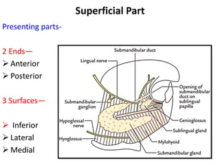
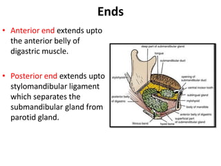
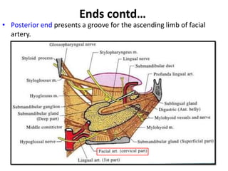
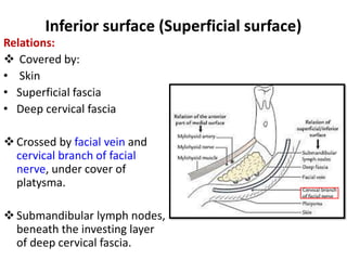
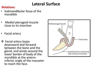
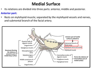
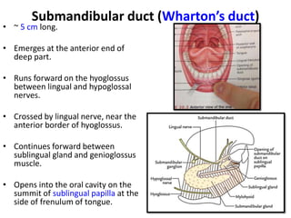
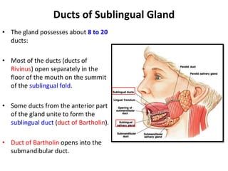
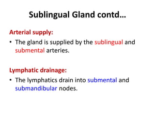
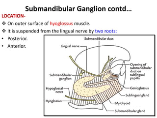
SUBMANDIBULAR
REGION
Presented by :- Dr. Sushma Tomar
Associate Professor
Department of Anatomy
21-1-2020
Introduction
• The region under cover of body of mandible.
• Extends between mylohyoid lines above and hyoid bone
below.
Deep Structures
 Suprahyoid muscles.
 Extrinsic muscles of the tongue.
 Salivary glands-
• Submandibular and Sublingual.
 Arteries-
• Facial and Lingual.
 Nerves-
• Lingual.
• Hypoglossal.
• Glossopharyngeal.
 Ganglion-
• Submandibular.
Key Muscle
• Hyoglossus
Muscular Planes
• 4
I- Digastric and Stylohyoid.
II-Mylohyoid.
III-Geniohyoid, Hyoglossus and
Styloglossus.
IV-Genioglossus and a part of
superior constrictor of pharynx.
Submandibular Gland
Introduction
• Size- about the size of a walnut.
PARTS
2
• Larger superficial part
• Smaller deep part.
 Two parts are continuous with each other around the posterior border of the
mylohyoid muscle.
Superficial Part
Presenting parts-
2 Ends—
 Anterior
 Posterior
3 Surfaces—
 Inferior
 Lateral
 Medial
Ends
• Anterior end extends upto
the anterior belly of
digastric muscle.
• Posterior end extends upto
stylomandibular ligament
which separates the
submandibular gland from
parotid gland.
Ends contd…
• Posterior end presents a groove for the ascending limb of facial
artery.
Inferior surface (Superficial surface)
Relations:
 Covered by:
• Skin
• Superficial fascia
• Deep cervical fascia
 Crossed by facial vein and
cervical branch of facial
nerve, under cover of
platysma.
 Submandibular lymph nodes,
beneath the investing layer
of deep cervical fascia.
Lateral Surface
Relations:
• Submandibular fossa of the
mandible
• Medial pterygoid muscle
close to its insertion
• Facial artery
 Facial artery loops
downward and forward
between the bone and the
gland, and winds around the
lower border of body of the
mandible at the antero-
inferior angle of the masseter
to reach the face.
Medial Surface
• Its relations are divided into three parts: anterior, middle and posterior.
Anterior part:
• Rests on mylohyoid muscle; separated by the mylohyoid vessels and nerves,
and submental branch of the facial artery.
Medial Surface contd…
Intermediate part:
Rests on:
• Hyoglossus muscle
• Lingual nerve
• Submandibular ganglion
• Hypoglossal nerve
Medial Surface contd…
Posterior part : related to
• Styloglossus muscle
• Stylohyoid ligament
• Glossopharyngeal nerve
• Wall of pharynx
Deep part
• Extends forward in the interval between the mylohyoid and
hyoglossus upto the posterior end of sublingual salivary
gland.
Relations of Deep part
Laterally-Mylohyoid
Medially-Hyoglossus
Above-Lingual nerve and submandibular ganglion
Below-Hypoglossal nerve accompanied by a pair of veins.
Submandibular duct (Wharton’s duct)
• ~ 5 cm long.
• Emerges at the anterior end of
deep part.
• Runs forward on the hyoglossus
between lingual and hypoglossal
nerves.
• Crossed by lingual nerve, near the
anterior border of hyoglossus.
• Continues forward between
sublingual gland and genioglossus
muscle.
• Opens into the oral cavity on the
summit of sublingual papilla at the
side of frenulum of tongue.
Nerve Supply
Clinical Correlates
 Formation of calculi-
 It is more common in submandibular gland and its duct for two reasons:
• Secretion is more viscid.
• Duct takes a tortuous and upward course (against gravity).
 Skin incision for removal of calculus or tumour from submandibular gland-
• 4 cm below the angle of mandible.
 Bimanual Palpation of Submandibular gland-
Sublingual Gland
• It is the smallest of the three large salivary
glands.
• Mostly mucous in nature.
SHAPE-almond shaped.
WEIGHT- 3 to 4 gm
LOCATION-
• In the floor of the mouth between the
mucous membrane and mylohyoid muscle.
• Lodges in the sublingual fossa of the
mandible.
Ducts of Sublingual Gland
• The gland possesses about 8 to 20
ducts:
• Most of the ducts (ducts of
Rivinus) open separately in the
floor of the mouth on the summit
of the sublingual fold.
• Some ducts from the anterior part
of the gland unite to form the
sublingual duct (duct of Bartholin).
• Duct of Bartholin opens into the
submandibular duct.
Sublingual Gland contd…
Arterial supply:
• The gland is supplied by the sublingual and
submental arteries.
Lymphatic drainage:
• The lymphatics drain into submental and
submandibular nodes.
Submandibular Ganglion (Langley’s Ganglion)
• It is a parasympathetic
ganglion.
• A relay station for
secretomotor fibers of
submandibular and
sublingual salivary glands.
CONNECTIONS-
Topographically- with the
lingual nerve.
Functionally- with the facial
nerve (through its chorda
tympani branch).
Submandibular Ganglion contd…
LOCATION-
 On outer surface of hyoglossus muscle.
 It is suspended from the lingual nerve by two roots:
• Posterior.
• Anterior.
Submandibular Ganglion contd…
ROOTS- 3
• Parasympathetic.
• Sympathetic.
• Sensory.
Parasympathetic or motor root:
• Derived from lingual nerve.
• The preganglionic fibres arise from
superior salivatory nucleus in the pons.
• Preganglionic fibres pass successively
through the facial, chorda tympani and
lingual nerves and reach the
submandibular ganglion.
• Preganglionic fibres relay in submandibular
ganglion.
Parasympathetic or motor root contd…
• Postganglionic fibres directly supply the submandibular gland by five
or more branches.
• Some postganglionic fibers join the lingual nerve through the anterior
root and supply the sublingual gland.
Submandibular Ganglion contd…
Sympathetic Root:
• It is derived from a plexus around
the facial artery which is formed
by postganglionic sympathetic
fibres.
• Postganglionic sympathetic fibres
arise from superior cervical
sympathetic ganglion of the
sympathetic trunk.
• These fibres pass through the
submandibular ganglion without
relay.
• These fibres supply the blood
vessels of submandibular and
sublingual salivary glands.
SUBMANDIBULAR-REGION.pdf

SUBMANDIBULAR-REGION.pdf

  • 1.
    SUBMANDIBULAR REGION Presented by :-Dr. Sushma Tomar Associate Professor Department of Anatomy 21-1-2020
  • 2.
    Introduction • The regionunder cover of body of mandible. • Extends between mylohyoid lines above and hyoid bone below.
  • 3.
    Deep Structures  Suprahyoidmuscles.  Extrinsic muscles of the tongue.  Salivary glands- • Submandibular and Sublingual.  Arteries- • Facial and Lingual.  Nerves- • Lingual. • Hypoglossal. • Glossopharyngeal.  Ganglion- • Submandibular.
  • 4.
  • 5.
    Muscular Planes • 4 I-Digastric and Stylohyoid. II-Mylohyoid. III-Geniohyoid, Hyoglossus and Styloglossus. IV-Genioglossus and a part of superior constrictor of pharynx.
  • 6.
  • 7.
    Introduction • Size- aboutthe size of a walnut.
  • 8.
    PARTS 2 • Larger superficialpart • Smaller deep part.  Two parts are continuous with each other around the posterior border of the mylohyoid muscle.
  • 9.
    Superficial Part Presenting parts- 2Ends—  Anterior  Posterior 3 Surfaces—  Inferior  Lateral  Medial
  • 11.
    Ends • Anterior endextends upto the anterior belly of digastric muscle. • Posterior end extends upto stylomandibular ligament which separates the submandibular gland from parotid gland.
  • 12.
    Ends contd… • Posteriorend presents a groove for the ascending limb of facial artery.
  • 13.
    Inferior surface (Superficialsurface) Relations:  Covered by: • Skin • Superficial fascia • Deep cervical fascia  Crossed by facial vein and cervical branch of facial nerve, under cover of platysma.  Submandibular lymph nodes, beneath the investing layer of deep cervical fascia.
  • 14.
    Lateral Surface Relations: • Submandibularfossa of the mandible • Medial pterygoid muscle close to its insertion • Facial artery  Facial artery loops downward and forward between the bone and the gland, and winds around the lower border of body of the mandible at the antero- inferior angle of the masseter to reach the face.
  • 15.
    Medial Surface • Itsrelations are divided into three parts: anterior, middle and posterior. Anterior part: • Rests on mylohyoid muscle; separated by the mylohyoid vessels and nerves, and submental branch of the facial artery.
  • 16.
    Medial Surface contd… Intermediatepart: Rests on: • Hyoglossus muscle • Lingual nerve • Submandibular ganglion • Hypoglossal nerve
  • 17.
    Medial Surface contd… Posteriorpart : related to • Styloglossus muscle • Stylohyoid ligament • Glossopharyngeal nerve • Wall of pharynx
  • 18.
    Deep part • Extendsforward in the interval between the mylohyoid and hyoglossus upto the posterior end of sublingual salivary gland.
  • 19.
    Relations of Deeppart Laterally-Mylohyoid Medially-Hyoglossus Above-Lingual nerve and submandibular ganglion Below-Hypoglossal nerve accompanied by a pair of veins.
  • 20.
    Submandibular duct (Wharton’sduct) • ~ 5 cm long. • Emerges at the anterior end of deep part. • Runs forward on the hyoglossus between lingual and hypoglossal nerves. • Crossed by lingual nerve, near the anterior border of hyoglossus. • Continues forward between sublingual gland and genioglossus muscle. • Opens into the oral cavity on the summit of sublingual papilla at the side of frenulum of tongue.
  • 21.
  • 22.
    Clinical Correlates  Formationof calculi-  It is more common in submandibular gland and its duct for two reasons: • Secretion is more viscid. • Duct takes a tortuous and upward course (against gravity).  Skin incision for removal of calculus or tumour from submandibular gland- • 4 cm below the angle of mandible.  Bimanual Palpation of Submandibular gland-
  • 23.
    Sublingual Gland • Itis the smallest of the three large salivary glands. • Mostly mucous in nature. SHAPE-almond shaped. WEIGHT- 3 to 4 gm LOCATION- • In the floor of the mouth between the mucous membrane and mylohyoid muscle. • Lodges in the sublingual fossa of the mandible.
  • 24.
    Ducts of SublingualGland • The gland possesses about 8 to 20 ducts: • Most of the ducts (ducts of Rivinus) open separately in the floor of the mouth on the summit of the sublingual fold. • Some ducts from the anterior part of the gland unite to form the sublingual duct (duct of Bartholin). • Duct of Bartholin opens into the submandibular duct.
  • 26.
    Sublingual Gland contd… Arterialsupply: • The gland is supplied by the sublingual and submental arteries. Lymphatic drainage: • The lymphatics drain into submental and submandibular nodes.
  • 27.
    Submandibular Ganglion (Langley’sGanglion) • It is a parasympathetic ganglion. • A relay station for secretomotor fibers of submandibular and sublingual salivary glands. CONNECTIONS- Topographically- with the lingual nerve. Functionally- with the facial nerve (through its chorda tympani branch).
  • 28.
    Submandibular Ganglion contd… LOCATION- On outer surface of hyoglossus muscle.  It is suspended from the lingual nerve by two roots: • Posterior. • Anterior.
  • 29.
    Submandibular Ganglion contd… ROOTS-3 • Parasympathetic. • Sympathetic. • Sensory. Parasympathetic or motor root: • Derived from lingual nerve. • The preganglionic fibres arise from superior salivatory nucleus in the pons. • Preganglionic fibres pass successively through the facial, chorda tympani and lingual nerves and reach the submandibular ganglion. • Preganglionic fibres relay in submandibular ganglion.
  • 30.
    Parasympathetic or motorroot contd… • Postganglionic fibres directly supply the submandibular gland by five or more branches. • Some postganglionic fibers join the lingual nerve through the anterior root and supply the sublingual gland.
  • 31.
    Submandibular Ganglion contd… SympatheticRoot: • It is derived from a plexus around the facial artery which is formed by postganglionic sympathetic fibres. • Postganglionic sympathetic fibres arise from superior cervical sympathetic ganglion of the sympathetic trunk. • These fibres pass through the submandibular ganglion without relay. • These fibres supply the blood vessels of submandibular and sublingual salivary glands.