SlideShare a Scribd company logo
1 of 103
TEMPOROMANDIBULAR
JOINT DISORDER AND ITS
MANAGEMENT
1
- Dr Priyanka Parihar
CONTENTS:
INTRODUCTION
TYPES OF JOINTS
COMPONENTS OF THE TMJ
MUSCLE OF THE JOINT
INNERVATIONS OF TMJ
VASCULARIZATION OF TMJ
CLASSIFICATION OF TMJ DISORDERS
ETIOLOGIC FACTORS
2
Treatment of Functional Disturbances of the Masticatory System
• Temporomandibular Disorders,
• Treatment of Masticatory Muscle Disorders
• Treatment of Temporomandibular Joint Disorders
• Treatment of Chronic Mandibular Hypomobility and Growth Disorders
• Occlusal Appliance Therapy
3
CONTENTS:
INTRODUCTION:
• Temporomandibular joint (TMJ) is
formed by the articulation of the
mandible and the temporal bone of
the cranium. It is located anteriorly
to the tragus of the ear, on the
lateral aspect of the face.
• It is a bicondylar joint in which the
condyles, located at the two ends of
the mandible.
4
TYPES OF JOINT:
• A joint is where two or more bones meet.
• Fibrous joint: Immovable,
Example: cranium, Radioulnar, Tibiofibular joint.
• Cartilaginous joint: slightly moveable
Example: vertebral column
• Synovial joint : freely moveable
example : knee joint , hip joint
5
• types of Synovial joint
1. Hinge joint
2. Pivot joint
3. Condyloid joint
4. Ball and socket joint
5. Gliding joint
6. Saddle joint
6
Craniomandibular articulation-TMJ
• Complex joint in the body
Hinging movements - Ginglymoid joint
Gliding movements –Arthroidal joint
 Hence called as Ginglymoarthroidal joint
• TMJ is a complex diarthroidal sliding- ginglymoid synovial joint,
which attaches the mandible to petrous part of temporal bone of
the cranium.
7
COMPONENTS OF TMJ
Hard tissue components
1. Mandibular condyles
2. Glenoid fossa of the
temporal bone
3. Articlar eminence
Soft tissue components
1. Articular capsule
2. Articular disc
3. Ligaments
4. Muscles
8
HARD TISSUE COMPONENTS
Mandibular condyle:
• Are ovoid and strongly convex anterioposterioly
and mildly convex mediolaterally.
• Perpendicular to ascending ramus of the mandible.
• The anterior surface shows a shallow depression
called as Lateral pterygoid fovea to which lateral
pterygoid muscle is attached.
• Bony surface of condyle and articular part of the
temporal bone covered with dense fibrous
connective tissue. 9
Glenoid fossa of the Temporal bone:
• The articular surface of the temporal bone is
situated on the inferior surface of the
squamous part of the temporal bone.
• This area which articulate with the
mandibular condyle is called the Glenoid
Fossa.
• The floor of the fossa is formed by a thin
plate of bone.
• It is also covered by a fibrous layer similar
to the condyle.
10
Articular eminence:
• The articular eminence is convex
anterioposteriorly and concave in the
transverse direction.
• It is enclosed by the articular capsule
of the TMJ.
11
SOFT TISSUE COMPONENTS:
Articular capsule:
• Articular capsule is a thin sleeve of dense
cartilaginous tissue enclosing the joint cavity.
• The articular capsule consists of two layers:
a) An outer layer (stratum fibrosum) composed of
avascular, white fibrous tissue.
b) An inner layer (stratum synoviale) which is a
secreting layer usually known as the synovial
membrane. 12
• Synovial membrane:
• It is a thin and flexible layer lining the inner
surface of the joint capsule
• The articulating surfaces and the
articulating disc are not covered by the
membrane
The synovial membrane consists of two
layers:
• Cellular intimal layer
• Vascular subintimal layer
13
• Function of synovial membrane
 Lubricating the joint
 Repairing wear
14
Articular disc:
• It is oval, firm, thick plate of dense fibrous cartilage
located between the condyle and the articulating
surface of the temporal bone.
• Disc divides the joint cavity in to two compartments
ie superior and inferior.
• Function of disc: due its viscoelasticity it acts as
shock absorber, stress distributor and provide gliding
motion during opening and closing of the jaw.
• The articular disc is predominantly avascular ,blood
vessels are seen at the margins of the disc. 15
Articular ligaments:
TMJ is associated with four ligaments, One
major and three minor ligaments.
Temporomandibular ligament : is fan
shaped present on lateral aspect of the
articular capsule. Hence also called as
lateral ligament.
TM ligament helps in preventing
displacement in posterior and inferior
direction.
Displacement of TMJ happens only in the
anterior or forward direction.
Temporomandibular Ligament (Lateral View).
There are two distinct parts: the outer oblique
portion (OOP) and the inner horizontal portion
(IHP). The OOP limits normal rotational
opening movement; the IHP limits posterior
movement of the condyle and disc.
16
 Sphenomandibular ligament:
• This ligament is a flat thin band which is attached to the
spine of the sphenoid superiorly and to the ligula
inferiorly.
• The main function of this ligament is to limit the
distention of the mandible in an inferior direction.
 Stylomandibular ligament:
• This ligament extends from the apex of the styloid
process of the temporal bone to the posterior border of
the angle of the mandible.
• Function: along with sphenomandibular ligament limits
excessive opening of the mandible.
17
 Capsular Ligament:
• the entire TMJ is surrounded and
encompassed by the capsular ligament.
• The capsular ligament acts to resist any
medial, lateral, or inferior forces that tend to
separate or dislocate the articular surfaces.
• Significant function to encompass the
thus
Retains the synovial fluid
Capsular Ligament (Lateral View).It
extends anterior to include the articular
eminence and encompass the entire
articular surface of the joint.
18
joint
• Collateral (discal) ligament
• The collateral ligaments attach the medial and lateral borders of
the articular disc to the poles of the condyle.
• They are commonly called the discal ligaments
• There are two Medial discal ligament
lateral discal ligament
lateral
19
MUSCLES OF THE JOINT
The muscle associated with the TMJ is the Lateral
pterygoid muscle.
• Function : it pull the coronoid process of the
mandible anteriorly, forcing the condylar process
out of the mandible fossa along the articular
eminence to protrude mandible. The upper head
influences the position of the articular disc
during movement of the TMJ.
20
INNERVATION OF TMJ
• The innervations of the TMJ involves four type of receptors:
1. Ruffin’s corpuscles function as static mechanoreceptors
which position the mandible
2. Pacinian’s corpuscles are mechanoreceptors which helps
in the perception of movement & firm pressure
(not light touch).
3. Golgi tendon protect the muscle from excessive or
damaging tension.
4. The nociceptors (along with the proprioceptors) primarily
function to monitor the condition, position, and movement
of the tissues in the masticatory system.
21
• These receptors are innervated by the
Auriculotemporal nerve which leaves the
mandibular nerve behind the joint and
ascends laterally and superiorly to wrap
around the posterior region of the joint.
• Additional innervations by Deep temporal
and Massetric nerve.
22
BLOOD SUPPLY TO TMJ
• The main source of blood supply of
TMJ is provided by the Superficial
temporal artery and maxillary artery
which are branches of the External
carotid artery
• Other branches of the external carotid
artery- The Deep auricular artery,
Anterior tympanic artery and
Ascending pharyngeal artery also
contribute to the arterial blood supply
to the joint.
23
• In brief the muscles of mastication helps in biomechanics/movements of
mandible:
Movement Muscle
Elevation (closing) Temporalis,
Masseter
Medial pterygoid
Depression ( opening) Lateral pterygoid
Superior infrahyoid
Protrusion (forward) Lateral pterygoid
Masseter
Medial pterygoid
Retrusion (backward) Temporalis
Masseter
Lateral movement Temporalis (same side)
Pterygoid (of Opposite side)
Masseter
24
CLASSIFICATION OF TMJ DISORDERS
25
•Pseudotumors
(synovial
chondromatosis)
•Benign
(chondroma,
osteotoma)
•Malignant
(primary,
metastatic)
•Synovitis
•Capsulitis
•Rheumatoid arthritis
•Juvenile rheumatoid arthritis
•Ankylosing spondylitis
•Psoriatic arthritis
•Reactive arthritis (bacterial, viral,
fungal)
PETERSON’S CLASSIFICATION
ARTICULAR
DISORDERS
Inflammatory
arthropathies
•Primary osteoarthrosis (no
clear predisposind factor)
•Secondary osteoarthrosis
(trauma,previous
surgery,avasular necrosis)
•Mechanical derangements
•Bone and cartilage
disorders with
articular manifestations
•
•Non-neoplastic:developmental
(hyperplasia, hypoplasia,
dysplasia)
•Non-neoplastic: acquired(ie,
condylolysis)
Growth Disorder
Noninflammatory
arthropathies
Neoplasm
Diffuse connective tissue
disorders
Miscellaneous articular
disorders
26
•Muscle spasm (strain)
•Myofascial pain and
dysfunction (MPD)
•Fibromyalgia
•Myotonic dystrophies
•Myositis ossificans
progressiva
NON-ARTICULAR
DISORDERS
Muscle disorders
Growth disorders
27
ETIOLOGIC FACTORS FOR
TEMPOROMANDIBULAR
DISORDERS
1. OCCLUSAL CONDITIONS
2. TRAUMA
3. EMOTIONAL STRESS
4. Deep Pain Input
5. Parafunctional Activity
28
TRAUMA
• Trauma can be divided into two general types: macrotrauma and
microtrauma.
• Macrotrauma is considered any sudden force that can result in structural
alterations, such as a direct blow to the face.
• Microtrauma refers to any small force that is repeatedly applied to the
structures over a long period of time. Activities such as bruxism or
clenching can produce microtrauma to the tissues that are being loaded
(i.e., teeth, joints, or muscles)
29
EMOTIONAL STRESS
• The emotional centers of the brain have an influence on muscle function.
• Stress affects the body by activating the hypothalamus-pituitary-adrenal
axis (HPA axis)
• The HPA axis, through complex neural pathways, increases the activity of
the gamma efferents, which cause the intrafusal fibers of the muscle
spindles to contract.
• This so sensitizes the spindle that any slight stretching of the muscle will
cause a reflex contraction. The overall effect is an increase in tonicity of
the muscle 30
DEEP PAIN INPUT
• Deep pain input can centrally excite the brainstem producing a muscle
response known as protective co-contraction.
• This represents a normal healthy manner in which the body responds to
injury or threat of injury.
• Therefore it is reasonable to find a patient who is suffering with pain, such
as toothache (i.e., necrotic pulp), to have limited mouth opening.
• This represents the body’s response to protect the injured part by limiting
its use.
31
32
33
34
35
TREATMENT OF
FUNCTIONAL
DISTURBANCES OF
THE MASTICATORY
SYSTEM
1. General Considerations In The Treatment Of
Temporomandibular Disorders
36
3. Treatment of Temporomandibular Joint Disorders
2. Treatment of Masticatory Muscle Disorders
4. Treatment of Chronic Mandibular Hypomobility
and Growth Disorders
5. Occlusal Appliance Therapy
• Definitive Treatment
• Definitive therapy is aimed directly toward the elimination or
alteration of the etiologic factors that are responsible for the
disorder.
• For example, definitive treatment for an anterior displacement of
the articular disc will reestablish the proper condyle-disc
relationship.
37
1. General Considerations In The Treatment Of
Temporomandibular Disorders
• Supportive Therapy
• Supportive therapy is directed toward altering the patient’s
symptoms and often has no effect on the etiology of the
disorder.
• A simple example is giving a patient aspirin for a headache
that is caused by hunger. The patient may feel relief from the
headache, but there is no change in the etiologic factor
(hunger) that created the symptom.
• Supportive therapy is directed toward the reduction of pain and
dysfunction.
38
• The two general types of supportive therapies are
Pharmacologic Therapy
 Physical Therapy
1. Pharmacologic Therapy
• The most common classes of pharmacologic agents used for the
management of TMD are analgesics, antiinflammatories, muscle
relaxants, anxiolytics, antidepressants, anticonvulsives,
injectables, and topicals.
39
2. Physical Therapy
• Physical therapy represents a group of supportive activities
that is usually instituted in conjunction with definitive treatment.
• Most physical therapy fits into one of two general categories:
modalities and manual techniques
40
41
a)THERMOTHERAPY
(moist heat)
b)COOLANT THERAPY,
(ice pack and Fluoromethane spray)
c)IONTOPHORESIS TREATMENT d)ULTRASOUND THERAPY
e)TRANSCUTANEOUS
ELECTRICAL NERVE
STIMULATION (TENS)
Physical Therapy Modalities- types:
f)ELECTROGALVANIC
STIMULATION THERAPY,
g)LASER.
h) PHONOPHORESIS,
• Manual Techniques
Manual techniques are the “hands-on” therapies provided by the
physical therapist for the reduction of pain and dysfunction.
• Manual techniques are divided into three categories:
soft tissue mobilization,
 joint mobilization, and
muscle conditioning.
42
43
MASSAGE THERAPY
SOFT TISSUE MOBILIZATION. JOINT MOBILIZATION
JOINT DISTRACTION OF THE TMJ
44
MUSCLE CONDITIONING
ASSISTED MUSCLE STRETCHING
PASSIVE MUSCLE STRETCHING.
RESISTANCE EXERCISES
A. The patient is instructed to open against gentle
resistance provided by the fingers. This will
promote relaxation in the elevator muscles,
thus allowing increased mandibular opening.
B. When eccentric movement is limited the
patient can be asked to move in the eccentric
position with gentle resistance from the fingers.
These exercises are repeated 10 times each
session, six sessions a day. If they elicit
pain,they are discontinued.
A B
Acupuncture-
• Stimulation of acupuncture points appears to
cause the release of endogenous opioids
(endorphins and enkephalins), which reduce
painful sensations by flooding the afferent
interneurons with subthreshold stimuli.
• These effectively block the transmission of
noxious impulses and thus reduce the
sensations of pain.
45
These needles are maintained in
place for approximately 30 minutes
during which they are twirled every
5 to 10 minutes.
• Protective Co-Contraction (Muscle Splinting)
• Local Myalgia (Noninflammatory Myalgia)
• Myospasms (Tonic Contraction Myalgia)
• Myofascial Pain (Trigger Point Myalgia)
• Centrally Mediated Myalgia (Chronic Myositis)
46
2. Treatment of Masticatory Muscle Disorders
PROTECTIVE CO-CONTRACTION
(MUSCLE SPLINTING)
• Protective co-contraction is the initial response of a muscle to
altered sensory or proprioceptive input or injury. This response
has been called protective muscle splinting .
• In the presence of altered sensory input or pain, antagonistic
muscle groups seem to activate during movement in an
attempt to protect the injured part. Therefore, pain felt in the
masticatory system can produce protective co-contraction of
masticatory muscles.
47
• protective co-contraction is not a pathologic condition but a normal
physiologic response of the musculoskeletal system.
Etiology
1. Altered sensory or proprioceptive input.
2. The presence of constant deep pain input.
3. Increased emotional stress.
• Protective co-contraction only remains a few hours or days. If it is not
resolved, local myalgia is likely to follow.
48
P. Co-Contraction
Clinical Characteristics
1. Structural dysfunction: Decreased range of
movement.
2. There is very minimal pain at rest.
3. There is increased pain to function.
4. The patient reports a feeling of muscle
weakness.
49
This patient bites his cheek
causing acute tissue injury
and pain. This pain led to
protective co-contraction.
P. Co-Contraction
TREATMENT
• Protective co-contraction is a normal CNS response and
therefore there is no indication to treat the muscle condition
itself. Treatment should instead be directed toward the reason
for the co-contraction.
• When co-contraction results from trauma, definitive treatment is
not indicated since the etiologic factor is no longer present.
• When co-contraction results from a poorly fitting restoration,
altering the restoration is advised.
• If an increase in emotional stress is the etiology then
appropriate stress management.
• Short-term pain medication NSAIDs may be indicated
50
P. Co-Contraction
TO BE CONTINUED……..
51
52
LOCAL MYALGIA
(NONINFLAMMATORY MYALGIA)
• It is noninflammatory, myogenous pain disorder. It is often the
first response of the muscle tissue to continued protective co-
contraction.
• Local myalgia represents a change in the local environment of
the muscle tissues. It represents the initial response to overuse
of muscle.
53
LOCAL MYALGIA (NON-INF. MYALGIA)
Etiology
1. Protracted protective co-contraction secondary to a recent
alteration in local structures.
2. A continued source of constant deep pain
3. Local tissue trauma.
4. Increased levels of emotional stress.
54
1. Structural dysfunction: There is marked decrease in the range
of mandibular movement.
2. There is minimum pain at rest and increased with function.
4. Actual muscle weakness present.
5. There is local tenderness when the involved muscles are
palpated
55
Clinical Characteristics
LOCAL MYALGIA (NON-INF. MYALGIA)
• The primary goal in treating local myalgia is to decrease sensory
input (such as pain)
• Achieved by-
1. Eliminate any ongoing altered sensory input.
2. Eliminate any ongoing source of deep pain input.
3. Provide patient education and information on self-management
i.e Advise the patient to restrict mandibular use to within painless
limits.
56
TREATMENT LOCAL MYALGIA (NON-INF. MYALGIA)
• If due to bruxism -fabricate an occlusal appliance
for nighttime use while for daytime educate
patient to control it.
• If the above therapies fail to resolve the pain
condition, the clinician may consider the use of a
mild analgesic.
• such as aspirin, acetaminophen, or an NSAID
(i.e., ibuprofen), can be helpful.
57
LOCAL MYALGIA (NON-INF. MYALGIA)
MYOSPASMS
(TONIC CONTRACTION MYALGIA)
• Myospasm is an involuntary CNS-induced tonic muscle
contraction often associated with local metabolic conditions
within the muscle tissues.
Etiology
1. Continued deep pain input.
2. overuse
3. Idiopathic myospasm mechanisms
58
CLINICAL CHARACTERISTICS
• The patient reports a sudden onset of restricted jaw movement
usually accompanied by muscle rigidity.
• Structural dysfunction: There is marked restriction in range of
mandibular movement.
• Acute malocclusion is common.
• There is pain at rest and pain is increased with function.
• The affected muscle is firm and painful to palpation.
59
• Two treatments are suggested for acute myospasms.
• The first is directed immediately toward reducing the spasm
itself while the other addresses the etiology.
1. Pain Reduction- can be achieved by manual massage,
vapocoolant spray, ice, or even an injection of local
anesthetic (2% lidocaine without a vasoconstrictor is
recommended.)
2. Etiology reduction When the myospasms are secondary to
fatigue and overuse, patient is advised to rest the muscle.
• Idiopathic - may represent an oromandibular dystonia
60
TREATMENT
MYOFASCIAL PAIN
(TRIGGER POINT MYALGIA)
• Myofascial pain is a regional myogenous
pain condition characterized by local areas
of firm, hypersensitive bands of muscle
tissue known as trigger points.
61
Etiology-
1. Continued source of deep pain input.
2. Increased levels of emotional stress.
3. The presence of sleep disturbances.
4. Local factors that influence muscle activity such as habits,
posture, muscle strains, or even chilling.
5. Systemic factors such as nutritional inadequacies, poor
physical conditioning, fatigue, and viral infections.
6. Idiopathic trigger point mechanism. 62
Myofascial Pain
63
TREATMENT
Myofascial Pain
1. Eliminate any source of ongoing deep pain.
2. If a sleep disorder is suspected, Often, low dosages
of a tricyclic antidepressant,such as 10 to 20 mg of
amitriptyline before bedtime, can be helpful.
3. elimination of the trigger points, by painlessly
stretching the muscle containing the trigger points.
• Spray and Stretch, Pressure and Massage,
Ultrasound and Electrogalvanic Stimulation,
Injection and Stretch.
• A medication such as cyclobenzaprine (Flexeril), 10 mg before
sleep can often reduce pain but the trigger points still need to
be treated.
• Muscle relaxants help convert an active trigger point into a
latent or dormant trigger point but may not necessarily
eliminate it.
• Analgesics may also be helpful in interrupting the cyclic effect
of pain.
64
TREATMENT
CENTRALLY MEDIATED MYALGIA
(CHRONIC MYOSITIS)
• Centrally mediated myalgia is a
chronic, continuous muscle pain
disorder originating predominantly
from CNS effects that are felt
peripherally in the muscle tissues.
65
The illustration depicts how central
activation of the hypothalamus, the
limbic structures and the cortex can
combine to produce an antidromic effect
to the muscle tissues.
Etiology
• Chronic centrally mediated myalgia may be caused by the
prolonged input of muscle pain associated with local myalgia or
myofascial pain.
• Chronic upregulation of the autonomic nervous system
• Chronic exposure to emotional stress
• Other sources of deep pain input.
66
CLINICAL CHARACTERISTICS
1. Structural dysfunction
2. There is significant pain at rest and pain is increased with
function.
4. There is a generalized feeling of muscle tightness.
5. There is significant pain on muscle palpation.
67
• Restrict mandibular use to within painless limits
• Avoid exercise and/or injections. Since the muscle tissue is
neurogenically inflamed, any use increases pain.
• The patient should rest the muscles as much as possible.
• Disengage the teeth by a stabilization appliance.
• Begin taking an antiinflammatory medication.
• If a sleep disorder is suspected, Often, low dosages of a tricyclic
antidepressant is advised at bedtime.
68
TREATMENT
• This internal physiology is only under the
control of the patient, not the clinician. That is
why the patient needs to understand this
relationship and work on changing behavior
that will downregulate the autonomic nervous
system.
• Physical self regulation plays important role
in its treatment
69
TREATMENT
Application of moist heat or
cold can be helpful for
chronic centrally mediated
myalgia
• Disc displacement and disc displacement with intermittent
locking
• Disc displacement without reduction
• Structural incompatibility Deviation in form and Adhesions
• Subluxation and Luxation
• Capsulitis, Retrodiscitis, and Acute Trauma to the TMJ
• Osteoarthritis
• Infectious arthritis 70
3.Treatment of Temporomandibular Joint Disorders
Inflammatory
disorders
Etiology
• Disc derangement disorders result from elongation of the capsular
and discal ligaments coupled with thinning of the articular disc.
Clinical Characteristics
• The clinical examination reveals a relatively normal range of
movement with restriction only associated with the pain.
• Discal movement can be felt by palpation of the joints during
opening and closing.
• Deviations in the opening pathway .
71
Disc displacement and disc displacement with
intermittent locking
• Anterior positioning appliance to reposition to
condyle back on the disc.
• It was originally suggested that this appliance be
worn 24 hours a day for as long as 3 to 6 months.
• The patient needs to be encouraged to decrease
loading of the joint whenever possible. Softer
foods, slower chewing,and smaller bites should be
promoted.
72
TREATMENT
• If inflammation is suspected, an NSAID should be prescribed.
• Moist heat or ice can be used.
• Active exercises are not advised.
73
SO YOU ALL MIGHT BE WONDERING
HOW DOES THIS WORKS ??
74
75
In the resting closed joint
position the disc is anteriorly
displaced from the condyle
A C
B
Maxillary occlusal
appliance is placed which
creates an occlusal
condition that requires the
mandible to shift slightly
forward.
Note that when the
appliance is in place & the
teeth are occluding, the
condyle is repositioned on
disc in normal condyle-disc
relationship.
Anterior Positioning Appliance
76
An anteriorly displaced disc
with the condyle articulating on
the retrodiscal tissues
producing pain.
APA is placed in mouth to
bring the condyle forward,off
of the retrodiscal tissues onto
the disc. This relationship
reduces the loading of the
retrodiscal tissues, which
decreases the pain.
Once the tissues have
adapted, the appliance is
removed. Condyle now
functions on the adaptive
fibrotic tissues resulting in
a painless functioning joint,
but because the disc is still
displaced, clicking may still
be present.
A C
B
DISC DISPLACEMENT WITHOUT REDUCTION
• Disc displacement without reduction is a clinical condition in
which the disc is totally displaced (dislocated) and does not
return to normal position with condylar movement.
Etiology
• Macrotrauma and microtrauma are the most common causes of
disc displacement without reduction.
77
• A sudden change in range of mandibular movement occurs.
• Examination reveals limited mandibular opening with some slight
defection to the ipsilateral side.
Treatment
• Fabricating an anterior positioning appliance is contraindicated for
this patient, as it will aggravate the condition by forcing the disc
even more forward.
• Recapture the disc by manual manipulation.
78
Clinical Characteristics
79
TREATMENT
• Technique for Manual Manipulation.
The clinician’s right thumb is placed intraorally over the
patient’s left mandibular second molar and the mandible is
grasped. With the left hand stabilizing the cranium, gentle but
firm force is applied downward on the molar and upward on the
chin to distract the joint.
• Once the joint is distracted, the mandible is brought forward and
to the right, enabling the condyle to move into the area of the
displaced disc. When this position is achieved, constant
distractive force is applied for 20 to 30 seconds while the patient
relaxes.
80
TREATMENT
• After the distraction, the thumb is removed and the patient is
asked to close on the anterior teeth, maintaining the jaw in a
slightly protrusive position.
• When the patient has rested a moment, he or she is instructed
to open maximally. If the disc has been reduced, a normal
range of movement (48 mm) will be possible.
81
TREATMENT
• Patients should be encouraged not to open too wide, especially
immediately following the disc displacement without reduction.
• The patient should also be told to decrease hard biting, no chewing
gum.
• If pain is present, heat or ice may be used. NSAIDs are indicated
for pain and inflammation.
• Joint distraction and phonophoresis over the joint area may be
helpful.
82
TREATMENT
• Surgical Considerations for Condyle-Disc Derangement Disorders
• Arthrocentesis- In this procedure, two needles are placed into the
joint and sterile saline solution is passed through lavaging the joint.
• Arthroscopy- With this technique, an arthroscope is placed into the
superior joint space and the intracapsular structures are visualized
on a monitor. (diagnostic)
• Arthrotomy- Open joint surgery
• Discal Repair Or Plication- During a plication procedure, a portion of
the retrodiscal tissue and inferior lamina is normally removed and
the disc is retracted posteriorly and secured with sutures
83
TREATMENT
STRUCTURAL INCOMPATIBILITY DEVIATION IN
FORM AND ADHESIONS
Deviation in Form-
• Includes a group of disorders that is created by changes in the
smooth articular surface of the joint and disc. These changes
produce an alteration in the normal pathway of condylar movement.
• The etiology of most deviations in form is trauma.
Adherences/Adhesions
• Adherences represent a temporary sticking of the articular surfaces
during normal joint movements. caused by a fibrosis attachment of
the articular surfaces. 84
85
Deviation in Form
and Adhesions
TREATMENT
SUBLUXATION AND LUXATION
Subluxation
• Subluxation or hypermobility is a clinical description of the condyle as
it moves anterior to the crest of the articular eminence.
• It is not a pathologic condition but a variation in anatomic form of the
fossa.
Luxation
• This condition is commonly referred to as an openlock since the
patient’s mouth is wide open and cannot reduce it.
• It can occur following wide-open mouth procedures such as having a
dental appointment.
86
87
Subluxation
and Luxation
TREATMENT
CAPSULITIS, RETRODISCITIS, AND ACUTE
TRAUMA TO THE TMJ
Capsulitis-
• An inflammatory condition of the capsular
tissues
Retrodiscitis-
• An inflammatory condition of the retrodiscal
tissues.
88
Retrodiscitis
89
Capsulitis,
Retrodiscitis,
and Acute
Trauma to the
TMJ.
TREATMENT
OSTEOARTHRITIS
• Arthritis means inflammation of the articular surfaces of the
joint.
• Osteoarthritis is one of most common arthritis affecting the
TMJ.
Etiology
• overloading of the articular structures of the joint.
90
91
Osteoarthritis
TREATMENT
A. Chronic Mandibular Hypomobility-
• The predominant feature of this disorder is the inability of the
patient to open the mouth to a normal range.
• Chronic mandibular hypomobility is rarely accompanied by
painful symptoms.
• subdivided into three categories according to etiology:
• ankylosis, muscle contracture, and coronoid impedance.
92
4. Treatment of Chronic Mandibular Hypomobility
and Growth Disorders
ANKYLOSIS
93
• Abnormal immobility of a joint.
• The two basic types of ankylosis are differentiated by
the tissues that limit the mobility: fibrous and bony.
• Fibrous ankylosis - most common and can occur
between the condyle and the disc or the disc and the
fossa.
• Bony ankylosis of TMJ- would occur between the
condyle and fossa and therefore the disc is mostly
absent from the discal space prior to the ankylosis.
• Etiology- The most common etiology of ankylosis is
hemarthrosis (bleeding within the joint).
• Chronic inflammation aggravates the disorder leading to the
development of more fibrous tissue.
• Management-
• If function is inadequate or the restriction is intolerable, surgery
is the only definitive treatment available.
94
MUSCLE CONTRACTURE
• MYOSTATIC CONTRACTURE
• Myostatic contracture results when a muscle is kept from
fully lengthening (stretching) for a prolonged time.
• For example if the mandible is fractured and wired
together with maxilla for 6 to 8 weeks the elevator muscles
cannot fully lengthen.
• Management -The resting length of the muscles can be
reestablished by two types of exercise: passive stretching
and resistant opening.
• Thermotherapy and ultrasound are also helpful.
95
Myofibrotic Contracture-
• Myofibrotic contracture occurs as a result of excessivev tissue
adhesions within the muscle or its sheaths.
• These fibrosis tissue adhesions prevent the muscle fibers from
sliding over themselves disallowing full lengthening of the
muscle.
• Etiology-myositis or trauma to the muscle tissues.
• Management - surgical detachment of the muscles involved.
96
CORONOID IMPEDANCE
• If the pathway of coronoid process is impeded,
it will not slide smoothly and the mouth will not
fully open.
• Etiology- Coronoid impedance is generally due
to either elongation of the process or the
encroachment of fibrous tissue.
• Management- 1. surgery, that either shortens
the coronoid process or eliminates the tissue
obstruction ( if function is severely impaired).
2. Gentle passive stretching
97
very long coronoid process
B. GROWTH DISORDERS
• Common growth disturbances of the bones are agenesis (no
growth), hypoplasia (insufficient growth), hyperplasia (too
much growth), or neoplasia (uncontrolled, destructive growth).
98
Congenital and Developmental Bone Disorders
Condylar Hyperplasia-
• Bone disease characterized by the increased
development of one mandibular condyle
• Management -Condylectomy and orthognathic
surgery, Mandibular ramus osteotomy
Condylar Hypoplasia-
• Known as underdevelopment of condyle
• Management - orthognathic surgery, distraction
osteogenesis, surgery with costochondral bone graft
99
Hyperplasia
Hypoplasia
• An occlusal appliance is a removal device made of hard acrylic
that fits over the occlusal surfaces of the teeth in one arch.
• It is commonly referred to as an occlusal splint, bite guard, night
guard, interocclusal appliance, or even orthopedic device .
• Occlusal appliances provides an occlusal condition that allows
the condyles to assume their most orthopedically stable joint
position.
• They are also used to protect the teeth and supportive structures
from abnormal forces that may create breakdown and tooth
wear 100
5. Occlusal Appliance Therapy
• The two most commonly used are the
stabilization appliance (muscle relaxation
appliance) and the anterior positioning appliance
(orthopedic repositioning appliance).
• Other types are the anterior bite plane, the
posterior bite plane, the pivoting appliance, and
the soft or resilient appliance
101
stabilization appliance
anterior positioning appliance
REFERENCES:
• B D Chaurasia; Human Anatomy For Dental Students 3rd
Edition.
• Jeffrey P. Okeson, DMD 8th Edition
• Peterson’s Principles Of Oral And Maxillofacial Surgery 2nd
Edition.
102
103

More Related Content

What's hot

Internal derangement of tmj
Internal derangement of tmjInternal derangement of tmj
Internal derangement of tmjDrKamini Dadsena
 
Temporomandibular joint /disorders /management / treatment
Temporomandibular joint /disorders /management / treatmentTemporomandibular joint /disorders /management / treatment
Temporomandibular joint /disorders /management / treatmentCairo University
 
Presentation temporomandibular joint anatomy
Presentation temporomandibular joint anatomyPresentation temporomandibular joint anatomy
Presentation temporomandibular joint anatomyLeena Gangil
 
Myofacial pain dysfunction syndrome anindya
Myofacial pain dysfunction syndrome anindyaMyofacial pain dysfunction syndrome anindya
Myofacial pain dysfunction syndrome anindyaDr. Anindya Chakrabarty
 
Mpds (Myofacial pain dysfunction syndrome)
Mpds (Myofacial pain dysfunction syndrome)Mpds (Myofacial pain dysfunction syndrome)
Mpds (Myofacial pain dysfunction syndrome)shayabu
 
Mandibular fractures
Mandibular fracturesMandibular fractures
Mandibular fracturesArjun Shenoy
 
Width of attached gingiva
Width of attached gingivaWidth of attached gingiva
Width of attached gingivanaren kumar
 
Mandibular Angle Fractures
Mandibular Angle FracturesMandibular Angle Fractures
Mandibular Angle FracturesAhmed Adawy
 
RESECTIVE OSSEOUS SURGERY
RESECTIVE OSSEOUS SURGERYRESECTIVE OSSEOUS SURGERY
RESECTIVE OSSEOUS SURGERYAnkita Dadwal
 
Gingival pigmentation
Gingival pigmentationGingival pigmentation
Gingival pigmentationNida Sumra
 

What's hot (20)

Tm j examination
Tm j examinationTm j examination
Tm j examination
 
Management of TMJ pain
Management of TMJ painManagement of TMJ pain
Management of TMJ pain
 
Internal derangement of tmj
Internal derangement of tmjInternal derangement of tmj
Internal derangement of tmj
 
Temporomandibular joint /disorders /management / treatment
Temporomandibular joint /disorders /management / treatmentTemporomandibular joint /disorders /management / treatment
Temporomandibular joint /disorders /management / treatment
 
Presentation temporomandibular joint anatomy
Presentation temporomandibular joint anatomyPresentation temporomandibular joint anatomy
Presentation temporomandibular joint anatomy
 
TMJ Ankylosis
TMJ AnkylosisTMJ Ankylosis
TMJ Ankylosis
 
Mpds
MpdsMpds
Mpds
 
Root planing
Root planingRoot planing
Root planing
 
Myofacial pain dysfunction syndrome anindya
Myofacial pain dysfunction syndrome anindyaMyofacial pain dysfunction syndrome anindya
Myofacial pain dysfunction syndrome anindya
 
Mpds (Myofacial pain dysfunction syndrome)
Mpds (Myofacial pain dysfunction syndrome)Mpds (Myofacial pain dysfunction syndrome)
Mpds (Myofacial pain dysfunction syndrome)
 
Tmj disorder
Tmj disorderTmj disorder
Tmj disorder
 
Mandibular fractures
Mandibular fracturesMandibular fractures
Mandibular fractures
 
Periodontal Sutures
 Periodontal Sutures Periodontal Sutures
Periodontal Sutures
 
Width of attached gingiva
Width of attached gingivaWidth of attached gingiva
Width of attached gingiva
 
Mandibular Angle Fractures
Mandibular Angle FracturesMandibular Angle Fractures
Mandibular Angle Fractures
 
RESECTIVE OSSEOUS SURGERY
RESECTIVE OSSEOUS SURGERYRESECTIVE OSSEOUS SURGERY
RESECTIVE OSSEOUS SURGERY
 
Periimplantitis
PeriimplantitisPeriimplantitis
Periimplantitis
 
Tmj ankylosis
Tmj ankylosisTmj ankylosis
Tmj ankylosis
 
Gingival pigmentation
Gingival pigmentationGingival pigmentation
Gingival pigmentation
 
Tmj
TmjTmj
Tmj
 

Similar to TMJ Disorder And its Management

Tmj by dr.meher moin
Tmj by dr.meher moinTmj by dr.meher moin
Tmj by dr.meher moinmehermoinkhan
 
Temporomandibular joint [Autosaved].pptx
Temporomandibular joint [Autosaved].pptxTemporomandibular joint [Autosaved].pptx
Temporomandibular joint [Autosaved].pptxAbhidha Tripathi
 
Temperomandibular joint Anatomy
Temperomandibular joint AnatomyTemperomandibular joint Anatomy
Temperomandibular joint AnatomyAshish Ranghani
 
Temporomandibular joint anatomy and function
Temporomandibular joint anatomy and functionTemporomandibular joint anatomy and function
Temporomandibular joint anatomy and functionDR POOJA
 
Surgical anatomy of tmj
Surgical anatomy of tmjSurgical anatomy of tmj
Surgical anatomy of tmjMinalSonare2
 
Muscles_of_mastication.pptx
Muscles_of_mastication.pptxMuscles_of_mastication.pptx
Muscles_of_mastication.pptxHelipatel56
 
temporomandibular joint.pptx
temporomandibular joint.pptxtemporomandibular joint.pptx
temporomandibular joint.pptxSumedhaThosar
 
Temporomandibular joint & disorders seminar.ppt
Temporomandibular joint & disorders seminar.pptTemporomandibular joint & disorders seminar.ppt
Temporomandibular joint & disorders seminar.pptSomyaJain603714
 
TMJ and its relation to periodontics
TMJ and its relation to periodonticsTMJ and its relation to periodontics
TMJ and its relation to periodonticsChittoor Deals
 
ANATOMY TEMPOROMANDIBULAR JUNCTION OF HUMAN
ANATOMY TEMPOROMANDIBULAR JUNCTION OF HUMANANATOMY TEMPOROMANDIBULAR JUNCTION OF HUMAN
ANATOMY TEMPOROMANDIBULAR JUNCTION OF HUMANDesiFitriani85
 
Examination of tmj &muscles of mastication (2)
Examination of tmj &muscles of mastication (2)Examination of tmj &muscles of mastication (2)
Examination of tmj &muscles of mastication (2)rachitajainr
 
Relationship of tmj anatomy and pathology and related
Relationship of tmj anatomy and pathology and relatedRelationship of tmj anatomy and pathology and related
Relationship of tmj anatomy and pathology and relatedDr. AJAY SRINIVAS
 
Temporomadibular joint and prosthodontic implications
Temporomadibular joint and prosthodontic implications Temporomadibular joint and prosthodontic implications
Temporomadibular joint and prosthodontic implications AniketShinde102
 
TMJ - Copy.pptx ANATOMY EXAMINITION DISORDER
TMJ - Copy.pptx ANATOMY EXAMINITION DISORDERTMJ - Copy.pptx ANATOMY EXAMINITION DISORDER
TMJ - Copy.pptx ANATOMY EXAMINITION DISORDERNitika588942
 
lec 15 T.M.J.Muscles-of-mustication.pptx
lec 15 T.M.J.Muscles-of-mustication.pptxlec 15 T.M.J.Muscles-of-mustication.pptx
lec 15 T.M.J.Muscles-of-mustication.pptxsiddhimeena3
 

Similar to TMJ Disorder And its Management (20)

Tmj by dr.meher moin
Tmj by dr.meher moinTmj by dr.meher moin
Tmj by dr.meher moin
 
Temporomandibular joint [Autosaved].pptx
Temporomandibular joint [Autosaved].pptxTemporomandibular joint [Autosaved].pptx
Temporomandibular joint [Autosaved].pptx
 
Temperomandibular joint Anatomy
Temperomandibular joint AnatomyTemperomandibular joint Anatomy
Temperomandibular joint Anatomy
 
Tmj final)
Tmj final)Tmj final)
Tmj final)
 
Temporomandibular joint anatomy and function
Temporomandibular joint anatomy and functionTemporomandibular joint anatomy and function
Temporomandibular joint anatomy and function
 
Surgical anatomy of tmj
Surgical anatomy of tmjSurgical anatomy of tmj
Surgical anatomy of tmj
 
Occlusion ppt
Occlusion pptOcclusion ppt
Occlusion ppt
 
Muscles_of_mastication.pptx
Muscles_of_mastication.pptxMuscles_of_mastication.pptx
Muscles_of_mastication.pptx
 
temporomandibular joint.pptx
temporomandibular joint.pptxtemporomandibular joint.pptx
temporomandibular joint.pptx
 
Tmj
TmjTmj
Tmj
 
Temporomandibular joint & disorders seminar.ppt
Temporomandibular joint & disorders seminar.pptTemporomandibular joint & disorders seminar.ppt
Temporomandibular joint & disorders seminar.ppt
 
tmj ana,phy,dys.pptx
tmj ana,phy,dys.pptxtmj ana,phy,dys.pptx
tmj ana,phy,dys.pptx
 
TMJ and its relation to periodontics
TMJ and its relation to periodonticsTMJ and its relation to periodontics
TMJ and its relation to periodontics
 
ANATOMY TEMPOROMANDIBULAR JUNCTION OF HUMAN
ANATOMY TEMPOROMANDIBULAR JUNCTION OF HUMANANATOMY TEMPOROMANDIBULAR JUNCTION OF HUMAN
ANATOMY TEMPOROMANDIBULAR JUNCTION OF HUMAN
 
TMD-FL.pdf
TMD-FL.pdfTMD-FL.pdf
TMD-FL.pdf
 
Examination of tmj &muscles of mastication (2)
Examination of tmj &muscles of mastication (2)Examination of tmj &muscles of mastication (2)
Examination of tmj &muscles of mastication (2)
 
Relationship of tmj anatomy and pathology and related
Relationship of tmj anatomy and pathology and relatedRelationship of tmj anatomy and pathology and related
Relationship of tmj anatomy and pathology and related
 
Temporomadibular joint and prosthodontic implications
Temporomadibular joint and prosthodontic implications Temporomadibular joint and prosthodontic implications
Temporomadibular joint and prosthodontic implications
 
TMJ - Copy.pptx ANATOMY EXAMINITION DISORDER
TMJ - Copy.pptx ANATOMY EXAMINITION DISORDERTMJ - Copy.pptx ANATOMY EXAMINITION DISORDER
TMJ - Copy.pptx ANATOMY EXAMINITION DISORDER
 
lec 15 T.M.J.Muscles-of-mustication.pptx
lec 15 T.M.J.Muscles-of-mustication.pptxlec 15 T.M.J.Muscles-of-mustication.pptx
lec 15 T.M.J.Muscles-of-mustication.pptx
 

Recently uploaded

Call Girls Service Surat Samaira ❤️🍑 8250192130 👄 Independent Escort Service ...
Call Girls Service Surat Samaira ❤️🍑 8250192130 👄 Independent Escort Service ...Call Girls Service Surat Samaira ❤️🍑 8250192130 👄 Independent Escort Service ...
Call Girls Service Surat Samaira ❤️🍑 8250192130 👄 Independent Escort Service ...CALL GIRLS
 
Call Girls Darjeeling Just Call 9907093804 Top Class Call Girl Service Available
Call Girls Darjeeling Just Call 9907093804 Top Class Call Girl Service AvailableCall Girls Darjeeling Just Call 9907093804 Top Class Call Girl Service Available
Call Girls Darjeeling Just Call 9907093804 Top Class Call Girl Service AvailableDipal Arora
 
Artifacts in Nuclear Medicine with Identifying and resolving artifacts.
Artifacts in Nuclear Medicine with Identifying and resolving artifacts.Artifacts in Nuclear Medicine with Identifying and resolving artifacts.
Artifacts in Nuclear Medicine with Identifying and resolving artifacts.MiadAlsulami
 
Call Girls Ooty Just Call 9907093804 Top Class Call Girl Service Available
Call Girls Ooty Just Call 9907093804 Top Class Call Girl Service AvailableCall Girls Ooty Just Call 9907093804 Top Class Call Girl Service Available
Call Girls Ooty Just Call 9907093804 Top Class Call Girl Service AvailableDipal Arora
 
Call Girls Varanasi Just Call 9907093804 Top Class Call Girl Service Available
Call Girls Varanasi Just Call 9907093804 Top Class Call Girl Service AvailableCall Girls Varanasi Just Call 9907093804 Top Class Call Girl Service Available
Call Girls Varanasi Just Call 9907093804 Top Class Call Girl Service AvailableDipal Arora
 
Bangalore Call Girls Hebbal Kempapura Number 7001035870 Meetin With Bangalor...
Bangalore Call Girls Hebbal Kempapura Number 7001035870  Meetin With Bangalor...Bangalore Call Girls Hebbal Kempapura Number 7001035870  Meetin With Bangalor...
Bangalore Call Girls Hebbal Kempapura Number 7001035870 Meetin With Bangalor...narwatsonia7
 
Call Girls Service Navi Mumbai Samaira 8617697112 Independent Escort Service ...
Call Girls Service Navi Mumbai Samaira 8617697112 Independent Escort Service ...Call Girls Service Navi Mumbai Samaira 8617697112 Independent Escort Service ...
Call Girls Service Navi Mumbai Samaira 8617697112 Independent Escort Service ...Call girls in Ahmedabad High profile
 
Lucknow Call girls - 8800925952 - 24x7 service with hotel room
Lucknow Call girls - 8800925952 - 24x7 service with hotel roomLucknow Call girls - 8800925952 - 24x7 service with hotel room
Lucknow Call girls - 8800925952 - 24x7 service with hotel roomdiscovermytutordmt
 
VIP Mumbai Call Girls Hiranandani Gardens Just Call 9920874524 with A/C Room ...
VIP Mumbai Call Girls Hiranandani Gardens Just Call 9920874524 with A/C Room ...VIP Mumbai Call Girls Hiranandani Gardens Just Call 9920874524 with A/C Room ...
VIP Mumbai Call Girls Hiranandani Gardens Just Call 9920874524 with A/C Room ...Garima Khatri
 
Call Girls Siliguri Just Call 9907093804 Top Class Call Girl Service Available
Call Girls Siliguri Just Call 9907093804 Top Class Call Girl Service AvailableCall Girls Siliguri Just Call 9907093804 Top Class Call Girl Service Available
Call Girls Siliguri Just Call 9907093804 Top Class Call Girl Service AvailableDipal Arora
 
VIP Call Girls Tirunelveli Aaradhya 8250192130 Independent Escort Service Tir...
VIP Call Girls Tirunelveli Aaradhya 8250192130 Independent Escort Service Tir...VIP Call Girls Tirunelveli Aaradhya 8250192130 Independent Escort Service Tir...
VIP Call Girls Tirunelveli Aaradhya 8250192130 Independent Escort Service Tir...narwatsonia7
 
VIP Service Call Girls Sindhi Colony 📳 7877925207 For 18+ VIP Call Girl At Th...
VIP Service Call Girls Sindhi Colony 📳 7877925207 For 18+ VIP Call Girl At Th...VIP Service Call Girls Sindhi Colony 📳 7877925207 For 18+ VIP Call Girl At Th...
VIP Service Call Girls Sindhi Colony 📳 7877925207 For 18+ VIP Call Girl At Th...jageshsingh5554
 
Call Girls Aurangabad Just Call 9907093804 Top Class Call Girl Service Available
Call Girls Aurangabad Just Call 9907093804 Top Class Call Girl Service AvailableCall Girls Aurangabad Just Call 9907093804 Top Class Call Girl Service Available
Call Girls Aurangabad Just Call 9907093804 Top Class Call Girl Service AvailableDipal Arora
 
Bangalore Call Girls Nelamangala Number 7001035870 Meetin With Bangalore Esc...
Bangalore Call Girls Nelamangala Number 7001035870  Meetin With Bangalore Esc...Bangalore Call Girls Nelamangala Number 7001035870  Meetin With Bangalore Esc...
Bangalore Call Girls Nelamangala Number 7001035870 Meetin With Bangalore Esc...narwatsonia7
 
Call Girls Nagpur Just Call 9907093804 Top Class Call Girl Service Available
Call Girls Nagpur Just Call 9907093804 Top Class Call Girl Service AvailableCall Girls Nagpur Just Call 9907093804 Top Class Call Girl Service Available
Call Girls Nagpur Just Call 9907093804 Top Class Call Girl Service AvailableDipal Arora
 
All Time Service Available Call Girls Marine Drive 📳 9820252231 For 18+ VIP C...
All Time Service Available Call Girls Marine Drive 📳 9820252231 For 18+ VIP C...All Time Service Available Call Girls Marine Drive 📳 9820252231 For 18+ VIP C...
All Time Service Available Call Girls Marine Drive 📳 9820252231 For 18+ VIP C...Arohi Goyal
 
Book Paid Powai Call Girls Mumbai 𖠋 9930245274 𖠋Low Budget Full Independent H...
Book Paid Powai Call Girls Mumbai 𖠋 9930245274 𖠋Low Budget Full Independent H...Book Paid Powai Call Girls Mumbai 𖠋 9930245274 𖠋Low Budget Full Independent H...
Book Paid Powai Call Girls Mumbai 𖠋 9930245274 𖠋Low Budget Full Independent H...Call Girls in Nagpur High Profile
 
Night 7k to 12k Chennai City Center Call Girls 👉👉 7427069034⭐⭐ 100% Genuine E...
Night 7k to 12k Chennai City Center Call Girls 👉👉 7427069034⭐⭐ 100% Genuine E...Night 7k to 12k Chennai City Center Call Girls 👉👉 7427069034⭐⭐ 100% Genuine E...
Night 7k to 12k Chennai City Center Call Girls 👉👉 7427069034⭐⭐ 100% Genuine E...hotbabesbook
 
Call Girl Number in Panvel Mumbai📲 9833363713 💞 Full Night Enjoy
Call Girl Number in Panvel Mumbai📲 9833363713 💞 Full Night EnjoyCall Girl Number in Panvel Mumbai📲 9833363713 💞 Full Night Enjoy
Call Girl Number in Panvel Mumbai📲 9833363713 💞 Full Night Enjoybabeytanya
 
Top Rated Bangalore Call Girls Mg Road ⟟ 8250192130 ⟟ Call Me For Genuine Sex...
Top Rated Bangalore Call Girls Mg Road ⟟ 8250192130 ⟟ Call Me For Genuine Sex...Top Rated Bangalore Call Girls Mg Road ⟟ 8250192130 ⟟ Call Me For Genuine Sex...
Top Rated Bangalore Call Girls Mg Road ⟟ 8250192130 ⟟ Call Me For Genuine Sex...narwatsonia7
 

Recently uploaded (20)

Call Girls Service Surat Samaira ❤️🍑 8250192130 👄 Independent Escort Service ...
Call Girls Service Surat Samaira ❤️🍑 8250192130 👄 Independent Escort Service ...Call Girls Service Surat Samaira ❤️🍑 8250192130 👄 Independent Escort Service ...
Call Girls Service Surat Samaira ❤️🍑 8250192130 👄 Independent Escort Service ...
 
Call Girls Darjeeling Just Call 9907093804 Top Class Call Girl Service Available
Call Girls Darjeeling Just Call 9907093804 Top Class Call Girl Service AvailableCall Girls Darjeeling Just Call 9907093804 Top Class Call Girl Service Available
Call Girls Darjeeling Just Call 9907093804 Top Class Call Girl Service Available
 
Artifacts in Nuclear Medicine with Identifying and resolving artifacts.
Artifacts in Nuclear Medicine with Identifying and resolving artifacts.Artifacts in Nuclear Medicine with Identifying and resolving artifacts.
Artifacts in Nuclear Medicine with Identifying and resolving artifacts.
 
Call Girls Ooty Just Call 9907093804 Top Class Call Girl Service Available
Call Girls Ooty Just Call 9907093804 Top Class Call Girl Service AvailableCall Girls Ooty Just Call 9907093804 Top Class Call Girl Service Available
Call Girls Ooty Just Call 9907093804 Top Class Call Girl Service Available
 
Call Girls Varanasi Just Call 9907093804 Top Class Call Girl Service Available
Call Girls Varanasi Just Call 9907093804 Top Class Call Girl Service AvailableCall Girls Varanasi Just Call 9907093804 Top Class Call Girl Service Available
Call Girls Varanasi Just Call 9907093804 Top Class Call Girl Service Available
 
Bangalore Call Girls Hebbal Kempapura Number 7001035870 Meetin With Bangalor...
Bangalore Call Girls Hebbal Kempapura Number 7001035870  Meetin With Bangalor...Bangalore Call Girls Hebbal Kempapura Number 7001035870  Meetin With Bangalor...
Bangalore Call Girls Hebbal Kempapura Number 7001035870 Meetin With Bangalor...
 
Call Girls Service Navi Mumbai Samaira 8617697112 Independent Escort Service ...
Call Girls Service Navi Mumbai Samaira 8617697112 Independent Escort Service ...Call Girls Service Navi Mumbai Samaira 8617697112 Independent Escort Service ...
Call Girls Service Navi Mumbai Samaira 8617697112 Independent Escort Service ...
 
Lucknow Call girls - 8800925952 - 24x7 service with hotel room
Lucknow Call girls - 8800925952 - 24x7 service with hotel roomLucknow Call girls - 8800925952 - 24x7 service with hotel room
Lucknow Call girls - 8800925952 - 24x7 service with hotel room
 
VIP Mumbai Call Girls Hiranandani Gardens Just Call 9920874524 with A/C Room ...
VIP Mumbai Call Girls Hiranandani Gardens Just Call 9920874524 with A/C Room ...VIP Mumbai Call Girls Hiranandani Gardens Just Call 9920874524 with A/C Room ...
VIP Mumbai Call Girls Hiranandani Gardens Just Call 9920874524 with A/C Room ...
 
Call Girls Siliguri Just Call 9907093804 Top Class Call Girl Service Available
Call Girls Siliguri Just Call 9907093804 Top Class Call Girl Service AvailableCall Girls Siliguri Just Call 9907093804 Top Class Call Girl Service Available
Call Girls Siliguri Just Call 9907093804 Top Class Call Girl Service Available
 
VIP Call Girls Tirunelveli Aaradhya 8250192130 Independent Escort Service Tir...
VIP Call Girls Tirunelveli Aaradhya 8250192130 Independent Escort Service Tir...VIP Call Girls Tirunelveli Aaradhya 8250192130 Independent Escort Service Tir...
VIP Call Girls Tirunelveli Aaradhya 8250192130 Independent Escort Service Tir...
 
VIP Service Call Girls Sindhi Colony 📳 7877925207 For 18+ VIP Call Girl At Th...
VIP Service Call Girls Sindhi Colony 📳 7877925207 For 18+ VIP Call Girl At Th...VIP Service Call Girls Sindhi Colony 📳 7877925207 For 18+ VIP Call Girl At Th...
VIP Service Call Girls Sindhi Colony 📳 7877925207 For 18+ VIP Call Girl At Th...
 
Call Girls Aurangabad Just Call 9907093804 Top Class Call Girl Service Available
Call Girls Aurangabad Just Call 9907093804 Top Class Call Girl Service AvailableCall Girls Aurangabad Just Call 9907093804 Top Class Call Girl Service Available
Call Girls Aurangabad Just Call 9907093804 Top Class Call Girl Service Available
 
Bangalore Call Girls Nelamangala Number 7001035870 Meetin With Bangalore Esc...
Bangalore Call Girls Nelamangala Number 7001035870  Meetin With Bangalore Esc...Bangalore Call Girls Nelamangala Number 7001035870  Meetin With Bangalore Esc...
Bangalore Call Girls Nelamangala Number 7001035870 Meetin With Bangalore Esc...
 
Call Girls Nagpur Just Call 9907093804 Top Class Call Girl Service Available
Call Girls Nagpur Just Call 9907093804 Top Class Call Girl Service AvailableCall Girls Nagpur Just Call 9907093804 Top Class Call Girl Service Available
Call Girls Nagpur Just Call 9907093804 Top Class Call Girl Service Available
 
All Time Service Available Call Girls Marine Drive 📳 9820252231 For 18+ VIP C...
All Time Service Available Call Girls Marine Drive 📳 9820252231 For 18+ VIP C...All Time Service Available Call Girls Marine Drive 📳 9820252231 For 18+ VIP C...
All Time Service Available Call Girls Marine Drive 📳 9820252231 For 18+ VIP C...
 
Book Paid Powai Call Girls Mumbai 𖠋 9930245274 𖠋Low Budget Full Independent H...
Book Paid Powai Call Girls Mumbai 𖠋 9930245274 𖠋Low Budget Full Independent H...Book Paid Powai Call Girls Mumbai 𖠋 9930245274 𖠋Low Budget Full Independent H...
Book Paid Powai Call Girls Mumbai 𖠋 9930245274 𖠋Low Budget Full Independent H...
 
Night 7k to 12k Chennai City Center Call Girls 👉👉 7427069034⭐⭐ 100% Genuine E...
Night 7k to 12k Chennai City Center Call Girls 👉👉 7427069034⭐⭐ 100% Genuine E...Night 7k to 12k Chennai City Center Call Girls 👉👉 7427069034⭐⭐ 100% Genuine E...
Night 7k to 12k Chennai City Center Call Girls 👉👉 7427069034⭐⭐ 100% Genuine E...
 
Call Girl Number in Panvel Mumbai📲 9833363713 💞 Full Night Enjoy
Call Girl Number in Panvel Mumbai📲 9833363713 💞 Full Night EnjoyCall Girl Number in Panvel Mumbai📲 9833363713 💞 Full Night Enjoy
Call Girl Number in Panvel Mumbai📲 9833363713 💞 Full Night Enjoy
 
Top Rated Bangalore Call Girls Mg Road ⟟ 8250192130 ⟟ Call Me For Genuine Sex...
Top Rated Bangalore Call Girls Mg Road ⟟ 8250192130 ⟟ Call Me For Genuine Sex...Top Rated Bangalore Call Girls Mg Road ⟟ 8250192130 ⟟ Call Me For Genuine Sex...
Top Rated Bangalore Call Girls Mg Road ⟟ 8250192130 ⟟ Call Me For Genuine Sex...
 

TMJ Disorder And its Management

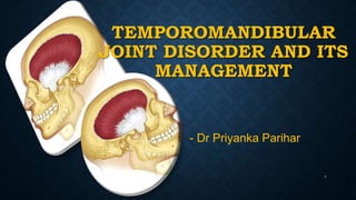
  • 1. TEMPOROMANDIBULAR JOINT DISORDER AND ITS MANAGEMENT 1 - Dr Priyanka Parihar
  • 2. CONTENTS: INTRODUCTION TYPES OF JOINTS COMPONENTS OF THE TMJ MUSCLE OF THE JOINT INNERVATIONS OF TMJ VASCULARIZATION OF TMJ CLASSIFICATION OF TMJ DISORDERS ETIOLOGIC FACTORS 2
  • 3. Treatment of Functional Disturbances of the Masticatory System • Temporomandibular Disorders, • Treatment of Masticatory Muscle Disorders • Treatment of Temporomandibular Joint Disorders • Treatment of Chronic Mandibular Hypomobility and Growth Disorders • Occlusal Appliance Therapy 3 CONTENTS:
  • 4. INTRODUCTION: • Temporomandibular joint (TMJ) is formed by the articulation of the mandible and the temporal bone of the cranium. It is located anteriorly to the tragus of the ear, on the lateral aspect of the face. • It is a bicondylar joint in which the condyles, located at the two ends of the mandible. 4
  • 5. TYPES OF JOINT: • A joint is where two or more bones meet. • Fibrous joint: Immovable, Example: cranium, Radioulnar, Tibiofibular joint. • Cartilaginous joint: slightly moveable Example: vertebral column • Synovial joint : freely moveable example : knee joint , hip joint 5
  • 6. • types of Synovial joint 1. Hinge joint 2. Pivot joint 3. Condyloid joint 4. Ball and socket joint 5. Gliding joint 6. Saddle joint 6
  • 7. Craniomandibular articulation-TMJ • Complex joint in the body Hinging movements - Ginglymoid joint Gliding movements –Arthroidal joint  Hence called as Ginglymoarthroidal joint • TMJ is a complex diarthroidal sliding- ginglymoid synovial joint, which attaches the mandible to petrous part of temporal bone of the cranium. 7
  • 8. COMPONENTS OF TMJ Hard tissue components 1. Mandibular condyles 2. Glenoid fossa of the temporal bone 3. Articlar eminence Soft tissue components 1. Articular capsule 2. Articular disc 3. Ligaments 4. Muscles 8
  • 9. HARD TISSUE COMPONENTS Mandibular condyle: • Are ovoid and strongly convex anterioposterioly and mildly convex mediolaterally. • Perpendicular to ascending ramus of the mandible. • The anterior surface shows a shallow depression called as Lateral pterygoid fovea to which lateral pterygoid muscle is attached. • Bony surface of condyle and articular part of the temporal bone covered with dense fibrous connective tissue. 9
  • 10. Glenoid fossa of the Temporal bone: • The articular surface of the temporal bone is situated on the inferior surface of the squamous part of the temporal bone. • This area which articulate with the mandibular condyle is called the Glenoid Fossa. • The floor of the fossa is formed by a thin plate of bone. • It is also covered by a fibrous layer similar to the condyle. 10
  • 11. Articular eminence: • The articular eminence is convex anterioposteriorly and concave in the transverse direction. • It is enclosed by the articular capsule of the TMJ. 11
  • 12. SOFT TISSUE COMPONENTS: Articular capsule: • Articular capsule is a thin sleeve of dense cartilaginous tissue enclosing the joint cavity. • The articular capsule consists of two layers: a) An outer layer (stratum fibrosum) composed of avascular, white fibrous tissue. b) An inner layer (stratum synoviale) which is a secreting layer usually known as the synovial membrane. 12
  • 13. • Synovial membrane: • It is a thin and flexible layer lining the inner surface of the joint capsule • The articulating surfaces and the articulating disc are not covered by the membrane The synovial membrane consists of two layers: • Cellular intimal layer • Vascular subintimal layer 13
  • 14. • Function of synovial membrane  Lubricating the joint  Repairing wear 14
  • 15. Articular disc: • It is oval, firm, thick plate of dense fibrous cartilage located between the condyle and the articulating surface of the temporal bone. • Disc divides the joint cavity in to two compartments ie superior and inferior. • Function of disc: due its viscoelasticity it acts as shock absorber, stress distributor and provide gliding motion during opening and closing of the jaw. • The articular disc is predominantly avascular ,blood vessels are seen at the margins of the disc. 15
  • 16. Articular ligaments: TMJ is associated with four ligaments, One major and three minor ligaments. Temporomandibular ligament : is fan shaped present on lateral aspect of the articular capsule. Hence also called as lateral ligament. TM ligament helps in preventing displacement in posterior and inferior direction. Displacement of TMJ happens only in the anterior or forward direction. Temporomandibular Ligament (Lateral View). There are two distinct parts: the outer oblique portion (OOP) and the inner horizontal portion (IHP). The OOP limits normal rotational opening movement; the IHP limits posterior movement of the condyle and disc. 16
  • 17.  Sphenomandibular ligament: • This ligament is a flat thin band which is attached to the spine of the sphenoid superiorly and to the ligula inferiorly. • The main function of this ligament is to limit the distention of the mandible in an inferior direction.  Stylomandibular ligament: • This ligament extends from the apex of the styloid process of the temporal bone to the posterior border of the angle of the mandible. • Function: along with sphenomandibular ligament limits excessive opening of the mandible. 17
  • 18.  Capsular Ligament: • the entire TMJ is surrounded and encompassed by the capsular ligament. • The capsular ligament acts to resist any medial, lateral, or inferior forces that tend to separate or dislocate the articular surfaces. • Significant function to encompass the thus Retains the synovial fluid Capsular Ligament (Lateral View).It extends anterior to include the articular eminence and encompass the entire articular surface of the joint. 18 joint
  • 19. • Collateral (discal) ligament • The collateral ligaments attach the medial and lateral borders of the articular disc to the poles of the condyle. • They are commonly called the discal ligaments • There are two Medial discal ligament lateral discal ligament lateral 19
  • 20. MUSCLES OF THE JOINT The muscle associated with the TMJ is the Lateral pterygoid muscle. • Function : it pull the coronoid process of the mandible anteriorly, forcing the condylar process out of the mandible fossa along the articular eminence to protrude mandible. The upper head influences the position of the articular disc during movement of the TMJ. 20
  • 21. INNERVATION OF TMJ • The innervations of the TMJ involves four type of receptors: 1. Ruffin’s corpuscles function as static mechanoreceptors which position the mandible 2. Pacinian’s corpuscles are mechanoreceptors which helps in the perception of movement & firm pressure (not light touch). 3. Golgi tendon protect the muscle from excessive or damaging tension. 4. The nociceptors (along with the proprioceptors) primarily function to monitor the condition, position, and movement of the tissues in the masticatory system. 21
  • 22. • These receptors are innervated by the Auriculotemporal nerve which leaves the mandibular nerve behind the joint and ascends laterally and superiorly to wrap around the posterior region of the joint. • Additional innervations by Deep temporal and Massetric nerve. 22
  • 23. BLOOD SUPPLY TO TMJ • The main source of blood supply of TMJ is provided by the Superficial temporal artery and maxillary artery which are branches of the External carotid artery • Other branches of the external carotid artery- The Deep auricular artery, Anterior tympanic artery and Ascending pharyngeal artery also contribute to the arterial blood supply to the joint. 23
  • 24. • In brief the muscles of mastication helps in biomechanics/movements of mandible: Movement Muscle Elevation (closing) Temporalis, Masseter Medial pterygoid Depression ( opening) Lateral pterygoid Superior infrahyoid Protrusion (forward) Lateral pterygoid Masseter Medial pterygoid Retrusion (backward) Temporalis Masseter Lateral movement Temporalis (same side) Pterygoid (of Opposite side) Masseter 24
  • 25. CLASSIFICATION OF TMJ DISORDERS 25
  • 26. •Pseudotumors (synovial chondromatosis) •Benign (chondroma, osteotoma) •Malignant (primary, metastatic) •Synovitis •Capsulitis •Rheumatoid arthritis •Juvenile rheumatoid arthritis •Ankylosing spondylitis •Psoriatic arthritis •Reactive arthritis (bacterial, viral, fungal) PETERSON’S CLASSIFICATION ARTICULAR DISORDERS Inflammatory arthropathies •Primary osteoarthrosis (no clear predisposind factor) •Secondary osteoarthrosis (trauma,previous surgery,avasular necrosis) •Mechanical derangements •Bone and cartilage disorders with articular manifestations • •Non-neoplastic:developmental (hyperplasia, hypoplasia, dysplasia) •Non-neoplastic: acquired(ie, condylolysis) Growth Disorder Noninflammatory arthropathies Neoplasm Diffuse connective tissue disorders Miscellaneous articular disorders 26
  • 27. •Muscle spasm (strain) •Myofascial pain and dysfunction (MPD) •Fibromyalgia •Myotonic dystrophies •Myositis ossificans progressiva NON-ARTICULAR DISORDERS Muscle disorders Growth disorders 27
  • 28. ETIOLOGIC FACTORS FOR TEMPOROMANDIBULAR DISORDERS 1. OCCLUSAL CONDITIONS 2. TRAUMA 3. EMOTIONAL STRESS 4. Deep Pain Input 5. Parafunctional Activity 28
  • 29. TRAUMA • Trauma can be divided into two general types: macrotrauma and microtrauma. • Macrotrauma is considered any sudden force that can result in structural alterations, such as a direct blow to the face. • Microtrauma refers to any small force that is repeatedly applied to the structures over a long period of time. Activities such as bruxism or clenching can produce microtrauma to the tissues that are being loaded (i.e., teeth, joints, or muscles) 29
  • 30. EMOTIONAL STRESS • The emotional centers of the brain have an influence on muscle function. • Stress affects the body by activating the hypothalamus-pituitary-adrenal axis (HPA axis) • The HPA axis, through complex neural pathways, increases the activity of the gamma efferents, which cause the intrafusal fibers of the muscle spindles to contract. • This so sensitizes the spindle that any slight stretching of the muscle will cause a reflex contraction. The overall effect is an increase in tonicity of the muscle 30
  • 31. DEEP PAIN INPUT • Deep pain input can centrally excite the brainstem producing a muscle response known as protective co-contraction. • This represents a normal healthy manner in which the body responds to injury or threat of injury. • Therefore it is reasonable to find a patient who is suffering with pain, such as toothache (i.e., necrotic pulp), to have limited mouth opening. • This represents the body’s response to protect the injured part by limiting its use. 31
  • 32. 32
  • 33. 33
  • 34. 34
  • 36. 1. General Considerations In The Treatment Of Temporomandibular Disorders 36 3. Treatment of Temporomandibular Joint Disorders 2. Treatment of Masticatory Muscle Disorders 4. Treatment of Chronic Mandibular Hypomobility and Growth Disorders 5. Occlusal Appliance Therapy
  • 37. • Definitive Treatment • Definitive therapy is aimed directly toward the elimination or alteration of the etiologic factors that are responsible for the disorder. • For example, definitive treatment for an anterior displacement of the articular disc will reestablish the proper condyle-disc relationship. 37 1. General Considerations In The Treatment Of Temporomandibular Disorders
  • 38. • Supportive Therapy • Supportive therapy is directed toward altering the patient’s symptoms and often has no effect on the etiology of the disorder. • A simple example is giving a patient aspirin for a headache that is caused by hunger. The patient may feel relief from the headache, but there is no change in the etiologic factor (hunger) that created the symptom. • Supportive therapy is directed toward the reduction of pain and dysfunction. 38
  • 39. • The two general types of supportive therapies are Pharmacologic Therapy  Physical Therapy 1. Pharmacologic Therapy • The most common classes of pharmacologic agents used for the management of TMD are analgesics, antiinflammatories, muscle relaxants, anxiolytics, antidepressants, anticonvulsives, injectables, and topicals. 39
  • 40. 2. Physical Therapy • Physical therapy represents a group of supportive activities that is usually instituted in conjunction with definitive treatment. • Most physical therapy fits into one of two general categories: modalities and manual techniques 40
  • 41. 41 a)THERMOTHERAPY (moist heat) b)COOLANT THERAPY, (ice pack and Fluoromethane spray) c)IONTOPHORESIS TREATMENT d)ULTRASOUND THERAPY e)TRANSCUTANEOUS ELECTRICAL NERVE STIMULATION (TENS) Physical Therapy Modalities- types: f)ELECTROGALVANIC STIMULATION THERAPY, g)LASER. h) PHONOPHORESIS,
  • 42. • Manual Techniques Manual techniques are the “hands-on” therapies provided by the physical therapist for the reduction of pain and dysfunction. • Manual techniques are divided into three categories: soft tissue mobilization,  joint mobilization, and muscle conditioning. 42
  • 43. 43 MASSAGE THERAPY SOFT TISSUE MOBILIZATION. JOINT MOBILIZATION JOINT DISTRACTION OF THE TMJ
  • 44. 44 MUSCLE CONDITIONING ASSISTED MUSCLE STRETCHING PASSIVE MUSCLE STRETCHING. RESISTANCE EXERCISES A. The patient is instructed to open against gentle resistance provided by the fingers. This will promote relaxation in the elevator muscles, thus allowing increased mandibular opening. B. When eccentric movement is limited the patient can be asked to move in the eccentric position with gentle resistance from the fingers. These exercises are repeated 10 times each session, six sessions a day. If they elicit pain,they are discontinued. A B
  • 45. Acupuncture- • Stimulation of acupuncture points appears to cause the release of endogenous opioids (endorphins and enkephalins), which reduce painful sensations by flooding the afferent interneurons with subthreshold stimuli. • These effectively block the transmission of noxious impulses and thus reduce the sensations of pain. 45 These needles are maintained in place for approximately 30 minutes during which they are twirled every 5 to 10 minutes.
  • 46. • Protective Co-Contraction (Muscle Splinting) • Local Myalgia (Noninflammatory Myalgia) • Myospasms (Tonic Contraction Myalgia) • Myofascial Pain (Trigger Point Myalgia) • Centrally Mediated Myalgia (Chronic Myositis) 46 2. Treatment of Masticatory Muscle Disorders
  • 47. PROTECTIVE CO-CONTRACTION (MUSCLE SPLINTING) • Protective co-contraction is the initial response of a muscle to altered sensory or proprioceptive input or injury. This response has been called protective muscle splinting . • In the presence of altered sensory input or pain, antagonistic muscle groups seem to activate during movement in an attempt to protect the injured part. Therefore, pain felt in the masticatory system can produce protective co-contraction of masticatory muscles. 47
  • 48. • protective co-contraction is not a pathologic condition but a normal physiologic response of the musculoskeletal system. Etiology 1. Altered sensory or proprioceptive input. 2. The presence of constant deep pain input. 3. Increased emotional stress. • Protective co-contraction only remains a few hours or days. If it is not resolved, local myalgia is likely to follow. 48 P. Co-Contraction
  • 49. Clinical Characteristics 1. Structural dysfunction: Decreased range of movement. 2. There is very minimal pain at rest. 3. There is increased pain to function. 4. The patient reports a feeling of muscle weakness. 49 This patient bites his cheek causing acute tissue injury and pain. This pain led to protective co-contraction. P. Co-Contraction
  • 50. TREATMENT • Protective co-contraction is a normal CNS response and therefore there is no indication to treat the muscle condition itself. Treatment should instead be directed toward the reason for the co-contraction. • When co-contraction results from trauma, definitive treatment is not indicated since the etiologic factor is no longer present. • When co-contraction results from a poorly fitting restoration, altering the restoration is advised. • If an increase in emotional stress is the etiology then appropriate stress management. • Short-term pain medication NSAIDs may be indicated 50 P. Co-Contraction
  • 52. 52
  • 53. LOCAL MYALGIA (NONINFLAMMATORY MYALGIA) • It is noninflammatory, myogenous pain disorder. It is often the first response of the muscle tissue to continued protective co- contraction. • Local myalgia represents a change in the local environment of the muscle tissues. It represents the initial response to overuse of muscle. 53
  • 54. LOCAL MYALGIA (NON-INF. MYALGIA) Etiology 1. Protracted protective co-contraction secondary to a recent alteration in local structures. 2. A continued source of constant deep pain 3. Local tissue trauma. 4. Increased levels of emotional stress. 54
  • 55. 1. Structural dysfunction: There is marked decrease in the range of mandibular movement. 2. There is minimum pain at rest and increased with function. 4. Actual muscle weakness present. 5. There is local tenderness when the involved muscles are palpated 55 Clinical Characteristics LOCAL MYALGIA (NON-INF. MYALGIA)
  • 56. • The primary goal in treating local myalgia is to decrease sensory input (such as pain) • Achieved by- 1. Eliminate any ongoing altered sensory input. 2. Eliminate any ongoing source of deep pain input. 3. Provide patient education and information on self-management i.e Advise the patient to restrict mandibular use to within painless limits. 56 TREATMENT LOCAL MYALGIA (NON-INF. MYALGIA)
  • 57. • If due to bruxism -fabricate an occlusal appliance for nighttime use while for daytime educate patient to control it. • If the above therapies fail to resolve the pain condition, the clinician may consider the use of a mild analgesic. • such as aspirin, acetaminophen, or an NSAID (i.e., ibuprofen), can be helpful. 57 LOCAL MYALGIA (NON-INF. MYALGIA)
  • 58. MYOSPASMS (TONIC CONTRACTION MYALGIA) • Myospasm is an involuntary CNS-induced tonic muscle contraction often associated with local metabolic conditions within the muscle tissues. Etiology 1. Continued deep pain input. 2. overuse 3. Idiopathic myospasm mechanisms 58
  • 59. CLINICAL CHARACTERISTICS • The patient reports a sudden onset of restricted jaw movement usually accompanied by muscle rigidity. • Structural dysfunction: There is marked restriction in range of mandibular movement. • Acute malocclusion is common. • There is pain at rest and pain is increased with function. • The affected muscle is firm and painful to palpation. 59
  • 60. • Two treatments are suggested for acute myospasms. • The first is directed immediately toward reducing the spasm itself while the other addresses the etiology. 1. Pain Reduction- can be achieved by manual massage, vapocoolant spray, ice, or even an injection of local anesthetic (2% lidocaine without a vasoconstrictor is recommended.) 2. Etiology reduction When the myospasms are secondary to fatigue and overuse, patient is advised to rest the muscle. • Idiopathic - may represent an oromandibular dystonia 60 TREATMENT
  • 61. MYOFASCIAL PAIN (TRIGGER POINT MYALGIA) • Myofascial pain is a regional myogenous pain condition characterized by local areas of firm, hypersensitive bands of muscle tissue known as trigger points. 61
  • 62. Etiology- 1. Continued source of deep pain input. 2. Increased levels of emotional stress. 3. The presence of sleep disturbances. 4. Local factors that influence muscle activity such as habits, posture, muscle strains, or even chilling. 5. Systemic factors such as nutritional inadequacies, poor physical conditioning, fatigue, and viral infections. 6. Idiopathic trigger point mechanism. 62 Myofascial Pain
  • 63. 63 TREATMENT Myofascial Pain 1. Eliminate any source of ongoing deep pain. 2. If a sleep disorder is suspected, Often, low dosages of a tricyclic antidepressant,such as 10 to 20 mg of amitriptyline before bedtime, can be helpful. 3. elimination of the trigger points, by painlessly stretching the muscle containing the trigger points. • Spray and Stretch, Pressure and Massage, Ultrasound and Electrogalvanic Stimulation, Injection and Stretch.
  • 64. • A medication such as cyclobenzaprine (Flexeril), 10 mg before sleep can often reduce pain but the trigger points still need to be treated. • Muscle relaxants help convert an active trigger point into a latent or dormant trigger point but may not necessarily eliminate it. • Analgesics may also be helpful in interrupting the cyclic effect of pain. 64 TREATMENT
  • 65. CENTRALLY MEDIATED MYALGIA (CHRONIC MYOSITIS) • Centrally mediated myalgia is a chronic, continuous muscle pain disorder originating predominantly from CNS effects that are felt peripherally in the muscle tissues. 65 The illustration depicts how central activation of the hypothalamus, the limbic structures and the cortex can combine to produce an antidromic effect to the muscle tissues.
  • 66. Etiology • Chronic centrally mediated myalgia may be caused by the prolonged input of muscle pain associated with local myalgia or myofascial pain. • Chronic upregulation of the autonomic nervous system • Chronic exposure to emotional stress • Other sources of deep pain input. 66
  • 67. CLINICAL CHARACTERISTICS 1. Structural dysfunction 2. There is significant pain at rest and pain is increased with function. 4. There is a generalized feeling of muscle tightness. 5. There is significant pain on muscle palpation. 67
  • 68. • Restrict mandibular use to within painless limits • Avoid exercise and/or injections. Since the muscle tissue is neurogenically inflamed, any use increases pain. • The patient should rest the muscles as much as possible. • Disengage the teeth by a stabilization appliance. • Begin taking an antiinflammatory medication. • If a sleep disorder is suspected, Often, low dosages of a tricyclic antidepressant is advised at bedtime. 68 TREATMENT
  • 69. • This internal physiology is only under the control of the patient, not the clinician. That is why the patient needs to understand this relationship and work on changing behavior that will downregulate the autonomic nervous system. • Physical self regulation plays important role in its treatment 69 TREATMENT Application of moist heat or cold can be helpful for chronic centrally mediated myalgia
  • 70. • Disc displacement and disc displacement with intermittent locking • Disc displacement without reduction • Structural incompatibility Deviation in form and Adhesions • Subluxation and Luxation • Capsulitis, Retrodiscitis, and Acute Trauma to the TMJ • Osteoarthritis • Infectious arthritis 70 3.Treatment of Temporomandibular Joint Disorders Inflammatory disorders
  • 71. Etiology • Disc derangement disorders result from elongation of the capsular and discal ligaments coupled with thinning of the articular disc. Clinical Characteristics • The clinical examination reveals a relatively normal range of movement with restriction only associated with the pain. • Discal movement can be felt by palpation of the joints during opening and closing. • Deviations in the opening pathway . 71 Disc displacement and disc displacement with intermittent locking
  • 72. • Anterior positioning appliance to reposition to condyle back on the disc. • It was originally suggested that this appliance be worn 24 hours a day for as long as 3 to 6 months. • The patient needs to be encouraged to decrease loading of the joint whenever possible. Softer foods, slower chewing,and smaller bites should be promoted. 72 TREATMENT
  • 73. • If inflammation is suspected, an NSAID should be prescribed. • Moist heat or ice can be used. • Active exercises are not advised. 73
  • 74. SO YOU ALL MIGHT BE WONDERING HOW DOES THIS WORKS ?? 74
  • 75. 75 In the resting closed joint position the disc is anteriorly displaced from the condyle A C B Maxillary occlusal appliance is placed which creates an occlusal condition that requires the mandible to shift slightly forward. Note that when the appliance is in place & the teeth are occluding, the condyle is repositioned on disc in normal condyle-disc relationship. Anterior Positioning Appliance
  • 76. 76 An anteriorly displaced disc with the condyle articulating on the retrodiscal tissues producing pain. APA is placed in mouth to bring the condyle forward,off of the retrodiscal tissues onto the disc. This relationship reduces the loading of the retrodiscal tissues, which decreases the pain. Once the tissues have adapted, the appliance is removed. Condyle now functions on the adaptive fibrotic tissues resulting in a painless functioning joint, but because the disc is still displaced, clicking may still be present. A C B
  • 77. DISC DISPLACEMENT WITHOUT REDUCTION • Disc displacement without reduction is a clinical condition in which the disc is totally displaced (dislocated) and does not return to normal position with condylar movement. Etiology • Macrotrauma and microtrauma are the most common causes of disc displacement without reduction. 77
  • 78. • A sudden change in range of mandibular movement occurs. • Examination reveals limited mandibular opening with some slight defection to the ipsilateral side. Treatment • Fabricating an anterior positioning appliance is contraindicated for this patient, as it will aggravate the condition by forcing the disc even more forward. • Recapture the disc by manual manipulation. 78 Clinical Characteristics
  • 79. 79 TREATMENT • Technique for Manual Manipulation. The clinician’s right thumb is placed intraorally over the patient’s left mandibular second molar and the mandible is grasped. With the left hand stabilizing the cranium, gentle but firm force is applied downward on the molar and upward on the chin to distract the joint.
  • 80. • Once the joint is distracted, the mandible is brought forward and to the right, enabling the condyle to move into the area of the displaced disc. When this position is achieved, constant distractive force is applied for 20 to 30 seconds while the patient relaxes. 80 TREATMENT
  • 81. • After the distraction, the thumb is removed and the patient is asked to close on the anterior teeth, maintaining the jaw in a slightly protrusive position. • When the patient has rested a moment, he or she is instructed to open maximally. If the disc has been reduced, a normal range of movement (48 mm) will be possible. 81 TREATMENT
  • 82. • Patients should be encouraged not to open too wide, especially immediately following the disc displacement without reduction. • The patient should also be told to decrease hard biting, no chewing gum. • If pain is present, heat or ice may be used. NSAIDs are indicated for pain and inflammation. • Joint distraction and phonophoresis over the joint area may be helpful. 82 TREATMENT
  • 83. • Surgical Considerations for Condyle-Disc Derangement Disorders • Arthrocentesis- In this procedure, two needles are placed into the joint and sterile saline solution is passed through lavaging the joint. • Arthroscopy- With this technique, an arthroscope is placed into the superior joint space and the intracapsular structures are visualized on a monitor. (diagnostic) • Arthrotomy- Open joint surgery • Discal Repair Or Plication- During a plication procedure, a portion of the retrodiscal tissue and inferior lamina is normally removed and the disc is retracted posteriorly and secured with sutures 83 TREATMENT
  • 84. STRUCTURAL INCOMPATIBILITY DEVIATION IN FORM AND ADHESIONS Deviation in Form- • Includes a group of disorders that is created by changes in the smooth articular surface of the joint and disc. These changes produce an alteration in the normal pathway of condylar movement. • The etiology of most deviations in form is trauma. Adherences/Adhesions • Adherences represent a temporary sticking of the articular surfaces during normal joint movements. caused by a fibrosis attachment of the articular surfaces. 84
  • 85. 85 Deviation in Form and Adhesions TREATMENT
  • 86. SUBLUXATION AND LUXATION Subluxation • Subluxation or hypermobility is a clinical description of the condyle as it moves anterior to the crest of the articular eminence. • It is not a pathologic condition but a variation in anatomic form of the fossa. Luxation • This condition is commonly referred to as an openlock since the patient’s mouth is wide open and cannot reduce it. • It can occur following wide-open mouth procedures such as having a dental appointment. 86
  • 88. CAPSULITIS, RETRODISCITIS, AND ACUTE TRAUMA TO THE TMJ Capsulitis- • An inflammatory condition of the capsular tissues Retrodiscitis- • An inflammatory condition of the retrodiscal tissues. 88 Retrodiscitis
  • 90. OSTEOARTHRITIS • Arthritis means inflammation of the articular surfaces of the joint. • Osteoarthritis is one of most common arthritis affecting the TMJ. Etiology • overloading of the articular structures of the joint. 90
  • 92. A. Chronic Mandibular Hypomobility- • The predominant feature of this disorder is the inability of the patient to open the mouth to a normal range. • Chronic mandibular hypomobility is rarely accompanied by painful symptoms. • subdivided into three categories according to etiology: • ankylosis, muscle contracture, and coronoid impedance. 92 4. Treatment of Chronic Mandibular Hypomobility and Growth Disorders
  • 93. ANKYLOSIS 93 • Abnormal immobility of a joint. • The two basic types of ankylosis are differentiated by the tissues that limit the mobility: fibrous and bony. • Fibrous ankylosis - most common and can occur between the condyle and the disc or the disc and the fossa. • Bony ankylosis of TMJ- would occur between the condyle and fossa and therefore the disc is mostly absent from the discal space prior to the ankylosis.
  • 94. • Etiology- The most common etiology of ankylosis is hemarthrosis (bleeding within the joint). • Chronic inflammation aggravates the disorder leading to the development of more fibrous tissue. • Management- • If function is inadequate or the restriction is intolerable, surgery is the only definitive treatment available. 94
  • 95. MUSCLE CONTRACTURE • MYOSTATIC CONTRACTURE • Myostatic contracture results when a muscle is kept from fully lengthening (stretching) for a prolonged time. • For example if the mandible is fractured and wired together with maxilla for 6 to 8 weeks the elevator muscles cannot fully lengthen. • Management -The resting length of the muscles can be reestablished by two types of exercise: passive stretching and resistant opening. • Thermotherapy and ultrasound are also helpful. 95
  • 96. Myofibrotic Contracture- • Myofibrotic contracture occurs as a result of excessivev tissue adhesions within the muscle or its sheaths. • These fibrosis tissue adhesions prevent the muscle fibers from sliding over themselves disallowing full lengthening of the muscle. • Etiology-myositis or trauma to the muscle tissues. • Management - surgical detachment of the muscles involved. 96
  • 97. CORONOID IMPEDANCE • If the pathway of coronoid process is impeded, it will not slide smoothly and the mouth will not fully open. • Etiology- Coronoid impedance is generally due to either elongation of the process or the encroachment of fibrous tissue. • Management- 1. surgery, that either shortens the coronoid process or eliminates the tissue obstruction ( if function is severely impaired). 2. Gentle passive stretching 97 very long coronoid process
  • 98. B. GROWTH DISORDERS • Common growth disturbances of the bones are agenesis (no growth), hypoplasia (insufficient growth), hyperplasia (too much growth), or neoplasia (uncontrolled, destructive growth). 98 Congenital and Developmental Bone Disorders
  • 99. Condylar Hyperplasia- • Bone disease characterized by the increased development of one mandibular condyle • Management -Condylectomy and orthognathic surgery, Mandibular ramus osteotomy Condylar Hypoplasia- • Known as underdevelopment of condyle • Management - orthognathic surgery, distraction osteogenesis, surgery with costochondral bone graft 99 Hyperplasia Hypoplasia
  • 100. • An occlusal appliance is a removal device made of hard acrylic that fits over the occlusal surfaces of the teeth in one arch. • It is commonly referred to as an occlusal splint, bite guard, night guard, interocclusal appliance, or even orthopedic device . • Occlusal appliances provides an occlusal condition that allows the condyles to assume their most orthopedically stable joint position. • They are also used to protect the teeth and supportive structures from abnormal forces that may create breakdown and tooth wear 100 5. Occlusal Appliance Therapy
  • 101. • The two most commonly used are the stabilization appliance (muscle relaxation appliance) and the anterior positioning appliance (orthopedic repositioning appliance). • Other types are the anterior bite plane, the posterior bite plane, the pivoting appliance, and the soft or resilient appliance 101 stabilization appliance anterior positioning appliance
  • 102. REFERENCES: • B D Chaurasia; Human Anatomy For Dental Students 3rd Edition. • Jeffrey P. Okeson, DMD 8th Edition • Peterson’s Principles Of Oral And Maxillofacial Surgery 2nd Edition. 102
  • 103. 103

Editor's Notes

  1. Good mornig respected mam staff my seniors and my collegues: today I am going to present a seminar on
  2. The contents are..
  3. By the name itself we can say that it is joint formed by the articulation of
  4. Type of joints
  5. the etiologic factor of occlusion is significant (perhaps a newly placed poorly fitting crown). If this factor exceeds the patient’s adaptability, temporomandibular disorder (TMD) symptoms are reported by the patient. In this instance, improvement of the occlusal condition (adjustment of the crown) would reduce this etiologic factor, thereby bringing the patient within adaptability thus resolving that TMD symptoms.