
Recommended
More Related Content
Similar to Larynx-III.pdf
Similar to Larynx-III.pdf (20)
More from snithiyuvarajayuvara
More from snithiyuvarajayuvara (17)
Recently uploaded
Recently uploaded (20)
Larynx-III.pdf
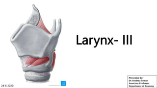
- 1. Larynx- III Presented by:- Dr. Sushma Tomar Associate Professor Department of Anatomy 24-6-2020
- 2. Lesson Plan Laryngeal Cavity: • Extent • Subdivisions Laryngeal Inlet & its Applied Aspects Subdivisions of Laryngeal Cavity: • Supraglottic compartment • Glottic compartment & its Applied Aspects • Infraglottic compartment Nerve supply of Larynx & its Applied aspects Arterial supply Venous drainage of Larynx Lymphatic drainage of Larynx Rima Glottidis Phonation Laryngoscopy Applied aspects: Singer’s nodules
- 3. Cavity of Larynx Extent- • From laryngeal inlet to lower border of cricoid cartilage. Communications- Posteriorly- with Laryngopharynx. Inferiorly- with lumen of Trachea. Anterior wall of laryngeal cavity is longer than the posterior wall.
- 4. Laryngeal Inlet • It is an opening (aperture) between laryngopharynx posteriorly and laryngeal cavity anteriorly. • It is obliquely placed. • Sloping downwards and backwards. • It is closed during deglutition to prevent entry of food into laryngeal cavity. Boundaries- Anterior- Epiglottis Posterior- Interarytenoid fold of mucous membrane. Lateral (on each side)- Aryepiglottic fold of
- 6. Applied Aspects Heimlich Maneuver- • It is a life saving maneuver, which is performed in case of laryngeal obstruction (choking). Method- • Stand behind the victim. • Pass your arms under his/her arms. • Put your hands in front of victim’s epigastrium. • With one hand, forms a fist and put the other hand over the fist. • Give 3-4 abdominal thrusts in backward and upward direction.
- 7. Cavity of Larynx contd… Vestibular Folds- • These are mucosal folds produced by Vestibular Ligaments ( False Vocal cords). • The space between these folds is called Rima Vestibuli. • When a person holds his breath, vestibular folds come together and prevent the air from leaving the lungs, simultaneously prevent the food and liquids from entering the larynx. Vocal Folds- • These are mucosal folds produced by Vocal Ligaments ( True Vocal cords) and Vocalis muscle. • The space between these folds is called Rima Glottidis.
- 8. Subdivisions of Laryngeal Cavity • Supraglottic compartment (Vestibule). • Glottic compartment (Ventricle/Sinus of Larynx). • Infraglottic compartment.
- 9. Supraglottic Compartment (Vestibule) • From laryngeal inlet to vestibular fold. Anterior wall- • Formed by mucous membrane covering the posterior surface of epiglottis. Posterior wall- • Formed by mucous membrane covering the apices of Arytenoid cartilages and Corniculate cartilages. On each side- Aryepiglottic fold.
- 10. Supraglottic compartment (Vestibule) contd… • Aryepiglottic folds separate the supraglottic compartment from piriform fossa.
- 11. Glottic Compartment (Ventricle/Sinus of Larynx) • It is an elliptical space between vestibular and vocal folds. • On each side, a blind diverticulum of mucous membrane, called Saccule is present. • Saccule is located between vestibular fold and lamina of Thyroid cartilage. • Saccule has mucous glands for lubrication of vocal cords. • Saccule is known as ‘Oil can of Larynx’.
- 12. Applied Aspects Laryngocele- It is an air filled cystic swelling of the saccule. Whenever air pressure in the laryngeal sinus is raised too much, the saccule dilates to produce an air-filled cystic swelling called Laryngocele. It may be: • Internal. • External. Internal Laryngocele- • When it is located within the larynx. External Laryngocele- • When distended saccule herniates Thyrohyoid Membrane
- 13. Laryngocele • Laryngocele is mostly formed in trumpet players, glass blowers and weight lifters. Trumpet Player Glass Blower Weight Lifter
- 14. Infraglottic Compartment • Extends from vocal folds to lower border of cricoid cartilage.
- 15. Nerve Supply of Larynx Motor Nerve Supply- • Recurrent Laryngeal Nerve. • External Laryngeal Nerve. Sensory Nerve Supply- Above the Vocal folds- • Internal Laryngeal Nerve. Below the Vocal folds- • Recurrent Laryngeal Nerve.
- 16. Applied Aspects Damage to External Laryngeal Nerve- • Some weakness of phonation. Unilateral damage to Recurrent Laryngeal Nerve- On the affected side- • Vocal cord lies in paramedian(between abduction and adduction) position. • Vocal cord does not vibrate. Normal vocal cord moves freely and even cross the midline to meet the paralyzed vocal cord. Normal vocal cord compensate and phonation is not Paralyzed Left
- 17. Applied Aspects contd… Bilateral damage to Recurrent Laryngeal Nerve- Both vocal cords lie in paramedian position. Loss of phonation. Difficulty in breathing. Bilateral damage to External and Recurrent Laryngeal Nerves- Both vocal cords lie in cadaveric position. Paramedian position of Vocal
- 18. Arterial Supply of Larynx Above the Vocal folds- • Superior Laryngeal artery. Below the Vocal folds- • Inferior Laryngeal artery.
- 19. Venous Drainage of Larynx Superior Laryngeal Vein- • Drains into Superior thyroid vein. Inferior Laryngeal Vein- • Drains into Inferior thyroid vein.
- 20. Lymphatic Drainage of Larynx Above the Vocal folds- • Upper deep cervical lymph nodes. Below the Vocal folds- • Prelaryngeal lymph nodes. • Pretracheal lymph nodes. • Lower deep cervical lymph nodes.
- 21. Rima Glottidis • It is the narrowest anteroposterior cleft of laryngeal cavity. Boundaries- Anterior- • Angle of Thyroid cartilage. Posterior- • Interarytenoid fold of mucous membrane. On each side- • Anterior 3/5th – Vocal fold. • Posterior 2/5th- Vocal process of Arytenoid cartilage. Subdivisions- Two: Intermembranous part- • Anterior 3/5th portion, between the two vocal folds. Intercartilaginous part- • Posterior 2/5th portion, between the
- 22. Phonation & Shape of Rima Glottidis In Quiet breathing- • Intermembranous part- triangular • Intercartilaginous part- rectangular. • Rima glottidis is pentagonal. In Full inspiration- • Rima glottidis is diamond shaped. • Abduction of vocal cords. During High Pitch Voice- • Rima glottidis is reduced to a linear chink. • Adduction of both intermembranous and intercartilaginous parts. During Whispering- • Rima glottidis is ‘inverted funnel’ shaped. • Vocal cords are highly adducted. • Vocal processes of Arytenoid cartilages are separated by a triangular gap.
- 23. Laryngoscopy • Inspection of interior of larynx. Types- • Direct • Indirect Direct Laryngoscopy- • With the help of a laryngoscope. Indirect Laryngoscopy- • With the help of a laryngeal mirror. Laryngoscope Laryngeal Mirror
- 24. Structures seen on Laryngoscopy • Base of Tongue. • Median Glosso-epiglottic fold. • Valleculae. • Epiglottis. • Aryepiglottic folds. • Piriform fossae. • Vestibular folds. • Vocal folds. • Sinus of larynx.
- 25. Applied Aspects Vocal Nodules ( Singer’s Nodules/Screamer’s Nodules)- • These are inflammatory nodules develop at the junction of anterior 1/3rd and posterior 2/3rd of vocal cords. • These are bilateral and symmetrical. • These nodules usually develop in individuals who overuse their voice such as teachers, pop singers etc.