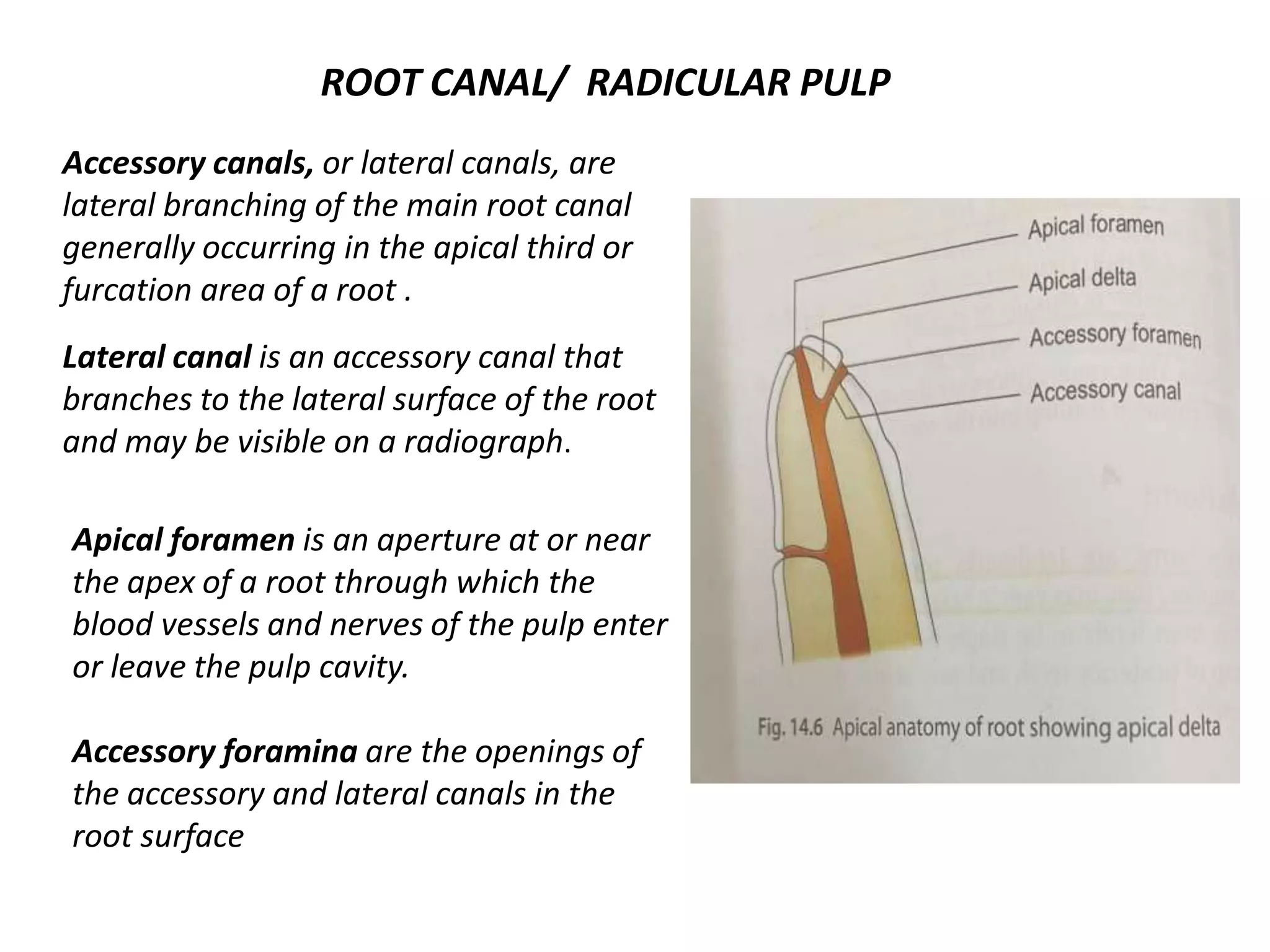
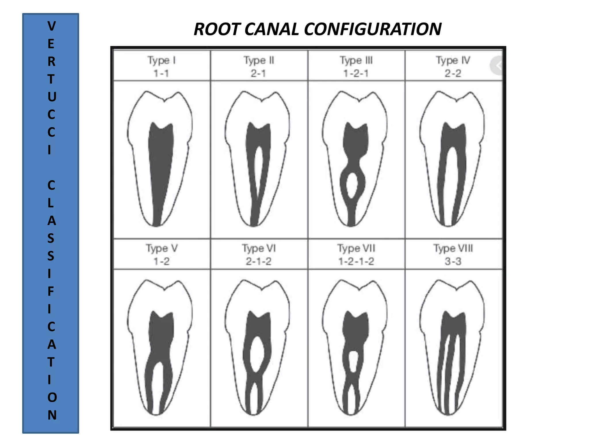
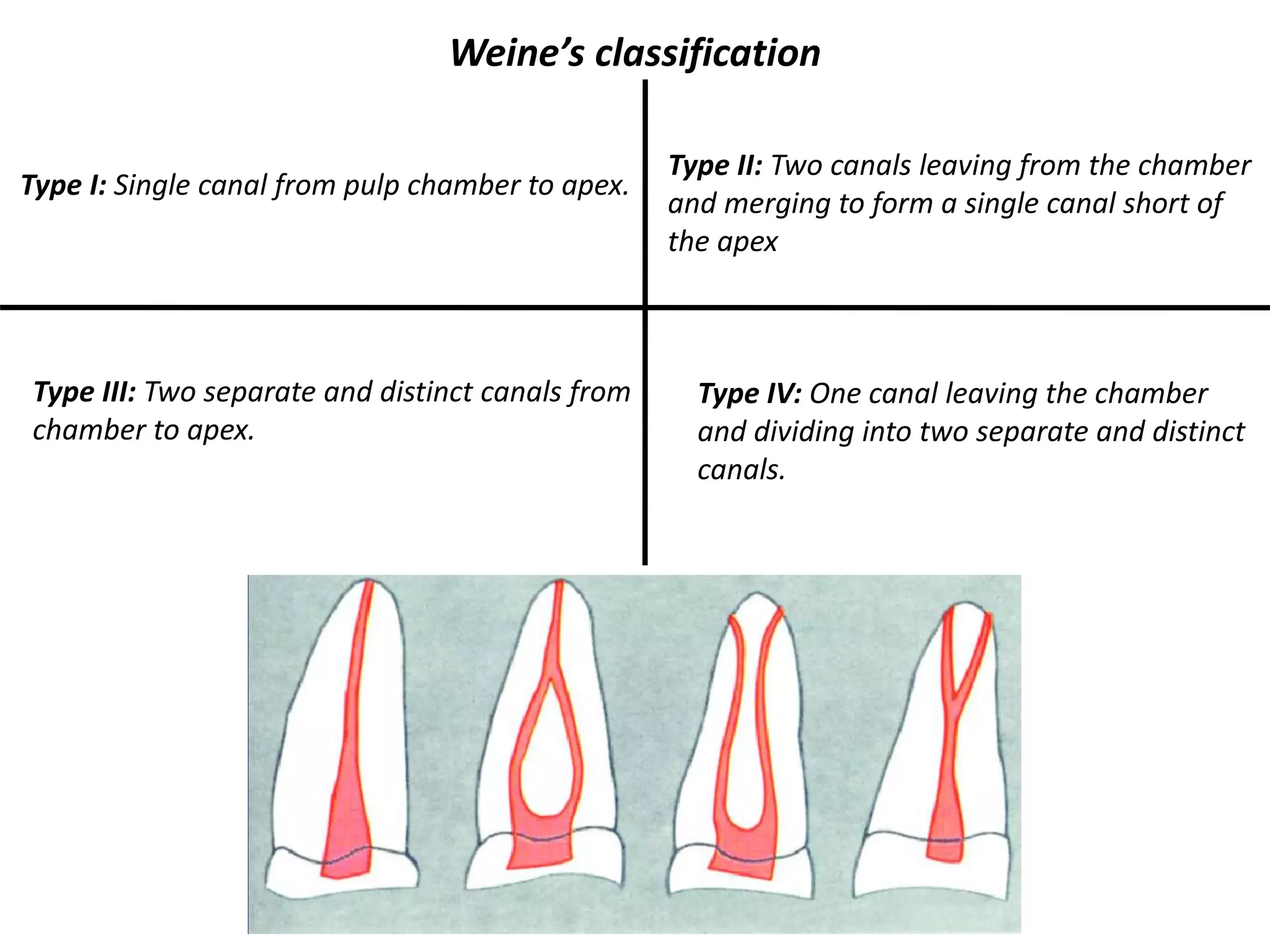
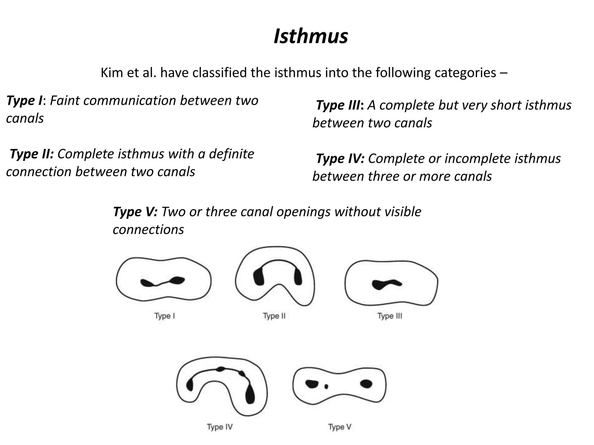
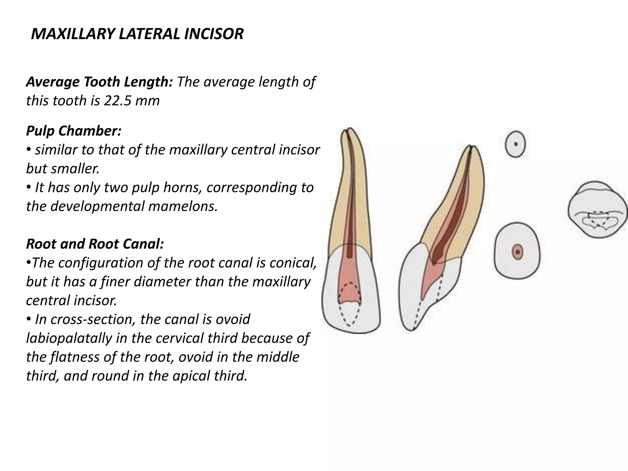
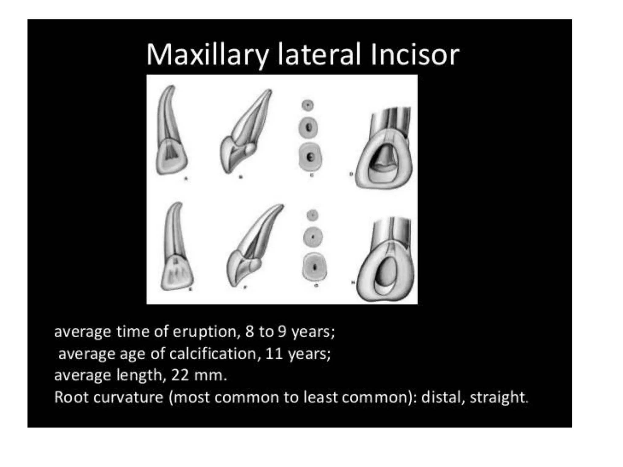
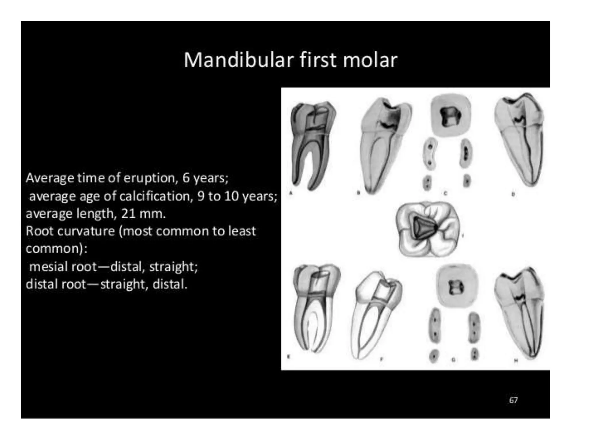
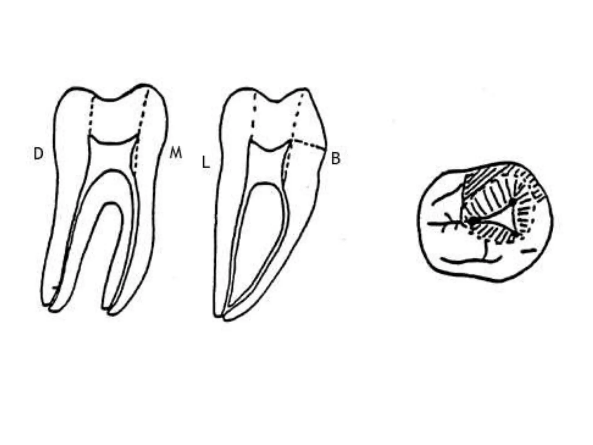
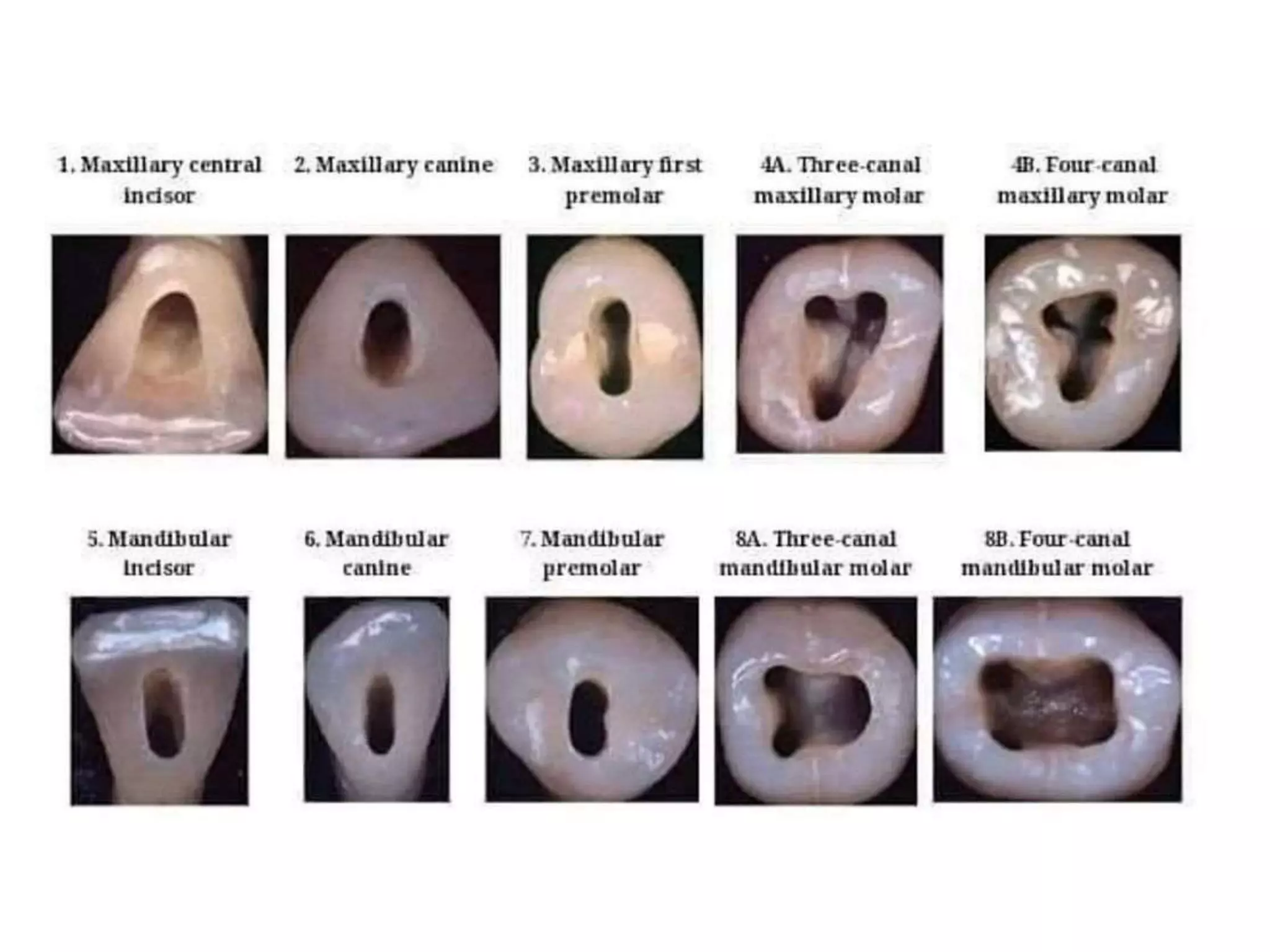
MORPHOLOGY AND INTERNAL
ANATOMY OF ROOT CANAL SYSTEM
Of all the phases of anatomic study in the
human system, one of the most complex is the
pulp cavity morphology. —M.T. Barrett
COMPONENTS OF ROOT CANAL
SYSTEM
ANATOMIC
COMPONENTS
PULP
CHAMBER/
CORONAL PULP
ROOTCANAL/
RADICULAR
PULP
Root canal system can be divided into two basic components
PULP CHAMBER: Coronal portion, that which is located in the
anatomic crown of tooth. Also called coronal pulp.
ROOT CANAL: Radicular portion, that which is located in anatomic
root of tooth. Also called radicular pulp.
PULP CHAMBER / CORONAL CHAMBER
Roof of pulp chamber- Dentin covering the pulp chamber
occlusally or incisally.
Pulp Horn- accentuation of roof of pulp chamber directly
under the cusp or a developmental lobe
Floor of pulp chamber- Runs parallel to roof and consist of
dentin bounding the pulp chamber near the cervical area of
tooth
Canal orifice – Opening in the floor of pulp chamber
leading into root canals
Accessory canals, or lateral canals, are
lateral branching of the main root canal
generally occurring in the apical third or
furcation area of a root .
Lateral canal is an accessory canal that
branches to the lateral surface of the root
and may be visible on a radiograph.
Apical foramen is an aperture at or near
the apex of a root through which the
blood vessels and nerves of the pulp enter
or leave the pulp cavity.
Accessory foramina are the openings of
the accessory and lateral canals in the
root surface
ROOT CANAL/ RADICULAR PULP
ROOT CANAL CONFIGURATIONV
E
R
T
U
C
C
I
C
L
A
S
S
I
F
I
C
A
T
I
O
N
Type I: Single canal from pulp chamber to apex.
Type III: Two separate and distinct canals from
chamber to apex.
Weine’s classification
Type II: Two canals leaving from the chamber
and merging to form a single canal short of
the apex
Type IV: One canal leaving the chamber
and dividing into two separate and distinct
canals.
Isthmus
Type I: Faint communication between two
canals
Type II: Complete isthmus with a definite
connection between two canals
Type III: A complete but very short isthmus
between two canals
Type IV: Complete or incomplete isthmus
between three or more canals
Kim et al. have classified the isthmus into the following categories –
Type V: Two or three canal openings without visible
connections
INDIVIDUAL TOOTH ANATOMY
MAXILLARY CENTRAL INCISOR
Average Tooth Length: The average length of
this tooth is 22.8 mm.
Pulp Chamber:
•Located in the center of the crown equidistant
from the dentinal walls.
• Broad mesiodistally
•The pulp chamber usually has three pulp
horns that correspond to the developmental
mamelons in a young tooth.
•The chamber is ovoid mesiodistally.
Root and Root Canal:
• One root with one root canal.
•The root canal is broad labiopalatally .
•Conical in shape, and centrally located.
• In cross-section, the canal is ovoid
mesiodistally in the cervical third, ovoid to
almost round in the middle third, and round in
the apical third.
MAXILLARY LATERAL INCISOR
Average Tooth Length: The average length of
this tooth is 22.5 mm
Pulp Chamber:
• similar to that of the maxillary central incisor
but smaller.
• It has only two pulp horns, corresponding to
the developmental mamelons.
Root and Root Canal:
•The configuration of the root canal is conical,
but it has a finer diameter than the maxillary
central incisor.
• In cross-section, the canal is ovoid
labiopalatally in the cervical third because of
the flatness of the root, ovoid in the middle
third, and round in the apical third.
Average Tooth Length: The average length of
this tooth is 26 mm, the longest of human
teeth. A specimen 33.5 mm in length has been
reported.
Pulp Chamber:
• The pulp chambers are the largest of any single-rooted teeth.
• Labiopalatally, the chamber is triangular in shape, with the
apex towards the single cusp and a broad base in the cervical
third of the crown.
• In cross-section, the chamber is ovoid in shape, with a greater
diameter labiopalatally.
• Only one pulp horn is present, corresponding to one cusp.
Root and Root Canal:
•The single root canal of the maxillary canine is larger than that
of the maxillary incisor.
• It is wider in labiopalatal than in mesiodistal dimension.
Maxillary Canine
Mandibular Central Incisor
Average Tooth Length: The average length of
this tooth is 20.8 mm.
Pulp Chamber:
•The mandibular central incisor is the smallest
tooth in the arch.
•The pulp chamber is small and flat
mesiodistally.
• Labiolingually, the pulp chamber is wide and
ovoid in cross-section in the cervical third of
the crown and tapers incisally.
•Root and Root Canals:
•The mandibular central incisor has one root,
which is flat and narrow mesiodistally but wide
labiolingually.
•From the labiolingual dimension, the canal is
broad in the cervical and middle thirds of the
root, tapers gradually toward the apex, and
forms a constriction in the apical third of the
root.
Average Tooth Length: This tooth averages
22.6 mm in length.
Pulp Chamber:
•The configuration of the pulp chamber is
similar to that of the mandibular central
incisor, but the lateral incisor has larger
dimensions.
Root and Root Canals:
• Although the root of the mandibular lateral
incisor is larger than that of the mandibular
central incisor, it has basically the same
configuration
Mandibular Lateral Incisor
•The majority of the roots are straight or
distally or labially curved, but the distal curve
of the lateral incisors is sharper.
•The incidence of double-root canals at the
apex is about 22%
Mandibular Canine
Average Tooth Length: The average length of
this tooth is 25 mm.
Pulp Chamber:
•The mandibular canine resembles the
maxillary canine, but it is smaller in all
dimensions.
•The pulp chamber is narrow mesiodistally.
Only one pulp horn is present in the adult
tooth. In cross-section, the chamber is ovoid in
the cervical third.
Root and Root Canals:
•Although the tooth usually has a single root, it
may have two (2.3%).
•The mandibular canine usually has one canal
exiting in one apical foramen (78%).
•It is ovoid in cross-section in the cervical and
middle thirds of the root and round in the
apical third.
Maxillary First Premolar
Average Tooth Length: The average length of
this tooth is 21.5 mm.
•Pulp Chamber:
•It has a pulp horn under each cusp.
• The buccal pulp horn is more prominent than
the palatal pulp horn.
•Usually it has two canal orifices, one buccal
and the other palatal.
• In cross-section, the pulp chamber is wide and
ovoid in a buccopalatal dimension.
Roots and Root Canals:
•The maxillary first premolar has two roots in
54.6% of cases.
• The buccal surface of a single- or double-
rooted maxillary first premolar is in close
proximity to the buccal cortical plate. The
proximity of these roots to the cortical plate
may produce a fenestration or a dehiscence in
that plate
Maxillary Second Premolar
Average Tooth Length: The average length of
this tooth is 21.6 mm.
Pulp Chamber:
It is wider buccopalatally than the maxillary
first premolar and shows two pulp horns in this
projection, a buccal and a palatal.
Root and Root Canals:
Maxillary second premolars have only a single
root in 90.3% of patients. Only 2% have two
well-developed roots. When two canals occur,
they may be distinct and separated along the
entire length of the root, or they may converge
to form a common canal as they approach the
apex. The majority of canals are curved.
Mandibular First Premolar
Average Tooth Length: This tooth averages 21.9 mm.
Pulp Chamber:
•The mesiodistal width of the pulp chamber is narrow.
• Buccolingually, the pulp chamber is wide, with a
prominent buccal pulp horn that extends under a well-
developed buccal cusp.
• The prominent buccal cusp and the smaller lingual cusp
give the crown of the mandibular first premolar about a
30° lingual tilt
Roots and Root Canals:
•The mandibular first premolar usually has a short, conical
root.
•The root is usually straight (48%), but some roots curve
distally (35%). Three canals exit in three foramina in
0.5% of cases. If one canal is present, it will be cone
shaped and simple in outline.
•In cross-section, the cervical and middle thirds are
ovoid, and the apical third is round.
Average Tooth Length: The length of this tooth
averages 22.3 mm.
Pulp Chamber:
•The pulp chamber is similar to that of the
mandibular first premolar, except that the
lingual horn is more prominent under a well-
developed lingual cusp.
Roots and Root Canals:
• The mandibular second premolar usually has
a single root, but on rare occasions two to
three roots are present.
•The root of the mandibular second premolar
may curve distally
•. Usually, one canal exits in one apical
foramen (97.5%), but in some roots (2.5%), a
single canal may bifurcate exiting in two
foramina.
Mandibular Second Premolar
MAXILLARY FIRST MOLAR
Average Tooth Length: The average length of this tooth is
21.3 mm.
Pulp Chamber:
• The pulp chamber of the maxillary first molar has four pulp
horns: mesiobuccal, distobuccal, mesiopalatal, and
distopalatal.
•The arrangement of the horns gives the pulpal roof a
rhomboidal shape in cross-section.
•The floor of the pulp chamber thus has a triangular form in
cross-section.
• The palatal orifice is the largest, round or oval in shape,
and easily accessible for exploration.
•The mesiobuccal orifice is under the mesiobuccal cusp, is
long buccopalatally, and may have a depression at the
palatal end in which the orifice of a fourth canal may be
present.
• This orifice is located by insinuating the tip of a long-
shank explorer in a mesiobucco-apical inclination into the
point angle created at the juncture of the buccal wall,
mesial wall, and subpulpal floor of the pulp chamber. The
distobuccal orifice is located slightly distal and palatal to
the mesiobuccal orifice and is accessible from the mesial
side for exploration.
Roots and Root Canals:
• The maxillary first molar has three roots
with, usually, three canals situated
mesiobuccally, distobuccally, and palatally.
•The mesiobuccal (MB) canal is not always
patent along its entire length and is divided
to form a second root canal known as the
Mb-2 canal.
•The distobuccal root is small and is more or
less round in shape.
•The distobuccal root usually has a single
root canal, which is a narrow and tapering
canal.
•The palatal root has the largest diameter
and is the longest root of the maxillary first
molar.
MAXILLARY SECOND MOLAR:
Average Tooth Length: The average length of this tooth is
21.7 mm.
Pulp Chamber:
•The pulp is similar to that of the maxillary first molar,
except it is narrower mesiodistally.
•The roof of the pulp chamber is more rhomboidal in
cross-section.
•The floor of the pulp chamber is an obtuse triangle in
cross-section.
Roots and Root Canals:
•The maxillary second molar usually has three roots.
•the buccal roots may fuse, and occasionally all three
roots fuse to form a single conical root.
• If the buccal roots fuse to form one buccal root, the
tooth may have only two canals, one buccal and one
palatal, although it is not unusual to find three canals
MANDIBULAR FIRST MOLAR:
•Average Tooth Length: The average length is 21.9 mm.
• Pulp Chamber:
•The roof of the pulp chamber of the mandibular first molar is often rectangular in shape.
•The roof of the pulp chamber has four pulp horns: mesiobuccal, mesiolingual,
distobuccal, and distolingual.
•Three distinct orifices are present in the pulpal floor: mesiobuccal, mesiolingual, and
distal.
•The mesiobuccal orifice is under the mesiobuccal cusp.
•The mesiolingual orifice is located in a depression formed by the mesial and lingual walls.
• The distal orifice, which is oval in shape, with the widest diameter present in a
buccolingual direction.
•The multiple orifices in the distal root are usually found in the buccal and lingual portion
of the ovoid coronal root canal.
•Roots and Root Canals: Usually, two well differentiated roots are present in the
mandibular first molar, one mesial and one distal.
•Both roots are wide and flat buccolingually. A third root is found in some cases, either
distally or mesially (5.3%), and is often referred to as the radix entomolaris.
• Although the mandibular first molar has two roots, three canals are usually present.
Average Tooth Length: This tooth averages
21.4 mm in length.
Pulp Chamber:
• The pulp chamber of the mandibular second
molar is smaller than that of the mandibular
first molar.
• the root canal orifices are smaller and closer
together.
Roots and Root Canals:
•The majority of the mandibular second molars
have two roots (71%), but teeth with one root
(27%) and teeth with three roots (2%) are also
seen.
•Three root canals are usually present in the
mandibular second molar. The most frequent
variation is the presence of only two canals.
MANDIBULAR SECOND MOLAR:
Morphology and internal anatomy of root canal system

Morphology and internal anatomy of root canal system

  • 1.
    MORPHOLOGY AND INTERNAL ANATOMYOF ROOT CANAL SYSTEM Of all the phases of anatomic study in the human system, one of the most complex is the pulp cavity morphology. —M.T. Barrett
  • 2.
    COMPONENTS OF ROOTCANAL SYSTEM ANATOMIC COMPONENTS PULP CHAMBER/ CORONAL PULP ROOTCANAL/ RADICULAR PULP
  • 3.
    Root canal systemcan be divided into two basic components PULP CHAMBER: Coronal portion, that which is located in the anatomic crown of tooth. Also called coronal pulp. ROOT CANAL: Radicular portion, that which is located in anatomic root of tooth. Also called radicular pulp.
  • 4.
    PULP CHAMBER /CORONAL CHAMBER
  • 5.
    Roof of pulpchamber- Dentin covering the pulp chamber occlusally or incisally. Pulp Horn- accentuation of roof of pulp chamber directly under the cusp or a developmental lobe Floor of pulp chamber- Runs parallel to roof and consist of dentin bounding the pulp chamber near the cervical area of tooth Canal orifice – Opening in the floor of pulp chamber leading into root canals
  • 6.
    Accessory canals, orlateral canals, are lateral branching of the main root canal generally occurring in the apical third or furcation area of a root . Lateral canal is an accessory canal that branches to the lateral surface of the root and may be visible on a radiograph. Apical foramen is an aperture at or near the apex of a root through which the blood vessels and nerves of the pulp enter or leave the pulp cavity. Accessory foramina are the openings of the accessory and lateral canals in the root surface ROOT CANAL/ RADICULAR PULP
  • 7.
  • 8.
    Type I: Singlecanal from pulp chamber to apex. Type III: Two separate and distinct canals from chamber to apex. Weine’s classification Type II: Two canals leaving from the chamber and merging to form a single canal short of the apex Type IV: One canal leaving the chamber and dividing into two separate and distinct canals.
  • 9.
    Isthmus Type I: Faintcommunication between two canals Type II: Complete isthmus with a definite connection between two canals Type III: A complete but very short isthmus between two canals Type IV: Complete or incomplete isthmus between three or more canals Kim et al. have classified the isthmus into the following categories – Type V: Two or three canal openings without visible connections
  • 10.
    INDIVIDUAL TOOTH ANATOMY MAXILLARYCENTRAL INCISOR Average Tooth Length: The average length of this tooth is 22.8 mm. Pulp Chamber: •Located in the center of the crown equidistant from the dentinal walls. • Broad mesiodistally •The pulp chamber usually has three pulp horns that correspond to the developmental mamelons in a young tooth. •The chamber is ovoid mesiodistally. Root and Root Canal: • One root with one root canal. •The root canal is broad labiopalatally . •Conical in shape, and centrally located. • In cross-section, the canal is ovoid mesiodistally in the cervical third, ovoid to almost round in the middle third, and round in the apical third.
  • 13.
    MAXILLARY LATERAL INCISOR AverageTooth Length: The average length of this tooth is 22.5 mm Pulp Chamber: • similar to that of the maxillary central incisor but smaller. • It has only two pulp horns, corresponding to the developmental mamelons. Root and Root Canal: •The configuration of the root canal is conical, but it has a finer diameter than the maxillary central incisor. • In cross-section, the canal is ovoid labiopalatally in the cervical third because of the flatness of the root, ovoid in the middle third, and round in the apical third.
  • 15.
    Average Tooth Length:The average length of this tooth is 26 mm, the longest of human teeth. A specimen 33.5 mm in length has been reported. Pulp Chamber: • The pulp chambers are the largest of any single-rooted teeth. • Labiopalatally, the chamber is triangular in shape, with the apex towards the single cusp and a broad base in the cervical third of the crown. • In cross-section, the chamber is ovoid in shape, with a greater diameter labiopalatally. • Only one pulp horn is present, corresponding to one cusp. Root and Root Canal: •The single root canal of the maxillary canine is larger than that of the maxillary incisor. • It is wider in labiopalatal than in mesiodistal dimension. Maxillary Canine
  • 18.
    Mandibular Central Incisor AverageTooth Length: The average length of this tooth is 20.8 mm. Pulp Chamber: •The mandibular central incisor is the smallest tooth in the arch. •The pulp chamber is small and flat mesiodistally. • Labiolingually, the pulp chamber is wide and ovoid in cross-section in the cervical third of the crown and tapers incisally. •Root and Root Canals: •The mandibular central incisor has one root, which is flat and narrow mesiodistally but wide labiolingually. •From the labiolingual dimension, the canal is broad in the cervical and middle thirds of the root, tapers gradually toward the apex, and forms a constriction in the apical third of the root.
  • 20.
    Average Tooth Length:This tooth averages 22.6 mm in length. Pulp Chamber: •The configuration of the pulp chamber is similar to that of the mandibular central incisor, but the lateral incisor has larger dimensions. Root and Root Canals: • Although the root of the mandibular lateral incisor is larger than that of the mandibular central incisor, it has basically the same configuration Mandibular Lateral Incisor •The majority of the roots are straight or distally or labially curved, but the distal curve of the lateral incisors is sharper. •The incidence of double-root canals at the apex is about 22%
  • 21.
    Mandibular Canine Average ToothLength: The average length of this tooth is 25 mm. Pulp Chamber: •The mandibular canine resembles the maxillary canine, but it is smaller in all dimensions. •The pulp chamber is narrow mesiodistally. Only one pulp horn is present in the adult tooth. In cross-section, the chamber is ovoid in the cervical third. Root and Root Canals: •Although the tooth usually has a single root, it may have two (2.3%). •The mandibular canine usually has one canal exiting in one apical foramen (78%). •It is ovoid in cross-section in the cervical and middle thirds of the root and round in the apical third.
  • 23.
    Maxillary First Premolar AverageTooth Length: The average length of this tooth is 21.5 mm. •Pulp Chamber: •It has a pulp horn under each cusp. • The buccal pulp horn is more prominent than the palatal pulp horn. •Usually it has two canal orifices, one buccal and the other palatal. • In cross-section, the pulp chamber is wide and ovoid in a buccopalatal dimension. Roots and Root Canals: •The maxillary first premolar has two roots in 54.6% of cases. • The buccal surface of a single- or double- rooted maxillary first premolar is in close proximity to the buccal cortical plate. The proximity of these roots to the cortical plate may produce a fenestration or a dehiscence in that plate
  • 25.
    Maxillary Second Premolar AverageTooth Length: The average length of this tooth is 21.6 mm. Pulp Chamber: It is wider buccopalatally than the maxillary first premolar and shows two pulp horns in this projection, a buccal and a palatal. Root and Root Canals: Maxillary second premolars have only a single root in 90.3% of patients. Only 2% have two well-developed roots. When two canals occur, they may be distinct and separated along the entire length of the root, or they may converge to form a common canal as they approach the apex. The majority of canals are curved.
  • 27.
    Mandibular First Premolar AverageTooth Length: This tooth averages 21.9 mm. Pulp Chamber: •The mesiodistal width of the pulp chamber is narrow. • Buccolingually, the pulp chamber is wide, with a prominent buccal pulp horn that extends under a well- developed buccal cusp. • The prominent buccal cusp and the smaller lingual cusp give the crown of the mandibular first premolar about a 30° lingual tilt Roots and Root Canals: •The mandibular first premolar usually has a short, conical root. •The root is usually straight (48%), but some roots curve distally (35%). Three canals exit in three foramina in 0.5% of cases. If one canal is present, it will be cone shaped and simple in outline. •In cross-section, the cervical and middle thirds are ovoid, and the apical third is round.
  • 29.
    Average Tooth Length:The length of this tooth averages 22.3 mm. Pulp Chamber: •The pulp chamber is similar to that of the mandibular first premolar, except that the lingual horn is more prominent under a well- developed lingual cusp. Roots and Root Canals: • The mandibular second premolar usually has a single root, but on rare occasions two to three roots are present. •The root of the mandibular second premolar may curve distally •. Usually, one canal exits in one apical foramen (97.5%), but in some roots (2.5%), a single canal may bifurcate exiting in two foramina. Mandibular Second Premolar
  • 31.
    MAXILLARY FIRST MOLAR AverageTooth Length: The average length of this tooth is 21.3 mm. Pulp Chamber: • The pulp chamber of the maxillary first molar has four pulp horns: mesiobuccal, distobuccal, mesiopalatal, and distopalatal. •The arrangement of the horns gives the pulpal roof a rhomboidal shape in cross-section. •The floor of the pulp chamber thus has a triangular form in cross-section. • The palatal orifice is the largest, round or oval in shape, and easily accessible for exploration. •The mesiobuccal orifice is under the mesiobuccal cusp, is long buccopalatally, and may have a depression at the palatal end in which the orifice of a fourth canal may be present. • This orifice is located by insinuating the tip of a long- shank explorer in a mesiobucco-apical inclination into the point angle created at the juncture of the buccal wall, mesial wall, and subpulpal floor of the pulp chamber. The distobuccal orifice is located slightly distal and palatal to the mesiobuccal orifice and is accessible from the mesial side for exploration.
  • 32.
    Roots and RootCanals: • The maxillary first molar has three roots with, usually, three canals situated mesiobuccally, distobuccally, and palatally. •The mesiobuccal (MB) canal is not always patent along its entire length and is divided to form a second root canal known as the Mb-2 canal. •The distobuccal root is small and is more or less round in shape. •The distobuccal root usually has a single root canal, which is a narrow and tapering canal. •The palatal root has the largest diameter and is the longest root of the maxillary first molar.
  • 35.
    MAXILLARY SECOND MOLAR: AverageTooth Length: The average length of this tooth is 21.7 mm. Pulp Chamber: •The pulp is similar to that of the maxillary first molar, except it is narrower mesiodistally. •The roof of the pulp chamber is more rhomboidal in cross-section. •The floor of the pulp chamber is an obtuse triangle in cross-section. Roots and Root Canals: •The maxillary second molar usually has three roots. •the buccal roots may fuse, and occasionally all three roots fuse to form a single conical root. • If the buccal roots fuse to form one buccal root, the tooth may have only two canals, one buccal and one palatal, although it is not unusual to find three canals
  • 37.
    MANDIBULAR FIRST MOLAR: •AverageTooth Length: The average length is 21.9 mm. • Pulp Chamber: •The roof of the pulp chamber of the mandibular first molar is often rectangular in shape. •The roof of the pulp chamber has four pulp horns: mesiobuccal, mesiolingual, distobuccal, and distolingual. •Three distinct orifices are present in the pulpal floor: mesiobuccal, mesiolingual, and distal. •The mesiobuccal orifice is under the mesiobuccal cusp. •The mesiolingual orifice is located in a depression formed by the mesial and lingual walls. • The distal orifice, which is oval in shape, with the widest diameter present in a buccolingual direction. •The multiple orifices in the distal root are usually found in the buccal and lingual portion of the ovoid coronal root canal. •Roots and Root Canals: Usually, two well differentiated roots are present in the mandibular first molar, one mesial and one distal. •Both roots are wide and flat buccolingually. A third root is found in some cases, either distally or mesially (5.3%), and is often referred to as the radix entomolaris. • Although the mandibular first molar has two roots, three canals are usually present.
  • 40.
    Average Tooth Length:This tooth averages 21.4 mm in length. Pulp Chamber: • The pulp chamber of the mandibular second molar is smaller than that of the mandibular first molar. • the root canal orifices are smaller and closer together. Roots and Root Canals: •The majority of the mandibular second molars have two roots (71%), but teeth with one root (27%) and teeth with three roots (2%) are also seen. •Three root canals are usually present in the mandibular second molar. The most frequent variation is the presence of only two canals. MANDIBULAR SECOND MOLAR: