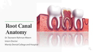
This document provides information on root canal anatomy, including:
- The root canal extends from the canal orifice in the pulp chamber to the apical foramen and is divided into coronal and radicular portions.
- Key anatomical landmarks in the apical third include the apical constriction, apical foramen, cementodentinal junction, accessory canals and lateral canals.
- Vertucci's and Weine's classifications describe different root canal configurations. Understanding root canal anatomy is important for successful root canal treatment.
The pulp cavity is the central cavity within a tooth and is entirely enclosed by dentin except at apical foramen.
It is divided into:
1. Coronal portion pulp chamber
2. Radicular portion root canal
PULP CHAMBER
ROOF OF PULP CAVITY: consists of dentin covering the pulp chamber occlussaly or incisally.
PULP HORN : Accentuation of the roof of pulp chamber directly under a cusp or developmental lobe.
FLOOR OF PULP CHAMBER: runs parallel to the roof and consists of dentin bounding the pulp chamber near cervical area of tooth, particularly dentin forming the furcation area.
CANAL ORIFICES: openings in the floor of pulp chamber leading to the root canals.
ROOT CANALS
Portion of the pulp cavity from the canal orifice to the apical foramen
Divided into 3 section( for convenience)
Coronal third
Middle third
Apical third
The root canal curvature
Straight canal extending with minimal apical curvature
Gradual curvature of canal with straight apical ending
Gradual curvature of entire canal
Sharp curvature of canal near the apex
Success of negotiating narrow curved canal depends on
Degree of curvature
Size and constriction of root canal
Size and flexibility of endodontic instrument blade
Skill of operator
Classification based on canal cross-section
Round/circular
Oval
Long oval
Flattened(flat/ribbon)
Irregular
Vertucci’s Classification
Weine’s Classification
ISTHMUS
A narrow passage or anatomic part connecting two larger structures (root canals)
APICAL FORAMEN
In young incompletely developed teeth the apical foramen is funnel shaped with wider portion extending outward
As root develops the apical foramen becomes narrower
Apical foramen is not the most constricted part of root apex\apical foramen is not always located at the centre of the root apex
LATERAL CANALS AND ACCESSARY FORAMINA
Lateral canals frequently occur in apical third of root
May occur in areas of bifurcation and trifurcation of multirooted teeth
With increasing age, number of accessory foramina reduce due to calcification of contained soft tissue
INFLUENCE OF AGING
METHODS OF DETERMINING PULP ANATOMY
CLINICAL METHODS
Anatomy studies
Radiographs
Explorations
High resolution compound tomography
Visualisation endogram
Fiberoptic endoscope
Magnetic resonance imaging
IN VITRO METHODS
sectioning of teeth
use of dyes
Contrasting media
Scanning electron microscope analysis
VARIATIONS IN INTERNAL ANATOMY
Variations in development
Gemination
Fusion
Concrescence
Taurodontism
Talon’s cusp
Dilaceration
Extra root canal
Dens invaginatus
Dens evaginatus
Maxillary Central Incisor
Maxillary Lateral Incisor
Maxillary Canine
Mandibular Central and Lateral Incisors
Mandibular Canine
Maxillary First Premolar
Maxillary Second Premolar
The typical second premolar has one
root and one canal and sometimes
has an apical distal curvature.
The Type I canal form is p
How many patients does case series should have In comparison to case reports.pdfpubrica101
Pubrica’s team of researchers and writers create scientific and medical research articles, which may be important resources for authors and practitioners. Pubrica medical writers assist you in creating and revising the introduction by alerting the reader to gaps in the chosen study subject. Our professionals understand the order in which the hypothesis topic is followed by the broad subject, the issue, and the backdrop.
https://pubrica.com/academy/case-study-or-series/how-many-patients-does-case-series-should-have-in-comparison-to-case-reports/
Global launch of the Healthy Ageing and Prevention Index 2nd wave – alongside...ILC- UK
The Healthy Ageing and Prevention Index is an online tool created by ILC that ranks countries on six metrics including, life span, health span, work span, income, environmental performance, and happiness. The Index helps us understand how well countries have adapted to longevity and inform decision makers on what must be done to maximise the economic benefits that comes with living well for longer.
Alongside the 77th World Health Assembly in Geneva on 28 May 2024, we launched the second version of our Index, allowing us to track progress and give new insights into what needs to be done to keep populations healthier for longer.
The speakers included:
Professor Orazio Schillaci, Minister of Health, Italy
Dr Hans Groth, Chairman of the Board, World Demographic & Ageing Forum
Professor Ilona Kickbusch, Founder and Chair, Global Health Centre, Geneva Graduate Institute and co-chair, World Health Summit Council
Dr Natasha Azzopardi Muscat, Director, Country Health Policies and Systems Division, World Health Organisation EURO
Dr Marta Lomazzi, Executive Manager, World Federation of Public Health Associations
Dr Shyam Bishen, Head, Centre for Health and Healthcare and Member of the Executive Committee, World Economic Forum
Dr Karin Tegmark Wisell, Director General, Public Health Agency of Sweden
CHAPTER 1 SEMESTER V - ROLE OF PEADIATRIC NURSE.pdfSachin Sharma
Pediatric nurses play a vital role in the health and well-being of children. Their responsibilities are wide-ranging, and their objectives can be categorized into several key areas:
1. Direct Patient Care:
Objective: Provide comprehensive and compassionate care to infants, children, and adolescents in various healthcare settings (hospitals, clinics, etc.).
This includes tasks like:
Monitoring vital signs and physical condition.
Administering medications and treatments.
Performing procedures as directed by doctors.
Assisting with daily living activities (bathing, feeding).
Providing emotional support and pain management.
2. Health Promotion and Education:
Objective: Promote healthy behaviors and educate children, families, and communities about preventive healthcare.
This includes tasks like:
Administering vaccinations.
Providing education on nutrition, hygiene, and development.
Offering breastfeeding and childbirth support.
Counseling families on safety and injury prevention.
3. Collaboration and Advocacy:
Objective: Collaborate effectively with doctors, social workers, therapists, and other healthcare professionals to ensure coordinated care for children.
Objective: Advocate for the rights and best interests of their patients, especially when children cannot speak for themselves.
This includes tasks like:
Communicating effectively with healthcare teams.
Identifying and addressing potential risks to child welfare.
Educating families about their child's condition and treatment options.
4. Professional Development and Research:
Objective: Stay up-to-date on the latest advancements in pediatric healthcare through continuing education and research.
Objective: Contribute to improving the quality of care for children by participating in research initiatives.
This includes tasks like:
Attending workshops and conferences on pediatric nursing.
Participating in clinical trials related to child health.
Implementing evidence-based practices into their daily routines.
By fulfilling these objectives, pediatric nurses play a crucial role in ensuring the optimal health and well-being of children throughout all stages of their development.
Telehealth Psychology Building Trust with Clients.pptxThe Harvest Clinic
Telehealth psychology is a digital approach that offers psychological services and mental health care to clients remotely, using technologies like video conferencing, phone calls, text messaging, and mobile apps for communication.
Explore our infographic on 'Essential Metrics for Palliative Care Management' which highlights key performance indicators crucial for enhancing the quality and efficiency of palliative care services.
This visual guide breaks down important metrics across four categories: Patient-Centered Metrics, Care Efficiency Metrics, Quality of Life Metrics, and Staff Metrics. Each section is designed to help healthcare professionals monitor and improve care delivery for patients facing serious illnesses. Understand how to implement these metrics in your palliative care practices for better outcomes and higher satisfaction levels.
Defecation
Normal defecation begins with movement in the left colon, moving stool toward the anus. When stool reaches the rectum, the distention causes relaxation of the internal sphincter and an awareness of the need to defecate. At the time of defecation, the external sphincter relaxes, and abdominal muscles contract, increasing intrarectal pressure and forcing the stool out
The Valsalva maneuver exerts pressure to expel faeces through a voluntary contraction of the abdominal muscles while maintaining forced expiration against a closed airway. Patients with cardiovascular disease, glaucoma, increased intracranial pressure, or a new surgical wound are at greater risk for cardiac dysrhythmias and elevated blood pressure with the Valsalva maneuver and need to avoid straining to pass the stool.
Normal defecation is painless, resulting in passage of soft, formed stool
CONSTIPATION
Constipation is a symptom, not a disease. Improper diet, reduced fluid intake, lack of exercise, and certain medications can cause constipation. For example, patients receiving opiates for pain after surgery often require a stool softener or laxative to prevent constipation. The signs of constipation include infrequent bowel movements (less than every 3 days), difficulty passing stools, excessive straining, inability to defecate at will, and hard feaces
IMPACTION
Fecal impaction results from unrelieved constipation. It is a collection of hardened feces wedged in the rectum that a person cannot expel. In cases of severe impaction the mass extends up into the sigmoid colon.
DIARRHEA
Diarrhea is an increase in the number of stools and the passage of liquid, unformed feces. It is associated with disorders affecting digestion, absorption, and secretion in the GI tract. Intestinal contents pass through the small and large intestine too quickly to allow for the usual absorption of fluid and nutrients. Irritation within the colon results in increased mucus secretion. As a result, feces become watery, and the patient is unable to control the urge to defecate. Normally an anal bag is safe and effective in long-term treatment of patients with fecal incontinence at home, in hospice, or in the hospital. Fecal incontinence is expensive and a potentially dangerous condition in terms of contamination and risk of skin ulceration
HEMORRHOIDS
Hemorrhoids are dilated, engorged veins in the lining of the rectum. They are either external or internal.
FLATULENCE
As gas accumulates in the lumen of the intestines, the bowel wall stretches and distends (flatulence). It is a common cause of abdominal fullness, pain, and cramping. Normally intestinal gas escapes through the mouth (belching) or the anus (passing of flatus)
FECAL INCONTINENCE
Fecal incontinence is the inability to control passage of feces and gas from the anus. Incontinence harms a patient’s body image
PREPARATION AND GIVING OF LAXATIVESACCORDING TO POTTER AND PERRY,
An enema is the instillation of a solution into the rectum and sig
Health Education on prevention of hypertensionRadhika kulvi
Hypertension is a chronic condition of concern due to its role in the causation of coronary heart diseases. Hypertension is a worldwide epidemic and important risk factor for coronary artery disease, stroke and renal diseases. Blood pressure is the force exerted by the blood against the walls of the blood vessels and is sufficient to maintain tissue perfusion during activity and rest. Hypertension is sustained elevation of BP. In adults, HTN exists when systolic blood pressure is equal to or greater than 140mmHg or diastolic BP is equal to or greater than 90mmHg. The
Antibiotic Stewardship by Anushri Srivastava.pptxAnushriSrivastav
Stewardship is the act of taking good care of something.
Antimicrobial stewardship is a coordinated program that promotes the appropriate use of antimicrobials (including antibiotics), improves patient outcomes, reduces microbial resistance, and decreases the spread of infections caused by multidrug-resistant organisms.
WHO launched the Global Antimicrobial Resistance and Use Surveillance System (GLASS) in 2015 to fill knowledge gaps and inform strategies at all levels.
ACCORDING TO apic.org,
Antimicrobial stewardship is a coordinated program that promotes the appropriate use of antimicrobials (including antibiotics), improves patient outcomes, reduces microbial resistance, and decreases the spread of infections caused by multidrug-resistant organisms.
ACCORDING TO pewtrusts.org,
Antibiotic stewardship refers to efforts in doctors’ offices, hospitals, long term care facilities, and other health care settings to ensure that antibiotics are used only when necessary and appropriate
According to WHO,
Antimicrobial stewardship is a systematic approach to educate and support health care professionals to follow evidence-based guidelines for prescribing and administering antimicrobials
In 1996, John McGowan and Dale Gerding first applied the term antimicrobial stewardship, where they suggested a causal association between antimicrobial agent use and resistance. They also focused on the urgency of large-scale controlled trials of antimicrobial-use regulation employing sophisticated epidemiologic methods, molecular typing, and precise resistance mechanism analysis.
Antimicrobial Stewardship(AMS) refers to the optimal selection, dosing, and duration of antimicrobial treatment resulting in the best clinical outcome with minimal side effects to the patients and minimal impact on subsequent resistance.
According to the 2019 report, in the US, more than 2.8 million antibiotic-resistant infections occur each year, and more than 35000 people die. In addition to this, it also mentioned that 223,900 cases of Clostridoides difficile occurred in 2017, of which 12800 people died. The report did not include viruses or parasites
VISION
Being proactive
Supporting optimal animal and human health
Exploring ways to reduce overall use of antimicrobials
Using the drugs that prevent and treat disease by killing microscopic organisms in a responsible way
GOAL
to prevent the generation and spread of antimicrobial resistance (AMR). Doing so will preserve the effectiveness of these drugs in animals and humans for years to come.
being to preserve human and animal health and the effectiveness of antimicrobial medications.
to implement a multidisciplinary approach in assembling a stewardship team to include an infectious disease physician, a clinical pharmacist with infectious diseases training, infection preventionist, and a close collaboration with the staff in the clinical microbiology laboratory
to prevent antimicrobial overuse, misuse and abuse.
to minimize the developme
One of the most developed cities of India, the city of Chennai is the capital of Tamilnadu and many people from different parts of India come here to earn their bread and butter. Being a metropolitan, the city is filled with towering building and beaches but the sad part as with almost every Indian city
2. Introduction
The entire internal space or central cavity
within a tooth is entirely enclosed by dentin
except at the apical foramen
It is divided into-
• Coronal Portion- PulpChamber
• Radicular Portion – RootCanal
Pulp
Chamber
Root
Canals
2
4. Coronal portion i.e pulp chamber
reflects the external form of crown
Pulp Horns : Pulp horns are landmarks
present occlusal to pulp chamber
The roof of pulp chamber consists of
dentin covering the pulp chamber
occlusally or incisally
The floor of pulp chamber merges into
the root canal at the orifices.Thus, canal
orifices are the openings in the floor of
pulp chamber leading into the root
canals
PulpChamber
4
5. Canal Orifice: Canal orifices are openings in
the floor of pulp chamber leading into root
canals
PulpChamber
5
6. The root canal extends from canal orifice to
the apical foramen
RootCanal
6
7. It is based on anatomic and histological
landmarks in the apical part of the root
canal
• ApicalConstriction ( Minor Diameter)
• Apical Foramen (Major Diameter)
• Cementodentinal junction
• Apical Delta
• Accessory Foramen
• LateralCanals
• Bifurcation/TrifurcationCanals
Apical Root Anatomy
7
8. BEST FOR You
O R G A N I C S C O M P A N Y
Apical Constriction
» It is the apical portion of the root
canal having the narrowest
diameter which is located 0.5-
1mm short of the apical foramen
8
9. BEST FOR You
O R G A N I C S C O M P A N Y
Apical Foramen
» It is the main apical opening on the
root surface through which blood
vessels enter into the root canal
»The shape of the space between
the major and minor diameter has
described as-
• Funnel shaped
• Hyperbolic
• Morning glory
9
10. BEST FOR You
O R G A N I C S C O M P A N Y
Cementodentinal
junction
» It is the point in the canal where
cementum and dentin are united.
» It is approximately 0.1mm away
from the apical foramen
1
0
11. BEST FOR You
O R G A N I C S C O M P A N Y
Apical Delta
» Opening of accessory and lateral
canals in the root surface
AccessoryForamen
» It is a triangular area of the root
surrounded by main canal, accessory
canal and periradicular tissue
11
12. BEST FOR You
O R G A N I C S C O M P A N Y
AccessoryCanal
» Canal that branches from the main root canal.
» Most commonly seen in the apical third
» May also occur in bifurcation and trifurcation
area of multirooted tooth which are known as
furcation canal
Lateral canal
» Canals that are located approximately at right
angle to the main root canal
1
2
13. Clinical Significance of ApicalThird
Most of the curvature occurs in the
apical third and so must be prepared
very carefully
Should be prepared adequately so that the
irrigant can chemically debride the accessory
canal as instruments cannot reach there
13
14. Clinical Significance of ApicalThird
During obturation, the filling should end at the
apical constriction otherwise periapical
healing will be impaired
During periapical surgery apical 3mm of root
should be resected to eliminate the accessory
canals which lodge microorganism
14
17. Weine’s Classification
A single canal extends
from the pulp chamber to
the apex
Two separate canals
leaving the pulp chamber
but exiting as one canal
Two separate canals leaving
the chamber and exiting as
two separate foramina
One canal leaving the
chamber but dividing into
two separate canals and
exiting in two separate
foramina 17
19. Vertucci’s Classification
A single canal extends
from the pulp chamber to
the apex
Two separate canals leave
the pulp chamber and join
short of the apex to form
one canal
One canal leave the pulp
chamber and divides into two
in the root, the two then
merge to exit as one canal
Two separate, distinct
canals extends from the
pulp chamber to the apex
19
20. Vertucci’s Classification
One canal leaves the pulp
chamber divides and then
rejoins in the body of the root
and finally redivides into two
distinct canals short of the apex
Three separate, distinct
canals extend from the
pulp chamber to the
apex
Two separate canals leave
the pulp chamber, merge in
the body of the root and
redivide short of the apex to
exit as two distinct canals
One canal leaves the pulp
chamber and divides short
of the apex two separate,
distinct canals with
separate apical foramina 20
21. BEST FOR You
O R G A N I C S C O M P A N Y
1. Clinical methods
• Anatomystudies
• Radiographs
• Exploration
Methodsof determining pulpanatomy
21
22. BEST FOR You
O R G A N I C S C O M P A N Y
Methodsof determining pulpanatomy
2. InVitro methods
• Sectioning of teeth byCBCT
• Use of dyes
Pulpal tissue remnants fluorescing under blue curing
light, marking the presence of the canal orifices
22
Sectioning of tooth byCBCT
23. BEST FOR You
O R G A N I C S C O M P A N Y
23
Variations of pulp space
1. Variations in development
Fusion Concrescence Taurodontism
Dilacerations
Dentogenesis imperfectas
2. Variations in shape of pulp cavity
C-shaped canal
Curved canal Bayonet-shaped canal
24. BEST FOR You
O R G A N I C S C O M P A N Y
Variations of pulp space
1. Variations in pulp cavity due to pathology 1. Variations in apical third
Pulpstones Calcifications
Internal resorption External resorption
Accessoryand lateral canals
24
25. BEST FOR You
O R G A N I C S C O M P A N Y
Maxillary Central Incisor
Length of tooth
(mm)
Canal Lateral canals Root Curvature (%)
Average length 22.5 One canal 99.4% 24% Straight 75
Maximum length
25.6
Two canals 0.6% Distal curved 8
Minimum length
21.0
Mesial curved 4
Range 4.6 Labial curved 9
Lingual curved 4
25
26. Maxillary Central
Incisor
Pulp Chamber
Located in the center of the crown with equal
distance from the dentinal walls
Mesiodistally,The pulp chamber is ovoid in
shape
Buccopalatally, it is narrow
In young patient,Central incisor has three pulp
horns
PulpCanal
Pulp horn
22.5mm
26
27. BEST FOR You
O R G A N I C S C O M P A N Y
Root Canal
» It has one root with one root canal
» Root canal is broad labio-palatally,
conical in shape and centrally
located
» 17% cases show labial or palatal
curvature of the root
» Lateral canals present in about 24% ,
usually in the apical third area
27
28. BEST FOR You
O R G A N I C S C O M P A N Y
In cross-section,
• Cervical level:Canal is ovoid mesiodistally
• Middle root level:Canal is ovoid to round
• Apical third level:Canal is generally round
in shape
28
29. BEST FOR You
O R G A N I C S C O M P A N Y
Maxillary Lateral Incisors
Length of tooth (mm) Canal Lateral canals Root Curvature (%)
Average length 21 One canal 93.4% 10% Straight 30
Maximum length 25.1 Two canals 6.6% Distal curved 53
Minimum length 20.5 Mesial curved 3
Range 4.6 Labial curved 4
Bayonet and gradual
curve 6
29
30. Maxillary Lateral
Incisor
Pulp Chamber
The shape of the pulp chamber is similar to the
maxillary central incisor
It has two pulp horns, corresponding to the
development mammelons
21mm
30
31. BEST FOR You
O R G A N I C S C O M P A N Y
Root Canal
» Root canal has finer diameter than that
of central incisor through shape is
similar to that
» The canal is wider labiopalatally
» Apical region of the canal is usually
curved in a palatal direction
31
32. BEST FOR You
O R G A N I C S C O M P A N Y
In cross-section,
• Cervical level:Canal is ovoid labiopalatally
• Middle third level:Canal is ovoid
• Apical third level:Canal is generally round
in shape
32
33. BEST FOR You
O R G A N I C S C O M P A N Y
Maxillary Canines
Length of tooth
(mm)
Canal Lateral canals Root Curvature (%)
Average length 26.5 One canal 96.5% 24% Straight 39
Maximum length 28.9 Two canals 3.5% Distal curved 32
Minimum length 23.1 Mesial curved 0
Range 5.8 Labial curved 13
Lingual curved 7
Bayonet and gradual
curve 7
33
34. Maxillary Canines
Pulp Chamber
Labiopalatally, the pulp chamber is
almost triangular shape
Mesiodistally, it is narrow
Usually one pulp horn is present
26.5mm
34
35. BEST FOR You
O R G A N I C S C O M P A N Y
Root Canal
» There is single root canal which is
wider labiopalatally than in
mesiodistal aspect
» Canal is usually straight but may
show a distal apical curvature
35
36. BEST FOR You
O R G A N I C S C O M P A N Y
In cross-section,
• Cervical and middle third level:Canal is
ovoid in shape
• Apical third level:At apex it becomes
circular
36
37. BEST FOR You
O R G A N I C S C O M P A N Y
Maxillary First Premolars
23.8 foramen 13
18.8 foramen 72
Curvature of roots
Length of Canal (%) Direction Double roots
tooth (mm) Single root Buccal Palatal
Average length One canal one Straight
21 foramen 9
38 28 45
Maximum length Two canalsOne Distal curved 37 14 14
Minimum length Two canalsTwo Mesial curved 0 0 0
Three canals
Range 5 Three foramen Labial curved
6
15 14 28
Lingual curved 3 36 9
Bayonet curve 0 8 0
37
38. Maxillary First
Premolars
Pulp Chamber
Pulp chamber is wider buccopalatally two pulp
horns; corresponding to buccal and palatal cusps
The roof of the pulp chamber is coronal to the
cervical line
Floor is convex generally with two canal orifices
21 mm
38
39. BEST FOR You
O R G A N I C S C O M P A N Y
Root Canal
» Two roots
»When fused roots, a groove running in
occlusoapical direction divides the root
buccal and palatal portions each
containing a single root canal
» The root canals are usually straight and
divergent
39
40. BEST FOR You
O R G A N I C S C O M P A N Y
In cross-section,
• Cervical level:Canal is ovoid in shape
• Middle and apical third level:Canals show
circular shape
40
41. BEST FOR You
O R G A N I C S C O M P A N Y
Maxillary Second Premolars
Length of tooth (mm) Canal (%) Root Curvature (%)
Average length 21.5 One canalOne foramen 75 Straight 9.5
Maximum length 23 Two canals Two foramen 24 Distal curved 27
Minimum length 19 Three canals 1 Mesial curved 1.6
Range 4 Buccal curved 12.7
Lingual curved 4.0
Bayonet curve 20.6
41
42. Maxillary Second
Premolars
Pulp Chamber
Pulp chamber is wider buccopalatally
Narrower mesiodistally
Pulp horn under each cusp, buccal pulp
horn more prominent
21.5 mm
42
43. BEST FOR You
O R G A N I C S C O M P A N Y
Root Canal
» In more than 60% cases, single root with
single canal is found
» If there are two canals, they may be
separated or distinct along the entire
length of the root
» Canal is wider buccopalatally forming
ribbon like shape
43
44. BEST FOR You
O R G A N I C S C O M P A N Y
In cross-section,
• Cervical level:Canal is ovoid and narrow in
shape
• Middle third level:Canal is ovoid
• Apical third level:At apex it becomes circular
44
45. R You
BEST FO
O R G A N I C S C O M P A N Y
Maxillary First Molars
Length
of tooth
(mm)`
Mesiob
uccal
(mm)
Distobu
ccal
(mm)
Palatal
(mm) Canal
(%)
Directio
n
Average
length
19.9 19.4 20.6
Three
41.1
Straight
Maximum
length
21.6 21.2 22.5 Four 56.5
Distal
curved
Minimum
length
Mesial
curved
Range
18.2 17.6 17.6 Five 2.4
3.4 3.6 3.8
Buccal
curved
Lingual
curved
Bayonet
curve
Curvature of roots
Mesial (%) Distal (%) Palatal (%)
Canals in
m
e
s
i
o
b
u
c
c
a
l
r
o
o
t
46. Maxillary First Molars
Pulp Chamber
Largest pulp chamber
Four pulp horns ; mesiobuccal, mesiopalatal,
distobuccal and distopalatal
Roof ; Rhomboidal in shape
Roof converges, palatal wall disappears and
forms a triangular form
21 mm
46
47. Maxillary First Molars
Pulp Chamber
Anatomic dark lines in the floor connect the
orifices
Orifices are located in the 3 angles of the floor
Mesiobuccal orifice under mesiobuccal cusp
May have depression in the palatal end of the
mesiobuccal orifice where a 4th canal may be
present
MB2 canal is located mesial to or directly on a
line between the MB1 and palatal orifice
47
48. BEST FOR You
O R G A N I C S C O M P A N Y
Root Canal
» Generally three roots with three or four
canals
» Two canals in mesiobuccal root are
closely interconnected and sometimes
merge into one canal
48
49. BEST FOR You
O R G A N I C S C O M P A N Y
Root Canal
» Mesiobuccal canal:
• Narrowest of the three canals
• Flattened in mesiodistal direction at cervix
but becomes round as it reaches apically
» Distobuccal canal:
• Narrow, tapering canal
• Flattened in mesiodistal direction but
generally it is round in cross- section
49
50. BEST FOR You
O R G A N I C S C O M P A N Y
Root Canal
» Palatal canal:
• Largest diameter
• In cross-section, rounded triangular
coronally and round apically
» Palatal canal can curve buccally in the
apical one-third
» Lateral canals are found in 40 percent of
the molars at apical third and at
trifurcation area
50
51. BEST FOR You
O R G A N I C S C O M P A N Y
Length
of tooth
(mm)`
Mesiob
uccal
(mm)
Distobu
ccal
(mm)
Palatal
(mm) Canal
(%)
Directio
n
Curvature of roots
Mesial (%) Distal (%) Palatal (%) Canals in
mesiobuccal
root
Average
length
20.2 19.4 20.8 Three 54 Straight 22 54 63
One canal one
foramen 63
Maximum
length
22.2 21.3 22.6 Fused 46
Distal
curved
54 0
Two canals
One foramen
13
Minimum
length
18.2 17.5 19.0
Mesial
curved
0 17 0
Two canals
Two foramen
24
Range 4.0 3.8 3.6
Buccal
curved
37
Lingual
curved
0
51
Maxillary Second Molars
52. Maxillary Second
molars
Pulp Chamber
Similar to maxillary 1st molar, except
narrower mesiodistally
Roof- Rhomboidal in shape
Floor-Obtuse triangle
Mesiobuccal and distobuccal canals closer
together
21 mm
52
53. BEST FOR You
O R G A N I C S C O M P A N Y
Root Canal
» Mesiobuccal root:
• Broad buccolingually
• Prominent depression in mesial and distal
surfaces
• 1 or 2 canals
» Distobuccal root:
• Rounded/Ovoid, single canal
• Orifice appears on same line joining
mesiobuccal and palatal canals
» Palatal root:
• Broad mesiodistally
• Ovoid ,single canal
53
54. BEST FOR You
O R G A N I C S C O M P A N Y
Conclusion
»Through knowledge of root canal anatomy and access cavity preparation will
enable the clinician to produce endodontic treatments of high quality and
considerable longevity
» A successful treatment outcome depends on the complete debridement and
disinfections of all canals
54
55. BEST FOR You
O R G A N I C S C O M P A N Y
You
Thank
For your attention. . .