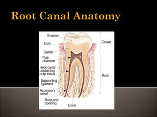
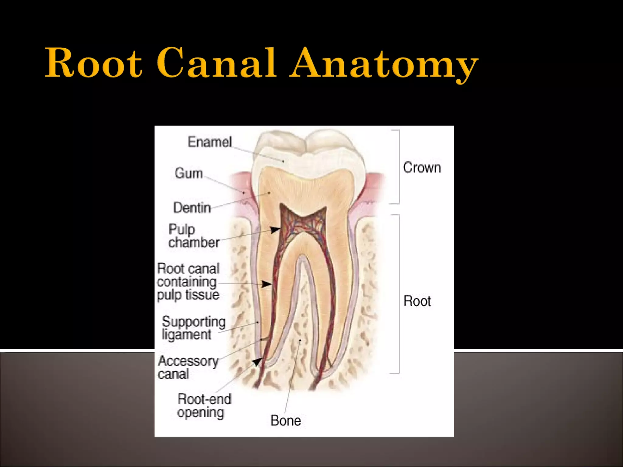
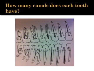
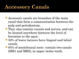
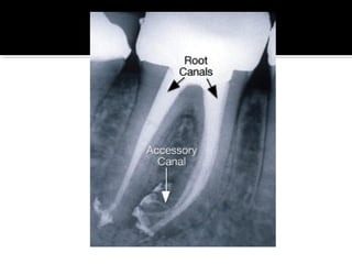
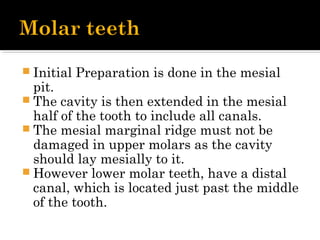
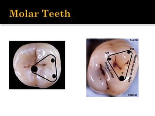
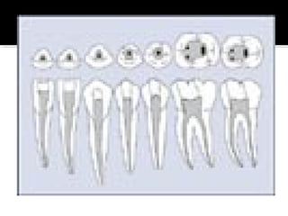
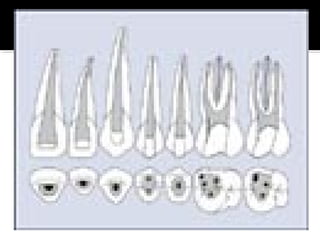
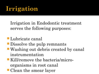
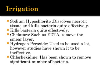
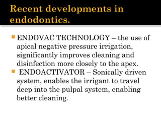
 Definition : Endodontics is the branch of
dentistry dealing with diseases of the
dental pulp.
 The number of canals in a tooth
correlates to the number of roots in a
tooth.
 The space inside the root canals is filled
with a highly vascularized and highly
innervated loose connective tissue,
called the dental pulp.
 The pulp tissue is in communication
with the periodontium and the rest of
the body through the apical foramen.
 Central region contains nerves and
blood vessels.
 Innermost layer, - contains fibroblasts
and undifferentiated mesenchymal cells.
 Zone of Weil – This is a cell free zone,
rich in capillaries and nerve fibres.
 Odontoblastic layer – outermost layer,
contains odontoblasts and is next to the
predetin and mature dentin.
 Primary Function is to form Dentin
 Nutritive – provides nutrients to the
organic surroundings
 Sensory – sensitive to temperature,
pressure or trauma
 Protective – forms secondary dentin,
when under attack from bacteria.
 Accessory canals are branches of the main
canal that form a communication between the
pulp and periodontum.
 They also contain vessels and nerves, and can
be located anywhere between the level of
furcation to the apex.
 30% of lower incisors have lingual and labial
canals.
 85% of mesiobuccal roots contain two canals
(MB1 and MB2), in upper molar teeth.
The shape of the preparation is dictated
by :
 The shape of the pulp chamber
 The morphology of the canals.
It is important that straight line access to
the apical foramen is created.
 The access cavity is initiated in the
middle of the palatal side of the tooth.
 Initial preparation should be at 90° to
the palatal aspect of the tooth.
 Once dentine has been reached the
angulation of the bur is changed to
follow a long axis of the tooth using a
slow handpiece.
 Initial preparation is made in the
middle of the fissure. The cavity is then
extended buccally and palatally.
 NB : Marginal ridges must not be
involved in this presentation, as this
would compromise the integrity of the
tooth.
 Initial Preparation is done in the mesial
pit.
 The cavity is then extended in the mesial
half of the tooth to include all canals.
 The mesial marginal ridge must not be
damaged in upper molars as the cavity
should lay mesially to it.
 However lower molar teeth, have a distal
canal, which is located just past the middle
of the tooth.
Irrigation in Endodontic treatment
serves the following purposes: 
 Lubricate canal
 Dissolve the pulp remnants
 Washing out debris created by canal
instrumentation
 Kill/remove the bacteria/micro-
organisms in root canal
 Clean the smear layer
 Sodium Hypochlorite :Dissolves necrotic
tissue and kills bacteria quite effectively.
 Kills bacteria quite effectively. 
 Chelators: Such as EDTA, remove the
smear layer.
 Hydrogen Peroxide: Used to be used a lot,
however studies have shown it to be
ineffective.
 Chlorhexidine: Has been shown to remove
significant number of bacteria.
 ENDOVAC TECHNOLOGY – the use of
apical negative pressure irrigation,
significantly improves cleaning and
disinfection more closely to the apex.
 ENDOACTIVATOR – Sonically driven
system, enables the irrigant to travel
deep into the pulpal system, enabling
better cleaning.
 Tooth Anatomy – Revision
 Root canal anatomy
 Accessory canals
 Access cavities
 Irrigants
 Recent Developments in Endodontics.
 http://www.dentistrytoday.com/endodon
tics/1043
 http://www.youtube.com/watch?
v=QrUgCNQs038
 Endodontics Lecture from Blackboard.
By Dr Qualtrough.
 http://www.dentistrytoday.info/content/i
rrigation-endo
 http://www.jendodon.com/article/S0099-
2399(07)00095-7/abstract
Root canal anatomy and access cavities

Root canal anatomy and access cavities

  • 2.
     Definition :Endodontics is the branch of dentistry dealing with diseases of the dental pulp.
  • 3.
     The numberof canals in a tooth correlates to the number of roots in a tooth.  The space inside the root canals is filled with a highly vascularized and highly innervated loose connective tissue, called the dental pulp.  The pulp tissue is in communication with the periodontium and the rest of the body through the apical foramen.
  • 4.
     Central regioncontains nerves and blood vessels.  Innermost layer, - contains fibroblasts and undifferentiated mesenchymal cells.  Zone of Weil – This is a cell free zone, rich in capillaries and nerve fibres.  Odontoblastic layer – outermost layer, contains odontoblasts and is next to the predetin and mature dentin.
  • 5.
     Primary Functionis to form Dentin  Nutritive – provides nutrients to the organic surroundings  Sensory – sensitive to temperature, pressure or trauma  Protective – forms secondary dentin, when under attack from bacteria.
  • 7.
     Accessory canalsare branches of the main canal that form a communication between the pulp and periodontum.  They also contain vessels and nerves, and can be located anywhere between the level of furcation to the apex.  30% of lower incisors have lingual and labial canals.  85% of mesiobuccal roots contain two canals (MB1 and MB2), in upper molar teeth.
  • 9.
    The shape ofthe preparation is dictated by :  The shape of the pulp chamber  The morphology of the canals. It is important that straight line access to the apical foramen is created.
  • 10.
     The accesscavity is initiated in the middle of the palatal side of the tooth.  Initial preparation should be at 90° to the palatal aspect of the tooth.  Once dentine has been reached the angulation of the bur is changed to follow a long axis of the tooth using a slow handpiece.
  • 11.
     Initial preparationis made in the middle of the fissure. The cavity is then extended buccally and palatally.  NB : Marginal ridges must not be involved in this presentation, as this would compromise the integrity of the tooth.
  • 12.
     Initial Preparationis done in the mesial pit.  The cavity is then extended in the mesial half of the tooth to include all canals.  The mesial marginal ridge must not be damaged in upper molars as the cavity should lay mesially to it.  However lower molar teeth, have a distal canal, which is located just past the middle of the tooth.
  • 16.
    Irrigation in Endodontictreatment serves the following purposes:   Lubricate canal  Dissolve the pulp remnants  Washing out debris created by canal instrumentation  Kill/remove the bacteria/micro- organisms in root canal  Clean the smear layer
  • 17.
     Sodium Hypochlorite:Dissolves necrotic tissue and kills bacteria quite effectively.  Kills bacteria quite effectively.   Chelators: Such as EDTA, remove the smear layer.  Hydrogen Peroxide: Used to be used a lot, however studies have shown it to be ineffective.  Chlorhexidine: Has been shown to remove significant number of bacteria.
  • 18.
     ENDOVAC TECHNOLOGY– the use of apical negative pressure irrigation, significantly improves cleaning and disinfection more closely to the apex.  ENDOACTIVATOR – Sonically driven system, enables the irrigant to travel deep into the pulpal system, enabling better cleaning.
  • 19.
     Tooth Anatomy– Revision  Root canal anatomy  Accessory canals  Access cavities  Irrigants  Recent Developments in Endodontics.
  • 20.
     http://www.dentistrytoday.com/endodon tics/1043  http://www.youtube.com/watch? v=QrUgCNQs038 Endodontics Lecture from Blackboard. By Dr Qualtrough.  http://www.dentistrytoday.info/content/i rrigation-endo  http://www.jendodon.com/article/S0099- 2399(07)00095-7/abstract