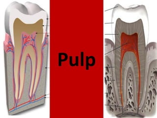
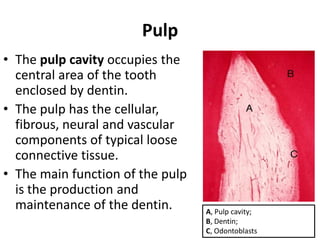
Pulp
• The pulpcavity occupies the
central area of the tooth
enclosed by dentin.
• The pulp has the cellular,
fibrous, neural and vascular
components of typical loose
connective tissue.
• The main function of the pulp
is the production and
maintenance of the dentin. A, Pulp cavity;
B, Dentin;
C, Odontoblasts
3.
Nerve Plexus ofRaschkow
• Sensory nerve fibers that originate from
inferior and superior alveolar nerves
innervate the odontoblastic layer of the
pulp cavity.
• These nerves enter the tooth through
the apical foramen as myelinated nerve
bundles.
• They branch to form the
subodontoblastic nerve plexus of
Raschkow which is separated from the
odontoblasts by a cell-free zone of
Weil.
• In addition to the sensory nerves,
sympathetic nerve bundles also enter
the tooth to innervate blood vessels.
A, Odontoblasts;
B, Cell-free zone of Weil;
C, Nerve plexus of Raschkow
4.
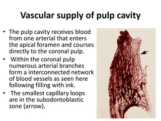
Vascular supply ofpulp cavity
• The pulp cavity receives blood
from one arterial that enters
the apical foramen and courses
directly to the coronal pulp.
• Within the coronal pulp
numerous arterial branches
form a interconnected network
of blood vessels as seen here
following filling with ink.
• The smallest capillary loops
are in the subodontoblastic
zone (arrow).
5.
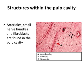
Structures within thepulp cavity
• Arterioles, small
nerve bundles
and fibroblasts
are found in the
pulp cavity
A, Nerve bundle;
B, Arteriole;
C, Fibroblasts
6.
Structures within thepulp cavity
• Arterioles can be
distinguished from
venules within the
pulp cavity by the
thickness and
contours of their
vascular walls.
A, Arteriole; B, Venule;
C, Nerve bundle (cut in cross-section)
7.
Subodontoblastic region
• Belowthe odontoblastic
layer is the cell-free zone
of Weil followed by a
cell-rich zone which is
thought to provide
replacement cells for
odontoblasts that die.
• Within these zones are
the nerve plexus of A, Cell-rich zone;
Raschkow and capillary B, Cell-free zone;
C, Odontoblastic layer;
network. D, Dentin
8.
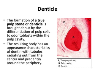
Denticle
• The formationof a true
pulp stone or denticle is
brought about by the
differentiation of pulp cells
to odontoblasts within the
pulp cavity.
• The resulting body has an
appearance characteristics
of dentin with tubules
radiating out from the
center and predentin A, True pulp stone;
around the periphery. B, Pulp cavity;
C, Dentin
9.
False pulp stone
•The formation of a false
pulp stone is caused by the
nonspecific calcification of
tissue around a central
nucleus within the pulp
cavity.
• This pulp stone is
characterized by concentric
layers of mineralization
rather than radiating
tubules as seen in true pulp
stones.
A, False pulp stone; B, Pulp cavity
10.
Regions of thepulp cavity
• The pulp cavity can be
divided into two main
regions: the coronal pulp
is located within the
crown of the tooth and
the radicular pulp is
located within the root.
A, Coronal pulp;
B, Radicular pulp
أدعيه قبل المذاكرهوبعدها...
قبل المذاكرة
اللهم أنً اسألك فهم النبٌٌن و حفظ المرسلٌن و المالئكة المقربٌن ، اللهم أجعل ألسنتنا عامرة بذكرك و قلوبنا بخشٌتك و أسرارنا
بطاعتك أنك على كل شًء قدٌر .. حسبنا هللا و نعم الوكٌل
بعد المذاكرة
اللهم أنً استودعتك ما قرأت و ما حفظت و ما تعلمت فرده عند حاجتً الٌه انك على كل شًء قدٌر ، حسبنا هللا و نعم الوكٌل
يوم اإلمتحان
اللهم أنً توكلت علٌك و سلمت امري الٌك ال ملجأ و منجا منك إال الٌك
دخول القاعة
رب أدخلنً مدخل صدق و أخرجنً مخرج صدق و أجعل لً من لدنك سلطانا نصٌرا
قبل البدء بالحل
رب أشرح لً صدري و ٌسر لً أمري و احلل عقدة من لسانً ٌفقه قولً بسم هللا الفتاح ، اللهم ال سهل أال ما جعلته سهال و انت تجعل الحزن اذا
شئت سهال ٌا ارحم الراحمٌن
أثناء األمتحان
ال إله اال انت سبحانك أنً كنت من الظالمٌن ٌا حً ٌا قٌوم برحمتك أستغٌث ، رب ان مسنً الضر أنك أرحم الراحمٌن
عند النسيان
اللهم ٌا جامع الناس فً ٌوم ال رٌب فٌه أجمعنً و ضالتً
بعد األنتهاء
الحمد هلل الذي هدانا لهذا و ما كنا لنهتدي لوال أن هدانا هللا