SlideShare a Scribd company logo
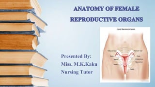
Presented By:
Miss. M.K.Kaku
Nursing Tutor
CONTENTS
 EXTERNAL GENITELIA
 INTERNAL GENITELIA
ASSESSORY REPRODUCTIVE ORGANS
INTRODUCTION
The reproductive organs in female is concerned with,
• Copulation
• Fertilization
• Growth & Development of fetus
• For exit of fetus to the outer world from womb
The organs are broadly Divided into:
External Genitelia
Internal Genitalia
Accessory reproductive Organs
EXTERNAL GENITALIA
The vulva or pudendum includes all visible external genital organs in the
perineum.
Consist of ,
 Mons pubis
 Labia majora
 Labia minora
 Clitoris
 Vestibule: Urethral opening
Vaginal orifice & Hymen
Opening of Bartholin’s Ducts
Skene’s Glands or paraurethral gland
MONS PUBIS
• It is the pad of subcutaneous adipose connective
tissue.
•Lying in front of the pubis.
•It is covered with hair.
LABIA MAJORA
• They are two skin folds of fat which makes the boundary of
vulva.
• It is covered with hair and contain sweat glands
• Where they join medially forms the postirior commissure in
front of the anus.
• The labia majora is homologous to scrotum in male.
LABIA MINORA
• They are two thin folds of skin on either side within the labia
majora.
• They divide to enclose the clitoris and unite with each other in
front and behind the clitoris to form prepuce and frenulum
respectively.
• The lower portion of labia minora fuses across the midline to form
a fold of skin known as fourchette.
• The labia minora has no fat , hair follicles and sweat glands.
CLITORIS
•It is a small cylindrical erectile body, measuring about
1.5 - 2 cm situated in the most anterior part of vulva.
•It is consist of glans, a body and two crura.
•Clitoris is homologous to penis in male.
VESTIBULE
• It is triangular space bounded anteriorly by clitoris, posteriorly by the
fourchette and either side by labia minora.
• There are four openings in vestibule:
1. Urethral opening : It is situated in front of vaginal orifice.
2. Vaginal orifice : Vaginal orifice lies in the postirior end of vestibule and
is of varying size and shape.
It is incompletely closed by a septum of mucous membrane, called
hymen. It is usually ruptured at consummation of marriage.
3. Openings of Bartholin’s ducts : There are two Bartholin's
glands ( Greater Vestibular glands ), one on each side of vagina.
 During sexual excitement it secretes alkaline mucus which
helps in lubrication.
4. Skene’s glands : Also known as paraurethral glands situated
either side of urethral orifice postirior. It is homologous to prostate
in male.
VESTIBULAR BULB
 These are bilateral
elongated erectile tissues
situated beneath the mucous
membrane of vestibule.
INTERNAL GENITALIA
The internal genital organs in female include,
• Vagina
• Uterus
• Fallopian tubes
• Ovaries
 RELATIONS :
 ANTIRIORLY : Upper 1/3 - Base of bladder
Lower 1/3 - Urethra and lower half firmly embedded with it.
 POSTIRIORLY : Upper 1/3 – Pouch of Duglas
Middle 1/3 – Rectal Wall
Lower 1/3 – Perineal body then anal canal.
 LATERAL WALLS : Upper 1/3 – Pelvic cellular tissue
Middle 1/3 – Jointed with lavator ani
Lower 1/3 – Vestibular bulb , Bartholin’s gland , Bulbocarveneous
Muscle.
VAGINA
The vagina is a fibro muscular canal extend from vestibule to cervix.
 It is directed upwards and backwards between the bladder in front and
rectum behind.
It is receptacle for the penis during sexual intercourse, excretory channel
for menstrual flow and passageway for childbirth.
WALLS:
1. ANTIRIOR WALL : 7 cm
2. POSTIRIOR WALL : 9 cm
3. LATERAL WALL
 FORNICES :
The fornices are the clefts formed at the top of vagina
due to projection of cervix through the anterior wall . There
are four fornices :
1. ANTERIOR FORNIX : deep
2. POSTIRIOR FORNIX : Shallow
3. TWO LATERAL FORNIX
 STUCTURE S:
• Muscular coat
• Submucosal layer
• Muscular layer - [ A ] Inner Circular [ B ] Outer longitudinal
• Fibrous Coat
 VAGINAL SECRETION:
 From the puberty to menopause the pH of vaginal secretion is ACIDIC (4-5)
because of presence of DODERLEINS BECILLI which produce lactic acid.
UTERUS
The uterus is hollow pyriform muscular organ situated in the pelvis between
the bladder and the rectum.
 POSITION:
• The normal position of uterus is ANTEVERSION & ANTEFLEXION
 MEASUREMENT AND PARTS:
• Length – 8 cm
• Width – 5cm
• Thickness – 1.25 cm
• Wight – 50 to 80 g.
Parts are:
1. FUNDUS : A dome shaped portion superior to the fallopian tubes.
2. BODY : The central portion between fundus and cervix is called Body of uterus.
3. CERVIX : An inferior portion is called cervix that opens into the vagina.
4. ISTHMUS : It is a constricted region between body and cervix.
CAVITY:
• The interior of the body of the uterus is called the UTERINE CAVITY. At the cervix it is called
CERVICAL CANAL. The fundus has no cavity within it.
• The cervix opens into the uterine cavity at internal os and exterior at vagina by external os.
 RELATIONS:
• ANTERIORLY : Above internal os – The body forms postirior wall of uterovesical pouch.
Bellow internal os – Separated from base of bladder by loose areola tissue.
• POSTIRIOR : It is covered with peritoneum & forms the anterior wall of the Pouch of Douglas.
• LATERALLY : The double fold of peritoneum of broad ligament.
STRUCTURE:
There are three layers of uterusfrom outsideto inside:
1. PARIMETRIUM : It is a part of visceral peritoneum laterallyit becomesbroad ligaments.
2. MAYOMETRIUM : It consistof thickbundlesof smooth muscle fibresarranged in inner circular
and outer longitudinalor oblique.
3. ENDOMATRIUM : The mucous lining of the cavity is called
endometrium.
 It is highly vascularised which allows the implantation after
fertilization
 The endometrium is further divided into two layers:
From inward to outward,
 Stratum functionalis ( Functional layer) : The compact and spongy
layer combinly called functional
layer. It lines the uterine cavity and slough off during menstruation.
 Stratum Basalis ( Basal layer) : It is permanent and give rise to anew
stratum functionalis.
 LIGAMENTS:
 Broad ligament
 Round ligament
 Uterosacral ligament
 Transverse/ Cardinal ligament
 Pubocervical fascia
FALLOPIAN TUBE
The fallopian tubes ( Uterine tubes) are hollow muscular tube, about 10 cm long
and extend from sides of uterus on each side between fundus and body in the uterine cavity.
They lies in the upper free border of broad ligament opening into the peritoneal cavity close to
the ovaries.
It has two openings:
1. Uterine opening which opens from uterine cavity
2. Pelvic opening which opens close to ovaries in peritoneal cavity.
PARTS:
 Interstital or Intramural : Lies in the wall of uterus. It is 1.25 cm long
 Isthumus : 2nd part after the interstital part ends in ampulla. It is 3-4 cm long
Ampulla : It is the largest part of fallopian tube which ios 5 cm long.
The fertilization normally occur at this part.
Infundibulum : It is the ending part of fallopian tube which is 1.25 cm
long . This part contains finger like projection
surrounding the pelvic opening called FIMBRIAE. It is attached to
the outer pole of the ovary, here it is called ovarian fimbriae.
STRUCTURE:
From outward to inward:
1. Serous layer
2. Muscular layer
3. Mucous membrane containing cillia.
OVARIES
The ovaries are paired sex glands in the female concerned
with germ cell maturation and steroidogenesis.
 Each gland is oval in shape and pinkish gray in colour.
It measures about 3cm in length , 2 cm in width and 1cm in thickness.
 STRUCTURE:
• Each ovary consist of the following parts: ( outward to inward )
Germinal epithelium – This is a layer of simple epithelium that covers
the surface of ovary.
Tunica albugenia – It is whitish capsule of dense irregular connective
tissue located intermediately deep to the germinal epithelium.
Germinal or
Ovarian follicles
 Ovarian cortex – It is situated just deep to the tunica albugenia which
contains Ovarian follicles
 Ovarian Medulla – It is situated deep to the ovarian cortex consist of
loosely arranged connective tissue. The medulla contains blood vessels ,
lymphatic vessels and nerves.
 Each ovary contains the hillum that is the point of entrance and exit for
blood vessels and nerves along which the mesovarium is attached.
 LIGAMENTS:
 Ovarian Ligament
Suspensory ligament
ACCESARY REPRODUCTIVE ORGANS
BREAST ( MAMMARY GLAND )
The breast are large modified sebaceous glands. The breast
are bilateral and concerned with lactation following childbirth.
 It usually extend from second to sixth rib in midclavicular line.
 It lies in the subcutaneous tissue .
 A lateral projection of the breast toward axilla is known as axillary tail of
spence.
 The weighs 200 – 300 g during childbearing age.
 STRUCTURE:
 AREOLA : The areola is placed at the centre of the breast and is pigmented. The diameter of
areola is 2.5 cm.
Montgomery glands are accessory glands located around the areola, they can secrete the milk.
 NIPPLE : The nipple is muscular projection covered by pigmented skin. It is vascular and
surrounded by unstriated muscles which make it erectile.
It accommodates about 15- 20 lactiferous ducts and their openings.
LOBE OF BREAST : The breast is composed of 12-20 lobes. Each lobe has one lactiferous duct
that opens in the nipple.
LOBULES : Each lobe has about 10 – 100 lobules that contains alveolar cell surrounded by
mayoepithelial cells and capillaries.
 Alveolar cells : Production, storage and secretion of milk.
 Mayoepithelial cells : They are placed at surrounding the
alveolar cells. Contraction of these cells squeezes the alveoli and
ejects the milk into larger duct.
 AMPULLA : Behind the nipple , the main duct dilates to form
ampulla here milk is stored.
 LIGAMENTS:
 Copper’ s ligament : This ligaments support the breast and
maintain the shape of breast.

More Related Content

What's hot

External genitalia -parts
External genitalia -partsExternal genitalia -parts
External genitalia -parts
Chandra Shekar
 
Female reproductive system
Female reproductive systemFemale reproductive system
Female reproductive system
msp4889
 
Anatomy of reproductive system
Anatomy of reproductive systemAnatomy of reproductive system
Anatomy of reproductive system
Arturo Decano
 
Female Reproductive System
Female Reproductive SystemFemale Reproductive System
Female Reproductive System
Osama Zahid
 
Mammary glands
Mammary glandsMammary glands
Pelvis By Dr. Fernandez
Pelvis By Dr. FernandezPelvis By Dr. Fernandez
Pelvis By Dr. Fernandez
MD Specialclass
 
Urinary bladder (Anatomy)
Urinary bladder (Anatomy)Urinary bladder (Anatomy)
Urinary bladder (Anatomy)
Dr. Prabin Kumar Bam
 
Fertilization (General Embryology)
Fertilization (General Embryology)Fertilization (General Embryology)
Fertilization (General Embryology)
Dr. Sherif Fahmy
 
The male reproductive system
The male reproductive systemThe male reproductive system
The male reproductive system
Merlyn Denesia
 
Perineum
Perineum Perineum
Perineum
Dr. Piyush Karn
 
Urinary bladder and urethra
Urinary bladder  and urethraUrinary bladder  and urethra
Urinary bladder and urethraLawrence James
 
Male reproductive system
Male reproductive systemMale reproductive system
Male reproductive system
Jegan Nadar
 
Male reproductive system
Male reproductive systemMale reproductive system
The female reproductive system
The female reproductive systemThe female reproductive system
The female reproductive system
Dr. Binu Babu Nursing Lectures Incredibly Easy
 
9 pelvic
9 pelvic9 pelvic
A&P Chapter 31 The Reproductive System
A&P Chapter 31 The Reproductive SystemA&P Chapter 31 The Reproductive System
A&P Chapter 31 The Reproductive Systemmarydelaney
 
Anatomy of The Female Genital Tract
Anatomy of The Female Genital TractAnatomy of The Female Genital Tract
Anatomy of The Female Genital Tract
Mohd Ikhwan Chacho
 
The perineum
The perineumThe perineum
The perineum
Idris Siddiqui
 
Anatomy of Ureter
Anatomy of UreterAnatomy of Ureter
Anatomy of Ureter
Dr. Vibhash Kumar Vaidya
 

What's hot (20)

External genitalia -parts
External genitalia -partsExternal genitalia -parts
External genitalia -parts
 
Reproductive physiology of male and female
Reproductive physiology of male and femaleReproductive physiology of male and female
Reproductive physiology of male and female
 
Female reproductive system
Female reproductive systemFemale reproductive system
Female reproductive system
 
Anatomy of reproductive system
Anatomy of reproductive systemAnatomy of reproductive system
Anatomy of reproductive system
 
Female Reproductive System
Female Reproductive SystemFemale Reproductive System
Female Reproductive System
 
Mammary glands
Mammary glandsMammary glands
Mammary glands
 
Pelvis By Dr. Fernandez
Pelvis By Dr. FernandezPelvis By Dr. Fernandez
Pelvis By Dr. Fernandez
 
Urinary bladder (Anatomy)
Urinary bladder (Anatomy)Urinary bladder (Anatomy)
Urinary bladder (Anatomy)
 
Fertilization (General Embryology)
Fertilization (General Embryology)Fertilization (General Embryology)
Fertilization (General Embryology)
 
The male reproductive system
The male reproductive systemThe male reproductive system
The male reproductive system
 
Perineum
Perineum Perineum
Perineum
 
Urinary bladder and urethra
Urinary bladder  and urethraUrinary bladder  and urethra
Urinary bladder and urethra
 
Male reproductive system
Male reproductive systemMale reproductive system
Male reproductive system
 
Male reproductive system
Male reproductive systemMale reproductive system
Male reproductive system
 
The female reproductive system
The female reproductive systemThe female reproductive system
The female reproductive system
 
9 pelvic
9 pelvic9 pelvic
9 pelvic
 
A&P Chapter 31 The Reproductive System
A&P Chapter 31 The Reproductive SystemA&P Chapter 31 The Reproductive System
A&P Chapter 31 The Reproductive System
 
Anatomy of The Female Genital Tract
Anatomy of The Female Genital TractAnatomy of The Female Genital Tract
Anatomy of The Female Genital Tract
 
The perineum
The perineumThe perineum
The perineum
 
Anatomy of Ureter
Anatomy of UreterAnatomy of Ureter
Anatomy of Ureter
 

Similar to Anatomy of female reproductive organs

Female reproductive system
Female reproductive system Female reproductive system
Female reproductive system
Rani Gurudasani
 
Anatomy of female reproductive organs
Anatomy of female reproductive organsAnatomy of female reproductive organs
Anatomy of female reproductive organs
Rehab Soliman Soliman
 
female reproductive system.pptx
female reproductive system.pptxfemale reproductive system.pptx
female reproductive system.pptx
KalanaDenuwan
 
Anatomy and embryology of female reproductive system
Anatomy and embryology of female reproductive systemAnatomy and embryology of female reproductive system
Anatomy and embryology of female reproductive system
Hussien Ali
 
female reproductive system.pptx
female reproductive system.pptxfemale reproductive system.pptx
female reproductive system.pptx
AnilDixit16
 
Female reproductive system.pdf
Female reproductive system.pdfFemale reproductive system.pdf
Female reproductive system.pdf
dipinbhandari
 
ANATOMY AND PHYSIOLOGY OF REPRODUCTIVE SYSTEM.pptx
ANATOMY AND PHYSIOLOGY OF REPRODUCTIVE SYSTEM.pptxANATOMY AND PHYSIOLOGY OF REPRODUCTIVE SYSTEM.pptx
ANATOMY AND PHYSIOLOGY OF REPRODUCTIVE SYSTEM.pptx
Swetaba Besh
 
anatomyphysiologyoffemalereproductivesystem-130506124715-phpapp02.pdf
anatomyphysiologyoffemalereproductivesystem-130506124715-phpapp02.pdfanatomyphysiologyoffemalereproductivesystem-130506124715-phpapp02.pdf
anatomyphysiologyoffemalereproductivesystem-130506124715-phpapp02.pdf
usman232639
 
anatomyphysiologyoffemalereproductivesystem-130506124715-phpapp02.ppt
anatomyphysiologyoffemalereproductivesystem-130506124715-phpapp02.pptanatomyphysiologyoffemalereproductivesystem-130506124715-phpapp02.ppt
anatomyphysiologyoffemalereproductivesystem-130506124715-phpapp02.ppt
ReshmaAnilKumar6
 
anatomyphysiologyoffemalereproductivesystem-130506124715-phpapp02.ppt
anatomyphysiologyoffemalereproductivesystem-130506124715-phpapp02.pptanatomyphysiologyoffemalereproductivesystem-130506124715-phpapp02.ppt
anatomyphysiologyoffemalereproductivesystem-130506124715-phpapp02.ppt
PriyankaSinha406376
 
anatomyphysiologyoffemalereproductivesystem-130506124715-phpapp02.ppt
anatomyphysiologyoffemalereproductivesystem-130506124715-phpapp02.pptanatomyphysiologyoffemalereproductivesystem-130506124715-phpapp02.ppt
anatomyphysiologyoffemalereproductivesystem-130506124715-phpapp02.ppt
PriyankaSinha406376
 
ADV MED-SURG REPROD. FUCTN.pptx
ADV MED-SURG REPROD. FUCTN.pptxADV MED-SURG REPROD. FUCTN.pptx
ADV MED-SURG REPROD. FUCTN.pptx
AdeojoAdewumiOlatooy
 
Anatomy physiology of female reproductive system
Anatomy  physiology of female reproductive systemAnatomy  physiology of female reproductive system
Anatomy physiology of female reproductive systemMonique Reyes
 
Prenatal Care
Prenatal CarePrenatal Care
Prenatal Care
Michelline Perez
 
reproductivesystem-170324071445 (1).pptx
reproductivesystem-170324071445 (1).pptxreproductivesystem-170324071445 (1).pptx
reproductivesystem-170324071445 (1).pptx
CharutaKunjeer1
 
reproductivesystem-170324071445 (1).pptx
reproductivesystem-170324071445 (1).pptxreproductivesystem-170324071445 (1).pptx
reproductivesystem-170324071445 (1).pptx
CharutaKunjeer1
 
female reproductive system.pptx
female reproductive system.pptxfemale reproductive system.pptx
female reproductive system.pptx
Liangkiuwiliu
 
Anatomy & physiology of female reproductive system
Anatomy & physiology of female reproductive systemAnatomy & physiology of female reproductive system
Anatomy & physiology of female reproductive system
Deeps Gupta
 
FEMALE REPRODUCTIVE SYSTEM.pptx
FEMALE REPRODUCTIVE SYSTEM.pptxFEMALE REPRODUCTIVE SYSTEM.pptx
FEMALE REPRODUCTIVE SYSTEM.pptx
ChinjuJoseSajith
 

Similar to Anatomy of female reproductive organs (20)

Female reproductive system
Female reproductive system Female reproductive system
Female reproductive system
 
Anatomy of female reproductive organs
Anatomy of female reproductive organsAnatomy of female reproductive organs
Anatomy of female reproductive organs
 
female reproductive system.pptx
female reproductive system.pptxfemale reproductive system.pptx
female reproductive system.pptx
 
Anatomy and embryology of female reproductive system
Anatomy and embryology of female reproductive systemAnatomy and embryology of female reproductive system
Anatomy and embryology of female reproductive system
 
female reproductive system.pptx
female reproductive system.pptxfemale reproductive system.pptx
female reproductive system.pptx
 
Female reproductive system.pdf
Female reproductive system.pdfFemale reproductive system.pdf
Female reproductive system.pdf
 
ANATOMY AND PHYSIOLOGY OF REPRODUCTIVE SYSTEM.pptx
ANATOMY AND PHYSIOLOGY OF REPRODUCTIVE SYSTEM.pptxANATOMY AND PHYSIOLOGY OF REPRODUCTIVE SYSTEM.pptx
ANATOMY AND PHYSIOLOGY OF REPRODUCTIVE SYSTEM.pptx
 
anatomyphysiologyoffemalereproductivesystem-130506124715-phpapp02.pdf
anatomyphysiologyoffemalereproductivesystem-130506124715-phpapp02.pdfanatomyphysiologyoffemalereproductivesystem-130506124715-phpapp02.pdf
anatomyphysiologyoffemalereproductivesystem-130506124715-phpapp02.pdf
 
anatomyphysiologyoffemalereproductivesystem-130506124715-phpapp02.ppt
anatomyphysiologyoffemalereproductivesystem-130506124715-phpapp02.pptanatomyphysiologyoffemalereproductivesystem-130506124715-phpapp02.ppt
anatomyphysiologyoffemalereproductivesystem-130506124715-phpapp02.ppt
 
anatomyphysiologyoffemalereproductivesystem-130506124715-phpapp02.ppt
anatomyphysiologyoffemalereproductivesystem-130506124715-phpapp02.pptanatomyphysiologyoffemalereproductivesystem-130506124715-phpapp02.ppt
anatomyphysiologyoffemalereproductivesystem-130506124715-phpapp02.ppt
 
anatomyphysiologyoffemalereproductivesystem-130506124715-phpapp02.ppt
anatomyphysiologyoffemalereproductivesystem-130506124715-phpapp02.pptanatomyphysiologyoffemalereproductivesystem-130506124715-phpapp02.ppt
anatomyphysiologyoffemalereproductivesystem-130506124715-phpapp02.ppt
 
ADV MED-SURG REPROD. FUCTN.pptx
ADV MED-SURG REPROD. FUCTN.pptxADV MED-SURG REPROD. FUCTN.pptx
ADV MED-SURG REPROD. FUCTN.pptx
 
Anatomy physiology of female reproductive system
Anatomy  physiology of female reproductive systemAnatomy  physiology of female reproductive system
Anatomy physiology of female reproductive system
 
Prenatal Care
Prenatal CarePrenatal Care
Prenatal Care
 
reproductivesystem-170324071445 (1).pptx
reproductivesystem-170324071445 (1).pptxreproductivesystem-170324071445 (1).pptx
reproductivesystem-170324071445 (1).pptx
 
reproductivesystem-170324071445 (1).pptx
reproductivesystem-170324071445 (1).pptxreproductivesystem-170324071445 (1).pptx
reproductivesystem-170324071445 (1).pptx
 
Female2
Female2Female2
Female2
 
female reproductive system.pptx
female reproductive system.pptxfemale reproductive system.pptx
female reproductive system.pptx
 
Anatomy & physiology of female reproductive system
Anatomy & physiology of female reproductive systemAnatomy & physiology of female reproductive system
Anatomy & physiology of female reproductive system
 
FEMALE REPRODUCTIVE SYSTEM.pptx
FEMALE REPRODUCTIVE SYSTEM.pptxFEMALE REPRODUCTIVE SYSTEM.pptx
FEMALE REPRODUCTIVE SYSTEM.pptx
 

Recently uploaded

Ophthalmology Clinical Tests for OSCE exam
Ophthalmology Clinical Tests for OSCE examOphthalmology Clinical Tests for OSCE exam
Ophthalmology Clinical Tests for OSCE exam
KafrELShiekh University
 
Flu Vaccine Alert in Bangalore Karnataka
Flu Vaccine Alert in Bangalore KarnatakaFlu Vaccine Alert in Bangalore Karnataka
Flu Vaccine Alert in Bangalore Karnataka
addon Scans
 
263778731218 Abortion Clinic /Pills In Harare ,
263778731218 Abortion Clinic /Pills In Harare ,263778731218 Abortion Clinic /Pills In Harare ,
263778731218 Abortion Clinic /Pills In Harare ,
sisternakatoto
 
Thyroid Gland- Gross Anatomy by Dr. Rabia Inam Gandapore.pptx
Thyroid Gland- Gross Anatomy by Dr. Rabia Inam Gandapore.pptxThyroid Gland- Gross Anatomy by Dr. Rabia Inam Gandapore.pptx
Thyroid Gland- Gross Anatomy by Dr. Rabia Inam Gandapore.pptx
Dr. Rabia Inam Gandapore
 
Hemodialysis: Chapter 4, Dialysate Circuit - Dr.Gawad
Hemodialysis: Chapter 4, Dialysate Circuit - Dr.GawadHemodialysis: Chapter 4, Dialysate Circuit - Dr.Gawad
Hemodialysis: Chapter 4, Dialysate Circuit - Dr.Gawad
NephroTube - Dr.Gawad
 
Basavarajeeyam - Ayurvedic heritage book of Andhra pradesh
Basavarajeeyam - Ayurvedic heritage book of Andhra pradeshBasavarajeeyam - Ayurvedic heritage book of Andhra pradesh
Basavarajeeyam - Ayurvedic heritage book of Andhra pradesh
Dr. Madduru Muni Haritha
 
Effective-Soaps-for-Fungal-Skin-Infections.pptx
Effective-Soaps-for-Fungal-Skin-Infections.pptxEffective-Soaps-for-Fungal-Skin-Infections.pptx
Effective-Soaps-for-Fungal-Skin-Infections.pptx
SwisschemDerma
 
Colonic and anorectal physiology with surgical implications
Colonic and anorectal physiology with surgical implicationsColonic and anorectal physiology with surgical implications
Colonic and anorectal physiology with surgical implications
Dr Maria Tamanna
 
Ozempic: Preoperative Management of Patients on GLP-1 Receptor Agonists
Ozempic: Preoperative Management of Patients on GLP-1 Receptor Agonists  Ozempic: Preoperative Management of Patients on GLP-1 Receptor Agonists
Ozempic: Preoperative Management of Patients on GLP-1 Receptor Agonists
Saeid Safari
 
How STIs Influence the Development of Pelvic Inflammatory Disease.pptx
How STIs Influence the Development of Pelvic Inflammatory Disease.pptxHow STIs Influence the Development of Pelvic Inflammatory Disease.pptx
How STIs Influence the Development of Pelvic Inflammatory Disease.pptx
FFragrant
 
Novas diretrizes da OMS para os cuidados perinatais de mais qualidade
Novas diretrizes da OMS para os cuidados perinatais de mais qualidadeNovas diretrizes da OMS para os cuidados perinatais de mais qualidade
Novas diretrizes da OMS para os cuidados perinatais de mais qualidade
Prof. Marcus Renato de Carvalho
 
Triangles of Neck and Clinical Correlation by Dr. RIG.pptx
Triangles of Neck and Clinical Correlation by Dr. RIG.pptxTriangles of Neck and Clinical Correlation by Dr. RIG.pptx
Triangles of Neck and Clinical Correlation by Dr. RIG.pptx
Dr. Rabia Inam Gandapore
 
Physiology of Chemical Sensation of smell.pdf
Physiology of Chemical Sensation of smell.pdfPhysiology of Chemical Sensation of smell.pdf
Physiology of Chemical Sensation of smell.pdf
MedicoseAcademics
 
The Electrocardiogram - Physiologic Principles
The Electrocardiogram - Physiologic PrinciplesThe Electrocardiogram - Physiologic Principles
The Electrocardiogram - Physiologic Principles
MedicoseAcademics
 
Dehradun #ℂall #gIRLS Oyo Hotel 9719300533 #ℂall #gIRL in Dehradun
Dehradun #ℂall #gIRLS Oyo Hotel 9719300533 #ℂall #gIRL in DehradunDehradun #ℂall #gIRLS Oyo Hotel 9719300533 #ℂall #gIRL in Dehradun
Dehradun #ℂall #gIRLS Oyo Hotel 9719300533 #ℂall #gIRL in Dehradun
chandankumarsmartiso
 
Non-respiratory Functions of the Lungs.pdf
Non-respiratory Functions of the Lungs.pdfNon-respiratory Functions of the Lungs.pdf
Non-respiratory Functions of the Lungs.pdf
MedicoseAcademics
 
Ocular injury ppt Upendra pal optometrist upums saifai etawah
Ocular injury  ppt  Upendra pal  optometrist upums saifai etawahOcular injury  ppt  Upendra pal  optometrist upums saifai etawah
Ocular injury ppt Upendra pal optometrist upums saifai etawah
pal078100
 
ARTHROLOGY PPT NCISM SYLLABUS AYURVEDA STUDENTS
ARTHROLOGY PPT NCISM SYLLABUS AYURVEDA STUDENTSARTHROLOGY PPT NCISM SYLLABUS AYURVEDA STUDENTS
ARTHROLOGY PPT NCISM SYLLABUS AYURVEDA STUDENTS
Dr. Vinay Pareek
 
ANATOMY AND PHYSIOLOGY OF URINARY SYSTEM.pptx
ANATOMY AND PHYSIOLOGY OF URINARY SYSTEM.pptxANATOMY AND PHYSIOLOGY OF URINARY SYSTEM.pptx
ANATOMY AND PHYSIOLOGY OF URINARY SYSTEM.pptx
Swetaba Besh
 
Role of Mukta Pishti in the Management of Hyperthyroidism
Role of Mukta Pishti in the Management of HyperthyroidismRole of Mukta Pishti in the Management of Hyperthyroidism
Role of Mukta Pishti in the Management of Hyperthyroidism
Dr. Jyothirmai Paindla
 

Recently uploaded (20)

Ophthalmology Clinical Tests for OSCE exam
Ophthalmology Clinical Tests for OSCE examOphthalmology Clinical Tests for OSCE exam
Ophthalmology Clinical Tests for OSCE exam
 
Flu Vaccine Alert in Bangalore Karnataka
Flu Vaccine Alert in Bangalore KarnatakaFlu Vaccine Alert in Bangalore Karnataka
Flu Vaccine Alert in Bangalore Karnataka
 
263778731218 Abortion Clinic /Pills In Harare ,
263778731218 Abortion Clinic /Pills In Harare ,263778731218 Abortion Clinic /Pills In Harare ,
263778731218 Abortion Clinic /Pills In Harare ,
 
Thyroid Gland- Gross Anatomy by Dr. Rabia Inam Gandapore.pptx
Thyroid Gland- Gross Anatomy by Dr. Rabia Inam Gandapore.pptxThyroid Gland- Gross Anatomy by Dr. Rabia Inam Gandapore.pptx
Thyroid Gland- Gross Anatomy by Dr. Rabia Inam Gandapore.pptx
 
Hemodialysis: Chapter 4, Dialysate Circuit - Dr.Gawad
Hemodialysis: Chapter 4, Dialysate Circuit - Dr.GawadHemodialysis: Chapter 4, Dialysate Circuit - Dr.Gawad
Hemodialysis: Chapter 4, Dialysate Circuit - Dr.Gawad
 
Basavarajeeyam - Ayurvedic heritage book of Andhra pradesh
Basavarajeeyam - Ayurvedic heritage book of Andhra pradeshBasavarajeeyam - Ayurvedic heritage book of Andhra pradesh
Basavarajeeyam - Ayurvedic heritage book of Andhra pradesh
 
Effective-Soaps-for-Fungal-Skin-Infections.pptx
Effective-Soaps-for-Fungal-Skin-Infections.pptxEffective-Soaps-for-Fungal-Skin-Infections.pptx
Effective-Soaps-for-Fungal-Skin-Infections.pptx
 
Colonic and anorectal physiology with surgical implications
Colonic and anorectal physiology with surgical implicationsColonic and anorectal physiology with surgical implications
Colonic and anorectal physiology with surgical implications
 
Ozempic: Preoperative Management of Patients on GLP-1 Receptor Agonists
Ozempic: Preoperative Management of Patients on GLP-1 Receptor Agonists  Ozempic: Preoperative Management of Patients on GLP-1 Receptor Agonists
Ozempic: Preoperative Management of Patients on GLP-1 Receptor Agonists
 
How STIs Influence the Development of Pelvic Inflammatory Disease.pptx
How STIs Influence the Development of Pelvic Inflammatory Disease.pptxHow STIs Influence the Development of Pelvic Inflammatory Disease.pptx
How STIs Influence the Development of Pelvic Inflammatory Disease.pptx
 
Novas diretrizes da OMS para os cuidados perinatais de mais qualidade
Novas diretrizes da OMS para os cuidados perinatais de mais qualidadeNovas diretrizes da OMS para os cuidados perinatais de mais qualidade
Novas diretrizes da OMS para os cuidados perinatais de mais qualidade
 
Triangles of Neck and Clinical Correlation by Dr. RIG.pptx
Triangles of Neck and Clinical Correlation by Dr. RIG.pptxTriangles of Neck and Clinical Correlation by Dr. RIG.pptx
Triangles of Neck and Clinical Correlation by Dr. RIG.pptx
 
Physiology of Chemical Sensation of smell.pdf
Physiology of Chemical Sensation of smell.pdfPhysiology of Chemical Sensation of smell.pdf
Physiology of Chemical Sensation of smell.pdf
 
The Electrocardiogram - Physiologic Principles
The Electrocardiogram - Physiologic PrinciplesThe Electrocardiogram - Physiologic Principles
The Electrocardiogram - Physiologic Principles
 
Dehradun #ℂall #gIRLS Oyo Hotel 9719300533 #ℂall #gIRL in Dehradun
Dehradun #ℂall #gIRLS Oyo Hotel 9719300533 #ℂall #gIRL in DehradunDehradun #ℂall #gIRLS Oyo Hotel 9719300533 #ℂall #gIRL in Dehradun
Dehradun #ℂall #gIRLS Oyo Hotel 9719300533 #ℂall #gIRL in Dehradun
 
Non-respiratory Functions of the Lungs.pdf
Non-respiratory Functions of the Lungs.pdfNon-respiratory Functions of the Lungs.pdf
Non-respiratory Functions of the Lungs.pdf
 
Ocular injury ppt Upendra pal optometrist upums saifai etawah
Ocular injury  ppt  Upendra pal  optometrist upums saifai etawahOcular injury  ppt  Upendra pal  optometrist upums saifai etawah
Ocular injury ppt Upendra pal optometrist upums saifai etawah
 
ARTHROLOGY PPT NCISM SYLLABUS AYURVEDA STUDENTS
ARTHROLOGY PPT NCISM SYLLABUS AYURVEDA STUDENTSARTHROLOGY PPT NCISM SYLLABUS AYURVEDA STUDENTS
ARTHROLOGY PPT NCISM SYLLABUS AYURVEDA STUDENTS
 
ANATOMY AND PHYSIOLOGY OF URINARY SYSTEM.pptx
ANATOMY AND PHYSIOLOGY OF URINARY SYSTEM.pptxANATOMY AND PHYSIOLOGY OF URINARY SYSTEM.pptx
ANATOMY AND PHYSIOLOGY OF URINARY SYSTEM.pptx
 
Role of Mukta Pishti in the Management of Hyperthyroidism
Role of Mukta Pishti in the Management of HyperthyroidismRole of Mukta Pishti in the Management of Hyperthyroidism
Role of Mukta Pishti in the Management of Hyperthyroidism
 

Anatomy of female reproductive organs

  • 2. CONTENTS  EXTERNAL GENITELIA  INTERNAL GENITELIA ASSESSORY REPRODUCTIVE ORGANS
  • 3. INTRODUCTION The reproductive organs in female is concerned with, • Copulation • Fertilization • Growth & Development of fetus • For exit of fetus to the outer world from womb The organs are broadly Divided into: External Genitelia Internal Genitalia Accessory reproductive Organs
  • 4. EXTERNAL GENITALIA The vulva or pudendum includes all visible external genital organs in the perineum. Consist of ,  Mons pubis  Labia majora  Labia minora  Clitoris  Vestibule: Urethral opening Vaginal orifice & Hymen Opening of Bartholin’s Ducts Skene’s Glands or paraurethral gland
  • 5. MONS PUBIS • It is the pad of subcutaneous adipose connective tissue. •Lying in front of the pubis. •It is covered with hair.
  • 6. LABIA MAJORA • They are two skin folds of fat which makes the boundary of vulva. • It is covered with hair and contain sweat glands • Where they join medially forms the postirior commissure in front of the anus. • The labia majora is homologous to scrotum in male.
  • 7. LABIA MINORA • They are two thin folds of skin on either side within the labia majora. • They divide to enclose the clitoris and unite with each other in front and behind the clitoris to form prepuce and frenulum respectively. • The lower portion of labia minora fuses across the midline to form a fold of skin known as fourchette. • The labia minora has no fat , hair follicles and sweat glands.
  • 8. CLITORIS •It is a small cylindrical erectile body, measuring about 1.5 - 2 cm situated in the most anterior part of vulva. •It is consist of glans, a body and two crura. •Clitoris is homologous to penis in male.
  • 9. VESTIBULE • It is triangular space bounded anteriorly by clitoris, posteriorly by the fourchette and either side by labia minora. • There are four openings in vestibule: 1. Urethral opening : It is situated in front of vaginal orifice. 2. Vaginal orifice : Vaginal orifice lies in the postirior end of vestibule and is of varying size and shape. It is incompletely closed by a septum of mucous membrane, called hymen. It is usually ruptured at consummation of marriage.
  • 10. 3. Openings of Bartholin’s ducts : There are two Bartholin's glands ( Greater Vestibular glands ), one on each side of vagina.  During sexual excitement it secretes alkaline mucus which helps in lubrication. 4. Skene’s glands : Also known as paraurethral glands situated either side of urethral orifice postirior. It is homologous to prostate in male.
  • 11. VESTIBULAR BULB  These are bilateral elongated erectile tissues situated beneath the mucous membrane of vestibule.
  • 12. INTERNAL GENITALIA The internal genital organs in female include, • Vagina • Uterus • Fallopian tubes • Ovaries
  • 13.  RELATIONS :  ANTIRIORLY : Upper 1/3 - Base of bladder Lower 1/3 - Urethra and lower half firmly embedded with it.  POSTIRIORLY : Upper 1/3 – Pouch of Duglas Middle 1/3 – Rectal Wall Lower 1/3 – Perineal body then anal canal.  LATERAL WALLS : Upper 1/3 – Pelvic cellular tissue Middle 1/3 – Jointed with lavator ani Lower 1/3 – Vestibular bulb , Bartholin’s gland , Bulbocarveneous Muscle.
  • 14. VAGINA The vagina is a fibro muscular canal extend from vestibule to cervix.  It is directed upwards and backwards between the bladder in front and rectum behind. It is receptacle for the penis during sexual intercourse, excretory channel for menstrual flow and passageway for childbirth. WALLS: 1. ANTIRIOR WALL : 7 cm 2. POSTIRIOR WALL : 9 cm 3. LATERAL WALL
  • 15.  FORNICES : The fornices are the clefts formed at the top of vagina due to projection of cervix through the anterior wall . There are four fornices : 1. ANTERIOR FORNIX : deep 2. POSTIRIOR FORNIX : Shallow 3. TWO LATERAL FORNIX
  • 16.  STUCTURE S: • Muscular coat • Submucosal layer • Muscular layer - [ A ] Inner Circular [ B ] Outer longitudinal • Fibrous Coat  VAGINAL SECRETION:  From the puberty to menopause the pH of vaginal secretion is ACIDIC (4-5) because of presence of DODERLEINS BECILLI which produce lactic acid.
  • 17. UTERUS The uterus is hollow pyriform muscular organ situated in the pelvis between the bladder and the rectum.  POSITION: • The normal position of uterus is ANTEVERSION & ANTEFLEXION  MEASUREMENT AND PARTS: • Length – 8 cm • Width – 5cm • Thickness – 1.25 cm • Wight – 50 to 80 g.
  • 18. Parts are: 1. FUNDUS : A dome shaped portion superior to the fallopian tubes. 2. BODY : The central portion between fundus and cervix is called Body of uterus. 3. CERVIX : An inferior portion is called cervix that opens into the vagina. 4. ISTHMUS : It is a constricted region between body and cervix. CAVITY: • The interior of the body of the uterus is called the UTERINE CAVITY. At the cervix it is called CERVICAL CANAL. The fundus has no cavity within it. • The cervix opens into the uterine cavity at internal os and exterior at vagina by external os.
  • 19.  RELATIONS: • ANTERIORLY : Above internal os – The body forms postirior wall of uterovesical pouch. Bellow internal os – Separated from base of bladder by loose areola tissue. • POSTIRIOR : It is covered with peritoneum & forms the anterior wall of the Pouch of Douglas. • LATERALLY : The double fold of peritoneum of broad ligament. STRUCTURE: There are three layers of uterusfrom outsideto inside: 1. PARIMETRIUM : It is a part of visceral peritoneum laterallyit becomesbroad ligaments. 2. MAYOMETRIUM : It consistof thickbundlesof smooth muscle fibresarranged in inner circular and outer longitudinalor oblique.
  • 20. 3. ENDOMATRIUM : The mucous lining of the cavity is called endometrium.  It is highly vascularised which allows the implantation after fertilization  The endometrium is further divided into two layers: From inward to outward,  Stratum functionalis ( Functional layer) : The compact and spongy layer combinly called functional layer. It lines the uterine cavity and slough off during menstruation.  Stratum Basalis ( Basal layer) : It is permanent and give rise to anew stratum functionalis.
  • 21.
  • 22.  LIGAMENTS:  Broad ligament  Round ligament  Uterosacral ligament  Transverse/ Cardinal ligament  Pubocervical fascia
  • 23. FALLOPIAN TUBE The fallopian tubes ( Uterine tubes) are hollow muscular tube, about 10 cm long and extend from sides of uterus on each side between fundus and body in the uterine cavity. They lies in the upper free border of broad ligament opening into the peritoneal cavity close to the ovaries. It has two openings: 1. Uterine opening which opens from uterine cavity 2. Pelvic opening which opens close to ovaries in peritoneal cavity. PARTS:  Interstital or Intramural : Lies in the wall of uterus. It is 1.25 cm long  Isthumus : 2nd part after the interstital part ends in ampulla. It is 3-4 cm long
  • 24.
  • 25. Ampulla : It is the largest part of fallopian tube which ios 5 cm long. The fertilization normally occur at this part. Infundibulum : It is the ending part of fallopian tube which is 1.25 cm long . This part contains finger like projection surrounding the pelvic opening called FIMBRIAE. It is attached to the outer pole of the ovary, here it is called ovarian fimbriae. STRUCTURE: From outward to inward: 1. Serous layer 2. Muscular layer 3. Mucous membrane containing cillia.
  • 26. OVARIES The ovaries are paired sex glands in the female concerned with germ cell maturation and steroidogenesis.  Each gland is oval in shape and pinkish gray in colour. It measures about 3cm in length , 2 cm in width and 1cm in thickness.  STRUCTURE: • Each ovary consist of the following parts: ( outward to inward ) Germinal epithelium – This is a layer of simple epithelium that covers the surface of ovary. Tunica albugenia – It is whitish capsule of dense irregular connective tissue located intermediately deep to the germinal epithelium.
  • 28.  Ovarian cortex – It is situated just deep to the tunica albugenia which contains Ovarian follicles  Ovarian Medulla – It is situated deep to the ovarian cortex consist of loosely arranged connective tissue. The medulla contains blood vessels , lymphatic vessels and nerves.  Each ovary contains the hillum that is the point of entrance and exit for blood vessels and nerves along which the mesovarium is attached.  LIGAMENTS:  Ovarian Ligament Suspensory ligament
  • 29. ACCESARY REPRODUCTIVE ORGANS BREAST ( MAMMARY GLAND ) The breast are large modified sebaceous glands. The breast are bilateral and concerned with lactation following childbirth.  It usually extend from second to sixth rib in midclavicular line.  It lies in the subcutaneous tissue .  A lateral projection of the breast toward axilla is known as axillary tail of spence.  The weighs 200 – 300 g during childbearing age.
  • 30.
  • 31.  STRUCTURE:  AREOLA : The areola is placed at the centre of the breast and is pigmented. The diameter of areola is 2.5 cm. Montgomery glands are accessory glands located around the areola, they can secrete the milk.  NIPPLE : The nipple is muscular projection covered by pigmented skin. It is vascular and surrounded by unstriated muscles which make it erectile. It accommodates about 15- 20 lactiferous ducts and their openings. LOBE OF BREAST : The breast is composed of 12-20 lobes. Each lobe has one lactiferous duct that opens in the nipple. LOBULES : Each lobe has about 10 – 100 lobules that contains alveolar cell surrounded by mayoepithelial cells and capillaries.
  • 32.  Alveolar cells : Production, storage and secretion of milk.  Mayoepithelial cells : They are placed at surrounding the alveolar cells. Contraction of these cells squeezes the alveoli and ejects the milk into larger duct.  AMPULLA : Behind the nipple , the main duct dilates to form ampulla here milk is stored.  LIGAMENTS:  Copper’ s ligament : This ligaments support the breast and maintain the shape of breast.