Anatomy of Eye by radhika kulvi, M.Sc nursingRadhika kulvi
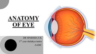
The eye is a paired organ, the organ of vision. The eye is made up of various components, which enable it to receive light stimuli from the environment, and deliver this stimuli to the brain in the form of an electrical signal. Vision involves all components of the eye.
DEFINITION:The human eye is a sensory organ,that reacts to visible light and allow to use visual information for various purposes including seeing things,keeping our balance and circadian rhythm.
STRUCTURE
The eye is contained within the bony orbit of the head. The bony orbit is a cavity, comprising parts of the lacrimal bone (includes fossa for nasolacrimal duct) and the maxilla (includes caudal foramen of infraorbital canal). It is continuous with the temporal bone and the pterygopalatine fossa caudally. It is situated in the orbital cavity and is supplied by 2nd cranial nerve.
The Atlas of the eye is a B.sc. degree research
It contains three parts:
- Anatomy & Physiology of the eye
- Pathology & errors in the eye
- Photography of the eye
enjoy it!
Anatomy of Eye by radhika kulvi, M.Sc nursingRadhika kulvi
The eye is a paired organ, the organ of vision. The eye is made up of various components, which enable it to receive light stimuli from the environment, and deliver this stimuli to the brain in the form of an electrical signal. Vision involves all components of the eye.
DEFINITION:The human eye is a sensory organ,that reacts to visible light and allow to use visual information for various purposes including seeing things,keeping our balance and circadian rhythm.
STRUCTURE
The eye is contained within the bony orbit of the head. The bony orbit is a cavity, comprising parts of the lacrimal bone (includes fossa for nasolacrimal duct) and the maxilla (includes caudal foramen of infraorbital canal). It is continuous with the temporal bone and the pterygopalatine fossa caudally. It is situated in the orbital cavity and is supplied by 2nd cranial nerve.
The Atlas of the eye is a B.sc. degree research
It contains three parts:
- Anatomy & Physiology of the eye
- Pathology & errors in the eye
- Photography of the eye
enjoy it!
these slide are modified or upgraded from the slid belonging to this website.i had added some of the content.hope that it will be more helpful to you all.
This lecture includes anatomy and Physiology of Cornea, if u like it kindly share it with colleagues and like it. I will share more lectures related to eye anatomy and optometry.
Thank You.
marna ppt includes the introduction, defination3, importance, types, vidda lakshana, pramana. modern co-relation of marmabhgata with shock, marma chiktsa point with acupressure
These lecture slides, by Dr Sidra Arshad, offer a quick overview of physiological basis of a normal electrocardiogram.
Learning objectives:
1. Define an electrocardiogram (ECG) and electrocardiography
2. Describe how dipoles generated by the heart produce the waveforms of the ECG
3. Describe the components of a normal electrocardiogram of a typical bipolar leads (limb II)
4. Differentiate between intervals and segments
5. Enlist some common indications for obtaining an ECG
Study Resources:
1. Chapter 11, Guyton and Hall Textbook of Medical Physiology, 14th edition
2. Chapter 9, Human Physiology - From Cells to Systems, Lauralee Sherwood, 9th edition
3. Chapter 29, Ganong’s Review of Medical Physiology, 26th edition
4. Electrocardiogram, StatPearls - https://www.ncbi.nlm.nih.gov/books/NBK549803/
5. ECG in Medical Practice by ABM Abdullah, 4th edition
6. ECG Basics, http://www.nataliescasebook.com/tag/e-c-g-basics
Prix Galien International 2024 Forum ProgramLevi Shapiro
June 20, 2024, Prix Galien International and Jerusalem Ethics Forum in ROME. Detailed agenda including panels:
- ADVANCES IN CARDIOLOGY: A NEW PARADIGM IS COMING
- WOMEN’S HEALTH: FERTILITY PRESERVATION
- WHAT’S NEW IN THE TREATMENT OF INFECTIOUS,
ONCOLOGICAL AND INFLAMMATORY SKIN DISEASES?
- ARTIFICIAL INTELLIGENCE AND ETHICS
- GENE THERAPY
- BEYOND BORDERS: GLOBAL INITIATIVES FOR DEMOCRATIZING LIFE SCIENCE TECHNOLOGIES AND PROMOTING ACCESS TO HEALTHCARE
- ETHICAL CHALLENGES IN LIFE SCIENCES
- Prix Galien International Awards Ceremony
Title: Sense of Smell
Presenter: Dr. Faiza, Assistant Professor of Physiology
Qualifications:
MBBS (Best Graduate, AIMC Lahore)
FCPS Physiology
ICMT, CHPE, DHPE (STMU)
MPH (GC University, Faisalabad)
MBA (Virtual University of Pakistan)
Learning Objectives:
Describe the primary categories of smells and the concept of odor blindness.
Explain the structure and location of the olfactory membrane and mucosa, including the types and roles of cells involved in olfaction.
Describe the pathway and mechanisms of olfactory signal transmission from the olfactory receptors to the brain.
Illustrate the biochemical cascade triggered by odorant binding to olfactory receptors, including the role of G-proteins and second messengers in generating an action potential.
Identify different types of olfactory disorders such as anosmia, hyposmia, hyperosmia, and dysosmia, including their potential causes.
Key Topics:
Olfactory Genes:
3% of the human genome accounts for olfactory genes.
400 genes for odorant receptors.
Olfactory Membrane:
Located in the superior part of the nasal cavity.
Medially: Folds downward along the superior septum.
Laterally: Folds over the superior turbinate and upper surface of the middle turbinate.
Total surface area: 5-10 square centimeters.
Olfactory Mucosa:
Olfactory Cells: Bipolar nerve cells derived from the CNS (100 million), with 4-25 olfactory cilia per cell.
Sustentacular Cells: Produce mucus and maintain ionic and molecular environment.
Basal Cells: Replace worn-out olfactory cells with an average lifespan of 1-2 months.
Bowman’s Gland: Secretes mucus.
Stimulation of Olfactory Cells:
Odorant dissolves in mucus and attaches to receptors on olfactory cilia.
Involves a cascade effect through G-proteins and second messengers, leading to depolarization and action potential generation in the olfactory nerve.
Quality of a Good Odorant:
Small (3-20 Carbon atoms), volatile, water-soluble, and lipid-soluble.
Facilitated by odorant-binding proteins in mucus.
Membrane Potential and Action Potential:
Resting membrane potential: -55mV.
Action potential frequency in the olfactory nerve increases with odorant strength.
Adaptation Towards the Sense of Smell:
Rapid adaptation within the first second, with further slow adaptation.
Psychological adaptation greater than receptor adaptation, involving feedback inhibition from the central nervous system.
Primary Sensations of Smell:
Camphoraceous, Musky, Floral, Pepperminty, Ethereal, Pungent, Putrid.
Odor Detection Threshold:
Examples: Hydrogen sulfide (0.0005 ppm), Methyl-mercaptan (0.002 ppm).
Some toxic substances are odorless at lethal concentrations.
Characteristics of Smell:
Odor blindness for single substances due to lack of appropriate receptor protein.
Behavioral and emotional influences of smell.
Transmission of Olfactory Signals:
From olfactory cells to glomeruli in the olfactory bulb, involving lateral inhibition.
Primitive, less old, and new olfactory systems with different path
- Video recording of this lecture in English language: https://youtu.be/lK81BzxMqdo
- Video recording of this lecture in Arabic language: https://youtu.be/Ve4P0COk9OI
- Link to download the book free: https://nephrotube.blogspot.com/p/nephrotube-nephrology-books.html
- Link to NephroTube website: www.NephroTube.com
- Link to NephroTube social media accounts: https://nephrotube.blogspot.com/p/join-nephrotube-on-social-media.html
Pulmonary Thromboembolism - etilogy, types, medical- Surgical and nursing man...VarunMahajani
Disruption of blood supply to lung alveoli due to blockage of one or more pulmonary blood vessels is called as Pulmonary thromboembolism. In this presentation we will discuss its causes, types and its management in depth.
Flu Vaccine Alert in Bangalore Karnatakaaddon Scans
As flu season approaches, health officials in Bangalore, Karnataka, are urging residents to get their flu vaccinations. The seasonal flu, while common, can lead to severe health complications, particularly for vulnerable populations such as young children, the elderly, and those with underlying health conditions.
Dr. Vidisha Kumari, a leading epidemiologist in Bangalore, emphasizes the importance of getting vaccinated. "The flu vaccine is our best defense against the influenza virus. It not only protects individuals but also helps prevent the spread of the virus in our communities," he says.
This year, the flu season is expected to coincide with a potential increase in other respiratory illnesses. The Karnataka Health Department has launched an awareness campaign highlighting the significance of flu vaccinations. They have set up multiple vaccination centers across Bangalore, making it convenient for residents to receive their shots.
To encourage widespread vaccination, the government is also collaborating with local schools, workplaces, and community centers to facilitate vaccination drives. Special attention is being given to ensuring that the vaccine is accessible to all, including marginalized communities who may have limited access to healthcare.
Residents are reminded that the flu vaccine is safe and effective. Common side effects are mild and may include soreness at the injection site, mild fever, or muscle aches. These side effects are generally short-lived and far less severe than the flu itself.
Healthcare providers are also stressing the importance of continuing COVID-19 precautions. Wearing masks, practicing good hand hygiene, and maintaining social distancing are still crucial, especially in crowded places.
Protect yourself and your loved ones by getting vaccinated. Together, we can help keep Bangalore healthy and safe this flu season. For more information on vaccination centers and schedules, residents can visit the Karnataka Health Department’s official website or follow their social media pages.
Stay informed, stay safe, and get your flu shot today!
HOT NEW PRODUCT! BIG SALES FAST SHIPPING NOW FROM CHINA!! EU KU DB BK substit...GL Anaacs
Contact us if you are interested:
Email / Skype : kefaya1771@gmail.com
Threema: PXHY5PDH
New BATCH Ku !!! MUCH IN DEMAND FAST SALE EVERY BATCH HAPPY GOOD EFFECT BIG BATCH !
Contact me on Threema or skype to start big business!!
Hot-sale products:
NEW HOT EUTYLONE WHITE CRYSTAL!!
5cl-adba precursor (semi finished )
5cl-adba raw materials
ADBB precursor (semi finished )
ADBB raw materials
APVP powder
5fadb/4f-adb
Jwh018 / Jwh210
Eutylone crystal
Protonitazene (hydrochloride) CAS: 119276-01-6
Flubrotizolam CAS: 57801-95-3
Metonitazene CAS: 14680-51-4
Payment terms: Western Union,MoneyGram,Bitcoin or USDT.
Deliver Time: Usually 7-15days
Shipping method: FedEx, TNT, DHL,UPS etc.Our deliveries are 100% safe, fast, reliable and discreet.
Samples will be sent for your evaluation!If you are interested in, please contact me, let's talk details.
We specializes in exporting high quality Research chemical, medical intermediate, Pharmaceutical chemicals and so on. Products are exported to USA, Canada, France, Korea, Japan,Russia, Southeast Asia and other countries.
8. Lens capsule
It is a thin, transparent, hyaline
membrane surrounding the lens
which is thicker over the anterior than
the posterior surface.
The lens capsule is thickest at pre-
equator regions (14 m)
thinnest at the posterior pole (3 m). 8
10. Anterior epithelium
It is a single layer of cuboidal
cells which lies deep to the
anterior capsule.
In the equatorial region these
cells become columnar
There is no posterior
epithelium,
10
13. Lens fibres
The epithelial cells elongate to form
lens fibres
Mature lens fibres are cells which have
lost their nuclei. As the lens fibres are
formed throughout the life, these are
arranged compactly as
A. nucleus
B. cortex
13
14. Nucleus
It is the central part containing the oldest fibres.
It consists of different zones,
In the beam of slit-lamp these are seen as zones of discontinuity.
14
15. Depending upon the period of development, the different zones
of the lens nucleus include
It is the innermost part of
nucleus which
corresponds to the lens
upto first 3 months of
gestation. It consists of
primary lens fibres which
are formed by elongation
of the cells of posterior
wall of lens vesicle
It lies around the
embryonic nucleus and
corresponds to the lens
from 3 months of
gestation till birth. Its
fibres meet around sutures
which are anteriorly Y-
shaped and posteriorly
inverted Y-shaped
Embryonic nucleus Fetal nucleus
corresponds to the lens
from birth to puberty
Infantile nucleus
15
Adult nucleus
lens fibres formed after
puberty to rest of the life
18. Suspensory ligaments of lens
Suspensory ligaments of lens
(Zonules of Zinn), also called as
ciliary zonules,
Consist essentially of a series of
fibres passing from ciliary body to
the lens.
These hold the lens in position
and enable the ciliary muscle to act
on it.
18
21. Lens transparency
Avascularity of the lens.
Characteristic of lens fibre:
– Tightly-packed nature of lens cells,
– Narrow lens fibre membranes,
– Loss of organelles
lens proteins.
Lens capsule: Semipermeable character.
Pump mechanism of lens fibre membranes that regulate the electrolyte and water
balance in the lens, maintaining relative dehydration
Auto-oxidation and high concentration of reduced glutathione in the lens maintains
the lens proteins in a reduced state and ensures the integrity of the cell membrane pump
21
23. CONGENITALANOMALIES OF LENS
a notch in the lower
quadrant of the equator
cone-shaped elevation
of the anterior pole
Coloboma of lens Lenticonus
lens is spherical in
shape (instead of
normal biconvex) and
small in size
Microspherophakia
23
25. Inert, transparent, jelly-like structure
that fills the posterior four-fifth of the
cavity of eyeball.
Spherical posteriorly and has a cup-
shaped depression (patellar fossa)
anteriorly.
It conforms to the contour of retina
behind and lens infront.
4 ml in volume
4 gm in weight.
25
26. Contains more than 98% of water
It has viscosity more than 2-3 times than that of water.
Refractive index 1.33
The word “vitreous” is derived from a Latin word “vitrum” which
means glass
26
27. Functions
It is a hydrophilic gel that mainly serves the optical functions.
It mechanically stabilizes the volume of the globe
Is a pathway for nutrients to reach the lens and retina.
27
28. Structure
The normal youthful vitreous gel is composed of a network of
randomly-oriented collagen fibrils interspersed with numerous
spheroidal macromolecules of hyaluronic acid.
The collapse of this structure with age or otherwise leads to
conversion of the gel into sol.
two parts: the cortex and
the medulla or nucleus (the main vitreous body)
28
29. Cortical vitreous
lies adjacent to the retina posteriorly
ciliary body, zonules and lens anteriorly.
The density of collagen fibrils is greater in this peripheral part.
The condensation of these fibrils form a false anatomic membrane
which is called as anterior hyaloid membrane anterior to ora serrata
and posterior hyaloid membrane posterior to ora.
29
30. Medulla or nucleus
has a less dense fibrillar structure and is a true
biological gel.
here where liquefactions of the vitreous gel start first.
Microscopically the vitreous body is homogenous, but
exhibits wavy lines as of watered silk in the slit-lamp
beams.
Running down the centre of the vitreous body from the
optic disc to the posterior pole of the lens
30
31. Attachments
vitreous base-vitreous about 4 mm across the
ora serrata
margins of the optic disc, foveal region and
back of the crystalline lens-hyaloidocapsular
ligament of Wiegert
31
32. Clinical Notes
Detachment of the Vitreous
the attachment to the retina is weak, and in
pathologic conditions the vitreous is easily
detached.
32
33. Accumulation of Fluid in the Retrolenticular
Space
retrolenticular space
Blood or exudates can accumulate in this space
in pathologic conditions.
Senile Changes in the Vitreous
degeneration and liquefaction.
vitreous detachment
predispose to retinal detachment.
33
34. Refference
Comprehensive Ophthalmology by AK Khurana
BD Chaurasia’s Applied Dissection and Clinical Head and
Neck 8th edition
Clinical Anatomy of the Eye by Richard S. Snell 2nd edition
www.eoptha.com
34