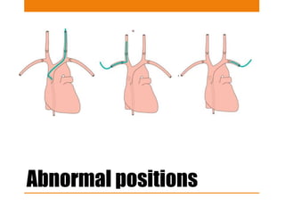
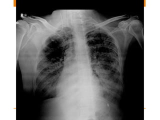
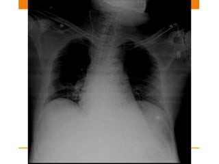
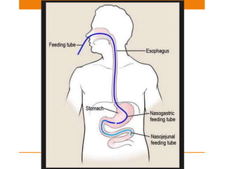
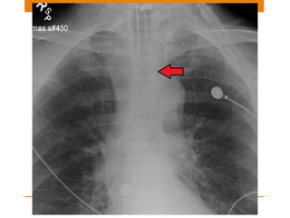
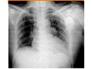
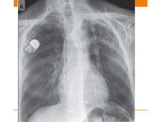
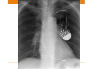
CHEST TUBES AND LINES
DR.SAHANA.S
COMMON LINES SEEN ON CXRY
• Central venous catheters
• Pulmonary artery catheter – Swartz Ganz catheter
• Nasogastric Tube
• Nasoenteric Tube
• Intercostal drain
• Endotracheal tube
• Tracheostomy tube
• Oesophageal Doppler tube
CENTRAL VENOUS CATHETER
Function
• To monitor right atrial pressure
• Fluid infusion/ nutrition
• Drug administration
Position
• SVC or Brachiocephalic vein
Normally placed CVC
Abnormal positions
Misplaced lines causes…
• Tip too High- Inaccurate right atrium monitoring
pressures
• Tip too low – In Right Atrium can cause arrhythmia and
Cardiac Tamponade
• Vessel wall perforation
• Infusion of fluid into mediastinum/pleural space
• Pneumothorax
Swartz Ganz Catheter
• Also known as pulmonary arterial line commonly
• To assess left atrial pressure and cardiac output
• Very important in ICU patients to distinguish between
cardiac and non cardiac pulmonary edema
• Now a days not much in use in regard with patient
outcome
Misplaced lines causes…
• Distally placed tip will cause Pulmonary infarction
• Proximallly placed in right ventricle -- Arrythmia
NASOGASTRIC TUBE
• Side holes of the tube in the NG tube extend around 5 cm
so the tube should be atleast 10 cm beyond the OG
junction
• Uses:
1. Gastric decompression
2. Gastric aspiration
3. Nutrition
Misplaced Tubes causes..
• If it enters trachea – ARDS
• If still in oesophagus – Can cause regurgitation
Nasoenteric Tube
• These feeding tubes are thin plastic catheters with a
mercury / tungsten filled tip.
• The optimum position for the tip is distal to the pyloric
sphincter.
Misplaced tube causes..
• The tube is very thin so caution should be taken while
inserting the tube
• It may coil itself pharynx , Oesophagus or stomach
• It may enter trachea or right main bronchus
Endotracheal tube
• USE: Assisted ventilation
• The tip of an ETT will be in a satisfactory position if it
approximates to the level of the medial ends of the
clavicles
• Ideal position is 5–7 cm above an adult’s carina when the
head is held in the neutral position.
• And if carina is not visible in 95% people it is situated
at T5-T7 vertebra
Position changes
• The ETT can move up or down
• Flex the neck and the tip can move 1.9 cm I,e ~2cm
downwards.
• Extend the neck and it can move 1.9 cm I,e ~2cm
upwards.
• Rotate the neck and it can move 0.7 cm upwards.
Malposition
• Tip of right main bronchus:
1. Left lung collapse
2. Right upper lobe collapse
3. Right lung overdistension/pneumothorax
• Tip in Oesophagus:
1. ETT lateral to tracheal air shadow
2. Oesophagus distended with air
3. Stomach distended with air
Tracheostomy tube
• Tracheostomy tube lies parallel to the long axis of the
trachea
• The tip lies several centimeters well above carina
• The inflated cuff should not extend lateral walls of
trachea
Misplaced position
• Mediastinum widening
• air in the mediastinum – leak is occurring
Oesophageal Doppler probe
• Position: Mid esophagus
• Use: To monitor cardiac output via measurement of blood
velocity in the descending aorta
Intercostal Drain
• Used in pneumothorax
• Position:
1. if its placed superiorly I,e towards apex – Pneumothorax
2. If tis placed inferiorly I,e towards cardiophrenic border
– Pleural drainage
• Correct position:
On entering the pleural cavity - gush of air is felt
• Misplacement - On connecting it with the bag – if bubble
is present then its in the lung parenchyma
Pacemaker
SINGLE OR DUAL CHAMBER
Single chamber –nowadays used less frequently
• used for atrial or ventricular dysarrythmia
• Atrial – positioned in right atrial appendage
• Ventricular – Electrode placed against myocardium at
apex of right ventricle
Pacemaker
Dual pacemaker
• Attempts to synchronize atrial and ventricular system
• One electrode @ Right atrium
• Other electrode @ apex of right ventricle
Sometimes a third lead is also noted
• Third –coronary sinus <Biventricular pacing>
Misplaced Leads
Complications
• Myocardial penetration – if electrode tip is within 3mm
of epicardial fat
• Myocardial perforation – if tip is in epicardial fat
• Pneumothorax
• Pleural effusion
Syndrome
• Twiddlers
• Subclavian crush
IABP
• Position – Approximately 2cm away from left subclavian
artery and counter pulsates
USES:
• Unstable angina
• Myocardial infarction
• Cardiopulmonary bypass

Chest tubes and lines

  • 1.
    CHEST TUBES ANDLINES DR.SAHANA.S
  • 2.
    COMMON LINES SEENON CXRY • Central venous catheters • Pulmonary artery catheter – Swartz Ganz catheter • Nasogastric Tube • Nasoenteric Tube • Intercostal drain • Endotracheal tube • Tracheostomy tube • Oesophageal Doppler tube
  • 3.
    CENTRAL VENOUS CATHETER Function •To monitor right atrial pressure • Fluid infusion/ nutrition • Drug administration Position • SVC or Brachiocephalic vein
  • 4.
  • 5.
  • 8.
    Misplaced lines causes… •Tip too High- Inaccurate right atrium monitoring pressures • Tip too low – In Right Atrium can cause arrhythmia and Cardiac Tamponade • Vessel wall perforation • Infusion of fluid into mediastinum/pleural space • Pneumothorax
  • 11.
    Swartz Ganz Catheter •Also known as pulmonary arterial line commonly • To assess left atrial pressure and cardiac output • Very important in ICU patients to distinguish between cardiac and non cardiac pulmonary edema • Now a days not much in use in regard with patient outcome
  • 13.
    Misplaced lines causes… •Distally placed tip will cause Pulmonary infarction • Proximallly placed in right ventricle -- Arrythmia
  • 14.
    NASOGASTRIC TUBE • Sideholes of the tube in the NG tube extend around 5 cm so the tube should be atleast 10 cm beyond the OG junction • Uses: 1. Gastric decompression 2. Gastric aspiration 3. Nutrition
  • 17.
    Misplaced Tubes causes.. •If it enters trachea – ARDS • If still in oesophagus – Can cause regurgitation
  • 18.
    Nasoenteric Tube • Thesefeeding tubes are thin plastic catheters with a mercury / tungsten filled tip. • The optimum position for the tip is distal to the pyloric sphincter.
  • 20.
    Misplaced tube causes.. •The tube is very thin so caution should be taken while inserting the tube • It may coil itself pharynx , Oesophagus or stomach • It may enter trachea or right main bronchus
  • 21.
    Endotracheal tube • USE:Assisted ventilation • The tip of an ETT will be in a satisfactory position if it approximates to the level of the medial ends of the clavicles • Ideal position is 5–7 cm above an adult’s carina when the head is held in the neutral position. • And if carina is not visible in 95% people it is situated at T5-T7 vertebra
  • 22.
    Position changes • TheETT can move up or down • Flex the neck and the tip can move 1.9 cm I,e ~2cm downwards. • Extend the neck and it can move 1.9 cm I,e ~2cm upwards. • Rotate the neck and it can move 0.7 cm upwards.
  • 26.
    Malposition • Tip ofright main bronchus: 1. Left lung collapse 2. Right upper lobe collapse 3. Right lung overdistension/pneumothorax • Tip in Oesophagus: 1. ETT lateral to tracheal air shadow 2. Oesophagus distended with air 3. Stomach distended with air
  • 27.
    Tracheostomy tube • Tracheostomytube lies parallel to the long axis of the trachea • The tip lies several centimeters well above carina • The inflated cuff should not extend lateral walls of trachea
  • 28.
    Misplaced position • Mediastinumwidening • air in the mediastinum – leak is occurring
  • 30.
    Oesophageal Doppler probe •Position: Mid esophagus • Use: To monitor cardiac output via measurement of blood velocity in the descending aorta
  • 32.
    Intercostal Drain • Usedin pneumothorax • Position: 1. if its placed superiorly I,e towards apex – Pneumothorax 2. If tis placed inferiorly I,e towards cardiophrenic border – Pleural drainage • Correct position: On entering the pleural cavity - gush of air is felt • Misplacement - On connecting it with the bag – if bubble is present then its in the lung parenchyma
  • 36.
    Pacemaker SINGLE OR DUALCHAMBER Single chamber –nowadays used less frequently • used for atrial or ventricular dysarrythmia • Atrial – positioned in right atrial appendage • Ventricular – Electrode placed against myocardium at apex of right ventricle
  • 37.
    Pacemaker Dual pacemaker • Attemptsto synchronize atrial and ventricular system • One electrode @ Right atrium • Other electrode @ apex of right ventricle Sometimes a third lead is also noted • Third –coronary sinus <Biventricular pacing>
  • 42.
  • 45.
    Complications • Myocardial penetration– if electrode tip is within 3mm of epicardial fat • Myocardial perforation – if tip is in epicardial fat • Pneumothorax • Pleural effusion
  • 46.
  • 48.
    IABP • Position –Approximately 2cm away from left subclavian artery and counter pulsates USES: • Unstable angina • Myocardial infarction • Cardiopulmonary bypass

Editor's Notes

  • #5 Svc commences at right first anterior intercostal space
  • #13 Postioned shud nt project 2cm beyond the mediastinal outline
  • #27 Right bronchus T5 Left T6